. 2014 Aug:462-463:363-76.
doi: 10.1016/j.virol.2014.06.006.

Eliciting neutralizing antibodies with gp120 outer domain constructs based on M-group consensus sequence

Affiliations

Eliciting neutralizing antibodies with gp120 outer domain constructs based on M-group consensus sequence

Yali Qin et al. Virology. 2014 Aug.

Abstract

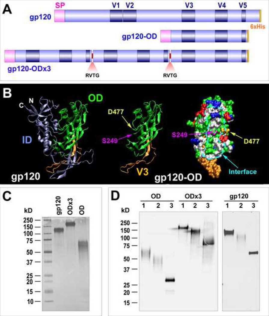
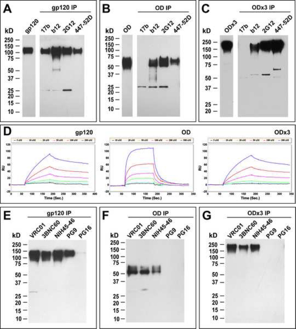
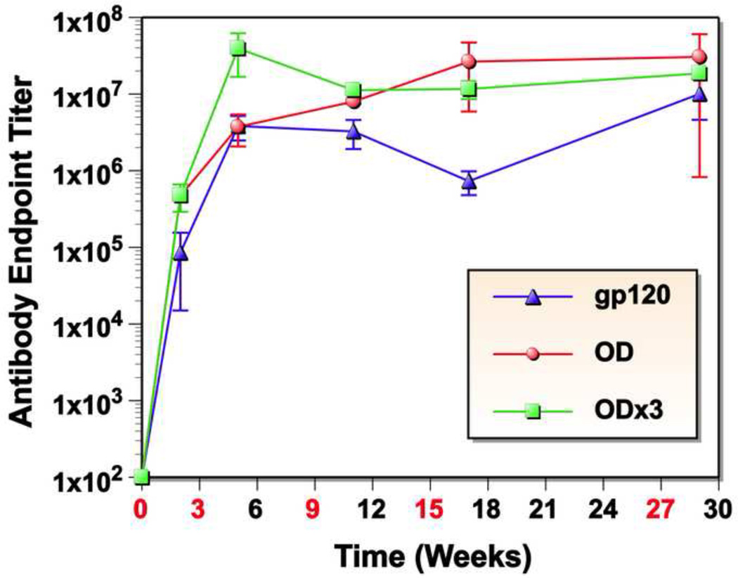
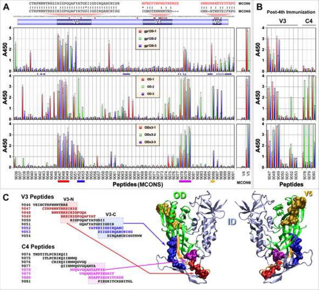
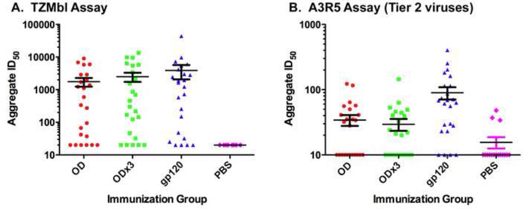
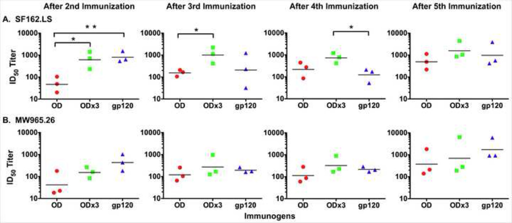
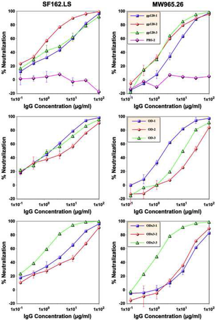
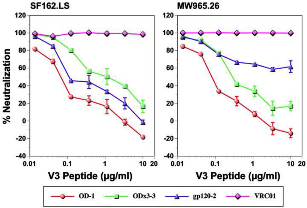
One strategy being evaluated for HIV-1 vaccine development is focusing immune responses towards neutralizing epitopes on the gp120 outer domain (OD) by removing the immunodominant, but non-neutralizing, inner domain. Previous OD constructs have not elicited strong neutralizing antibodies (nAbs). We constructed two immunogens, a monomeric gp120-OD and a trimeric gp120-OD×3, based on an M group consensus sequence (MCON6). Their biochemical and immunological properties were compared with intact gp120. Results indicated better preservation of critical neutralizing epitopes on gp120-OD×3. In contrast to previous studies, our immunogens induced potent, cross-reactive nAbs in rabbits. Although nAbs primarily targeted Tier 1 viruses, they exhibited significant breadth. Epitope mapping analyses indicated that nAbs primarily targeted conserved V3 loop elements. Although the potency and breadth of nAbs were similar for all three immunogens, nAb induction kinetics indicated that gp120-OD×3 was superior to gp120-OD, suggesting that gp120-OD×3 is a promising prototype for further gp120 OD-based immunogen development.

Keywords: HIV-1; Neutralizing antibody; Outer domain; V3; Vaccine; gp120.


Figures

Fig. 1
Fig. 1
Schematic diagram, expression and purification of gp120, gp120-OD and -OD×3. (A) A schematic diagram of three immunogens. All proteins contain N-terminal signal peptide (SP) and C-terminal 6xHis tag. For gp120-OD×3, the OD segments are connected by a four amino acid linker (RVTG). (B) Structural representations of gp120 and gp120-OD. The OD and ID are shown in green and ice blue, respectively. The V3 loop is shown in orange. The N- and C-terminal ends of gp120-OD (S249 and D477, respectively), as well as the interface region between the ID and OD are indicated. The crystal structure of trimeric gp140 SOSIP (4NCO) was used (Khayat et al., 2013). (C) Silver stain of purified proteins expressed in HEK293 cells. (D) Western Blot analyses of deglycosylated proteins. Lane 1: untreated; lane 2: EndoH; lane 3: PNGaseF.
Fig. 2
Fig. 2
Evaluation of antigenic properties. Immunoprecipitation analyses of gp120 (A), gp120-OD (B) and gp120-OD×3 (C) with mAbs 17b, b12, 2G12 and 447-52D. SPR analyses of interactions between IgG b12 and gp120, gp120-OD and -OD×3. Immunoprecipitation analyses of gp120 (E), gp120-OD (F) and gp120-OD×3 (G) with more recently isolated bnAbs VRC01, 3BNC60, NIH45-46, PG9 and PG16.
Fig. 3
Fig. 3
Antibody endpoint titers determined by ELISA. Sera obtained before or 2 weeks after each immunization were monitored for autologous antigen-specific antibodies by ELISA. Data are presented as average endpoint titers of three rabbits with standard deviation (SD).
Fig. 4
Fig. 4
Comparison of immunogenic linear epitopes by ELISA using overlapping peptides. (A) ELISA using immune sera after the fifth immunization. Peptide numbers represent catalog numbers from the NIH AIDS RRRP. On the top of the figure shows a schematic diagram of gp120-OD, MCONS envelope sequence, identification of amino acid residues different from MCON6 (small red square), and sequence of MCON6 peptides covering the V4 and V5 region (red text). Blue circles below gp120 ELISA data indicates amino acid residues that makes contact with CD4 and/or VRC01. (B) ELISA of select peptides from the V3 and C4 regions with immune sera after the fourth immunization. (C) Plotting of four immunogenic epitopes onto a crystal structure of trimeric gp140 SOSIP (4NCO). Two segments within V3 loop (red and blue), one in C4 (magenta) and one in V5 (gold). Only the likely cores of the V3 and C4 epitopes (boxed in) are plotted on the crystal structure.
Fig. 5
Fig. 5
Aggregate analyses of ID50 values of neutralizing activity. (A) Data from neutralization assay using TZM-bl cells. Data shown in Table 1 are presented. (B). Neutralization assay against Tier 2 pseudoviruses using A3R5 cells. Neutralization assays were done using immune sera collected after the fifth immunization.
Fig. 6
Fig. 6
Kinetic analyses of neutralizing antibodies induced by gp120, gp120-OD, and -OD×3. (A) Neutralizing activity against clade B SF162.LS with immune sera collected after the second, third, fourth, and fifth immunization. The geometric mean titer for each group of sera is shown as a horizontal bar. Statistical analysis was done with Prism GraphPad 5 using a two-tailed t-test. **P<0.01; *0.01
Fig. 7
Fig. 7
Neutralizing activity of purified serum IgG. Serial dilutions of purified IgG from gp120, gp120-OD, and -OD×3 immunized rabbits and negative control (PBS only) were tested for neutralizing activity against the SF162.LS and MW965.26 viruses.
Fig. 8
Fig. 8
Inhibition of neutralizing activity by V3 loop peptide. Neutralizing activity of purified IgG (50 µg/ml) from one rabbit from each immunogen group was evaluated in the absence or the presence of various concentrations of MCON6 V3 peptide (TRPNNNTRKSIHIGPGQAFYATGEIIGDIRQAH). VRC01 bnAb (5 µg/ml) that targets the CD4BS was used as a negative control.

References

    1. Barbas CF, Björling E, Chiodi F, Dunlop N, Cababa D, Jones TM, Zebedee SL, Persson MA, Nara PL, Norrby E. Recombinant human Fab fragments neutralize human type 1 immunodeficiency virus in vitro. Proc Natl Acad Sci USA. 1992;89:9339–9343. - PMC - PubMed
    1. Bhattacharyya S, Rajan RE, Swarupa Y, Rathore U, Verma A, Udaykumar R, Varadarajan R. Design of a non-glycosylated outer domain-derived HIV-1 gp120 immunogen that binds to CD4 and induces neutralizing antibodies. J Biol Chem. 2010;285:27100–27110. - PMC - PubMed
    1. Blattner C, Lee JH, Sliepen K, Derking R, Falkowska E, la Peña de AT, Cupo A, Julien J-P, van Gils M, Lee PS, Peng W, Paulson JC, Poignard P, Burton DR, Moore JP, Sanders RW, Wilson IA, Ward AB. Structural Delineation of a Quaternary, Cleavage-Dependent Epitope at the gp41-gp120 Interface on Intact HIV-1 Env Trimers. Immunity. 2014;40:669–680. - PMC - PubMed
    1. Buchacher A, Predl R, Strutzenberger K, Steinfellner W, Trkola A, Purtscher M, Gruber G, Tauer C, Steindl F, Jungbauer A. Generation of human monoclonal antibodies against HIV-1 proteins; electrofusion and Epstein-Barr virus transformation for peripheral blood lymphocyte immortalization. AIDS Res Hum Retroviruses. 1994;10:359–369. - PubMed
    1. Burton DR, Barbas CF, Persson MA, Koenig S, Chanock RM, Lerner RA. A large array of human monoclonal antibodies to type 1 human immunodeficiency virus from combinatorial libraries of asymptomatic seropositive individuals. Proc Natl Acad Sci USA. 1991;88:10134–10137. - PMC - PubMed

Publication types