. 2014 Nov;11(11):2064-72.
doi: 10.1016/j.hrthm.2014.07.022. Epub 2014 Jul 18.

Cellular mechanism of premature ventricular contraction-induced cardiomyopathy

Affiliations

Cellular mechanism of premature ventricular contraction-induced cardiomyopathy

Yuhong Wang et al. Heart Rhythm. 2014 Nov.

Abstract

Background: Frequent premature ventricular contractions (PVCs) are associated with increased risk of sudden cardiac death and can cause secondary cardiomyopathy.

Objective: We sought to determine the mechanism(s) responsible for prolonged refractory period and left ventricular (LV) dysfunction demonstrated in our canine model of PVC-induced cardiomyopathy.

Methods: Single myocytes were isolated from LV free wall of PVC and control canines and used for patch-clamp recording, intracellular Ca(2+) measurements, and immunocytochemistry/confocal microscopy. LV tissues adjacent to the area of myocyte isolation were used for the immunoblot quantification of protein expression.

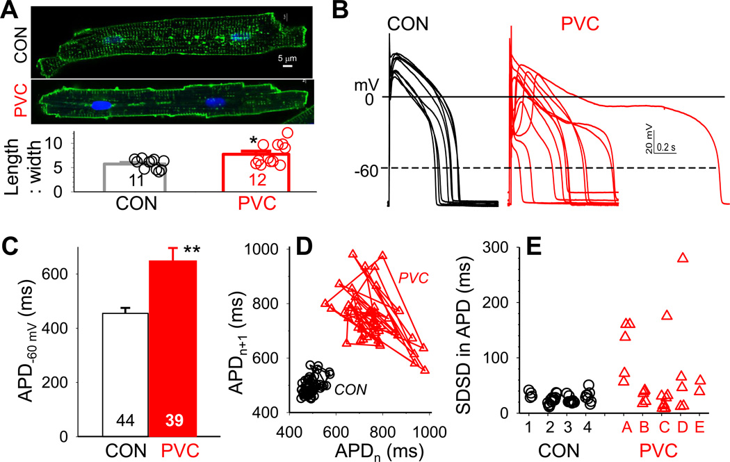
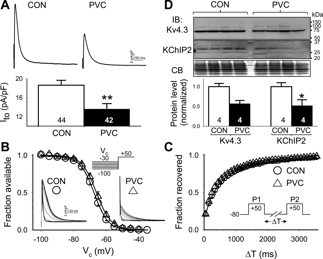
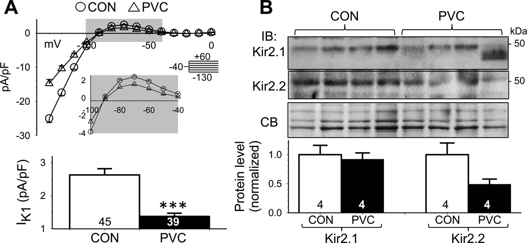
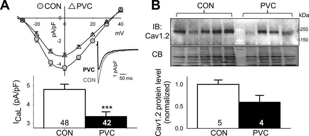
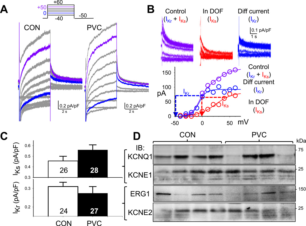
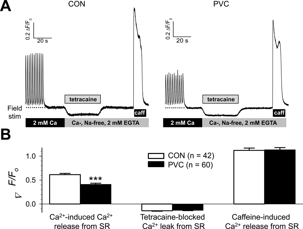
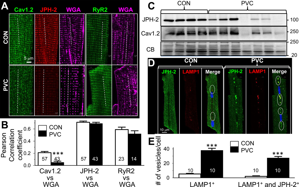
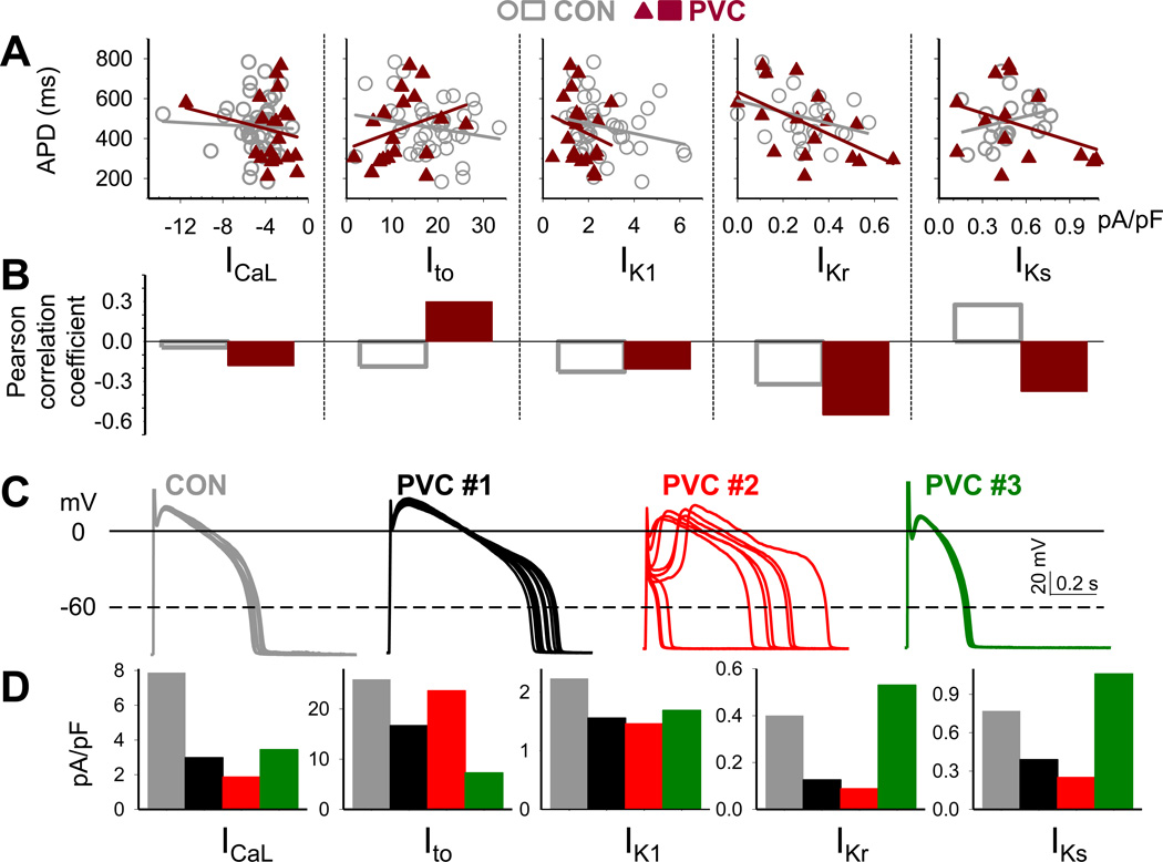
Results: In the PVC group, LV ejection fraction decreased from 57.6% ± 1.5% to 30.4% ± 3.1% after ≥4 months of ventricular bigeminy. Compared to control myocytes, PVC myocytes had decreased densities of both outward (transient outward current [Ito] and inward rectifier current [IK1]) and inward (L-type Ca current [ICaL]) currents, but no consistent changes in rapid or slow delayed rectifier currents. The reduction in Ito, IK1, and ICaL was accompanied by decreased protein levels of their channel subunits. The extent of reduction in Ito, IK1, and ICaL varied among PVC myocytes, creating marked heterogeneity in action potential configurations and durations. PVC myocytes showed impaired Ca-induced Ca release from the sarcoplasmic reticulum (SR), without increase in SR Ca leak or decrease in SR Ca store. This was accompanied by a decrease in dyad scaffolding protein, junctophilin-2, and loss of Cav1.2 registry with Ca-releasing channels (ryanodine receptor 2).

Conclusion: PVCs increase dispersion of action potential configuration/duration, a risk factor for sudden cardiac death, because of the heterogeneous reduction in Ito, IK1, and ICaL. The excitation-contraction coupling is impaired because of the decrease in ICaL and Cav1.2 misalignment with respect to ryanodine receptor 2.

Keywords: Cardiomyopathy; Electrical remodeling; Excitation-contraction coupling; Premature ventricular contraction.


Figures

Fig. 1
Fig. 1. PVC myocytes had prolonged action potential durations (APD) with exaggerated beat-tobeat variations
(A) Top: Images of CON and PVC myocytes with cell membrane/t-tubules stained by fluorescent WGA or di-8-ANEPPS. Bottom: Length:width ratios of CON and PVC myocytes. Data from individual myocytes shown as open circles; values of Mean±SE shown as histogram bars. (B) Superimposed AP traces recorded from ten CON and ten PVC myocytes. The cycle length (CL) was 1 s, except the PVC myocyte with markedly prolonged APD (CL = 2 s). (C) APDs quantified as time between upstroke and when membrane repolarized to −60 mV (APD−60 mV). (D) APDn+1 plotted against APDn for fifty consecutive action potentials recorded from a CON and a PVC myocyte (corresponding to data points marked by solid symbols in ‘D’). (E) Standard deviations of successive differences (SDSD) in APD. Data points from individual myocytes are grouped by the hearts they were isolated from (CON hearts 1–4, PVC hearts A–E). For this and the following figures, details of voltage-clamp protocols and data analysis are provided in on-line Detailed Methods. Numerical data values are listed in Tables S1–S3. Numbers of myocytes or samples analyzed are listed in histogram bars or insets. CON and PVC myocytes were isolated from 4 and 5 animals, respectively.
Fig. 2
Fig. 2. PVC myocytes had reduced transient outward (Ito) current density but unaltered Ito gating kinetics, accompanied by a downregulation of Ito channel subunits
(A) Top: Representative Ito traces recorded from CON and PVC myocytes. Bottom: peak Ito densities in CON and PVC myocytes. (B) Voltage-dependence of Ito inactivation. (C) Time course of recovery from inactivation (Ito restitution). (D) Immunoblot quantification of protein levels of pore-forming and auxiliary subunits of Ito channels (Kv4.3 and KChIP2). Top: Images of immunoblots and Coomassie blue stain of the gel (CB, as loading control). Size marker bands (in kDa) are shown on the right. Bottom: Densitometry quantification of Kv4.3 and KChIP2 protein levels.
Fig. 3
Fig. 3. PVC myocytes had reduced inward rectifier (IK1) current densities, accompanied by a downregulation of Kir2.2
(A) Top: Average steady-state current-voltage (I-V) relationships of CON and PVC myocytes. The enlarged view of gray shading area highlights the outward portion of IK1. Bottom: outward IK1 densities at −70 mV in CON and PVC myocytes. (B) Immunoblot quantification of IK1 channel subunits, Kir2.1 and Kir2.2, in LV samples. Data analysis and presentation are the same as Fig. 2D.
Fig. 4
Fig. 4. PVC myocytes had reduced peak L-type Ca (ICaL) current density, accompanied by a downregulation of pore-forming subunit (Cav1.2)
(A) Top: I–V of ICaL, with representative ICaL traces recorded at 0 mV from CON and PVC myocytes shown (inset). Bottom: peak ICaL densities at 0 mV in CON and PVC myocytes. (B) Immunoblot quantification of Cav1.2. Data analysis and presentation are the same as Fig. 2D.
Fig. 5
Fig. 5. No consistent or statistically significant differences in rapid and slow delayed rectifier currents (IKr and IKs) between PVC and CON myocytes
(A) Combined ‘IKr+IKs’ traces in a CON and a PVC myocyte, elicited by the diagrammed voltage clamp protocol. (B) Validation of IKr and IKs quantification. The rationale, experimental protocols and data analysis are provided in on-line Detailed Methods. (C) Comparison of IKs and IKr tail current densities between CON and PVC myocytes. (D) Immunoblot quantification of pore-forming and auxiliary subunits of IKs (KCNQ1 and KCNE1) and IKr (ERG1 and KCNE2). Densitometry quantification as described for Fig. 2D.
Fig. 6
Fig. 6. PVC myocytes had reduced amplitudes of Ca-induced Ca release from SR, but no significant change in either SR Ca leak or SR Ca store
(A) Representative fluorescence signals from fluo-4-loaded CON and PVC myocytes. (B) Data summary for CON and PVC myocytes. Details of experimental protocols and data analysis are provided in on-line Detailed Methods.
Fig. 7
Fig. 7. PVC myocytes showed dyad remodeling with disarray in Cav1.2 distribution
(A) Confocal images of Cav1.2, junctophilin-2 (JPH-2) immunofluorescence signals from the same myocytes, and RyR2 from 2 other myocytes. Alexa647-WGA signals served as a reference for t-tubule locations. Dotted lines depict how colocalization between WGA and Cav1.2, JPH-2 or RyR2 was analyzed (more in on-lineDetailed Methods). (B) Summary of Pearson correlation coefficients between WGA and Cav1.2 (left), JPH-2 (middle) or RyR2 (right) in CON and PVC myocytes (*** p < 0.001). (C) JPH-2 and Cav1.2 immunoblots of the same set of CON and PVC hearts. (D) JPH-2 and LAMP1 signals in a CON and a PVC myocytes. Open circles in the ‘Merge’ panel highlight JPH-2/LAMP1 colocalization. (E) Quantification of vesicles per cell that are positive for LAMP1 (LAMP1+) or both LAMP1+ and JPH-2+.
Fig. 8
Fig. 8. Correlation between action potential duration (APD), beat-to-beat variability of APD, and current densities of ICaL, Ito, IK1, IKr and IKs of individual CON and PVC myocytes
(A) Linear regression between APDs (quantified as described for Fig. 1) and current densities (quantified as described for Fig. 2 – 5). (B) Pearson correlation coefficient between APDs and current densities. (C) Exemplar action potentials of CON and PVC myocytes and (D) corresponding ICaL, Ito, IK1, IKr and IKs current densities, color-coded as dark gray (CON), black (PVC #1), red (PVC #2) and dark green (PVC #3).

Comment in

References

    1. Huizar JF, Kaszala K, Potfay J, et al. Left ventricular systolic dysfunction induced by ventricular ectopy. A novel model for premature ventricular contraction-induced cardiomyopathy. Circ Arrhythm Electrophysiol. 2011;4:543–549. - PMC - PubMed
    1. Liu X-S, Jiang M, Zhang M, Tang D, Higgins RSD, Tseng G-N. Electrical remodeling in a canine model of ischemic cardiomyopathy. Am J Physiol. 2007;292:H560–H571. - PubMed
    1. Heijman J, Zaza A, Johnson DM, Rudy Y, Peeters RLM, Volders PGA, Westra RL. Determinants of beat-to-beat variability of repolarization duration in the canine ventricular myocyte: a computational analysis. PLoS-One. 2013;9:e1003202. - PMC - PubMed
    1. Brennan M, Palaniswami M, Kamen P. Do existing measures of Poincare plot geometry reflect nonlinear features of heart rate variability? IEEE Transactions on Biomedical Engineering. 2001;48:1342–1347. - PubMed
    1. Greenstein JL, Wu R, Po S, Tomaselli GF, Winslow RL. Role of the calcium-independent transient outward current Ito1 in shaping action potential morphology and duration. Circ Res. 2000;87:1026–1033. - PubMed

Publication types