

Kappa and lambda light chain mRNA in situ hybridization compared to flow cytometry and immunohistochemistry in B cell lymphomas
- PMID: 25047073
- PMCID: PMC4223387
- DOI: 10.1186/1746-1596-9-144
Kappa and lambda light chain mRNA in situ hybridization compared to flow cytometry and immunohistochemistry in B cell lymphomas
Abstract
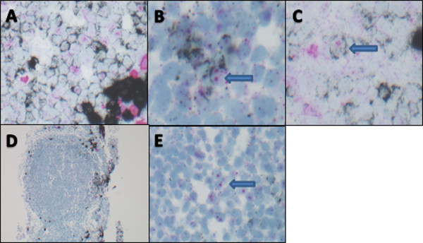
Background: Detection of B cell clonality is useful for assisting in the diagnosis of B cell lymphomas. Clonality assessment can be accomplished through evaluation of KAPPA and LAMBDA light chain expression. Currently, only slide based methods are available for the majority of patient biopsies and do not detect light chain protein or mRNA in many B-cell lymphomas. Herein we evaluated a new method, known as colorimetric in situ hybridization (CISH), with improved sensitivity and multiplexing capacity, for its usefulness in clonality detection in mature B cell malignancies.
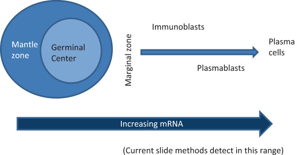
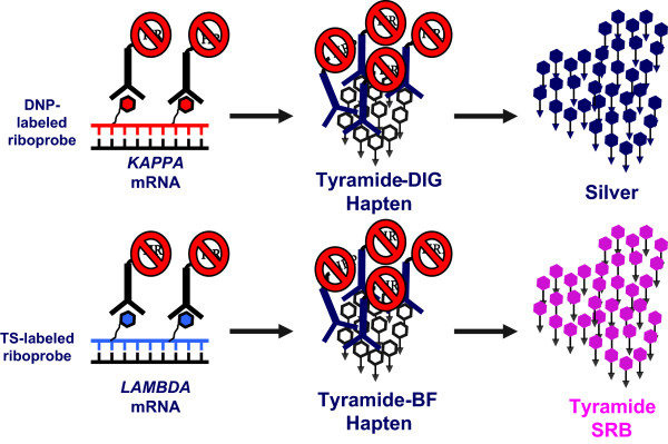
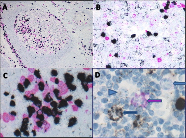
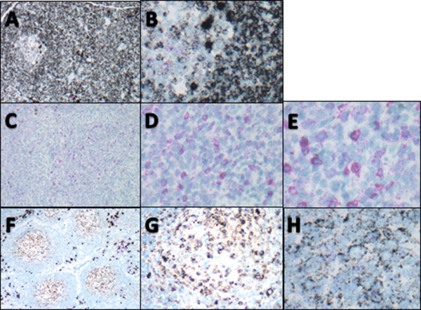
Methods: The KAPPA and LAMBDA ISH was performed on a Ventana Benchmark XT utilizing two color chromogenetic detection. The probes comprised 2 haptenated riboprobes each approximately 500 base pairs long directed against the conserved regions of either KAPPA or LAMBDA mRNA. The dual colors consisted of silver deposition (black) for KAPPA light chain and a novel (pink) chromogen for LAMBDA light chain. Following optimization, CISH allowed visualization of mRNA in benign B cells in reactive tissues including germinal center, mantle zone, and post-germinal center cells. We then identified 79 cases of B cell lymphoma with formalin-fixed paraffin-embedded (FFPE) biopsies including: follicular (36 cases), mantle cell (6 cases), marginal zone (12 cases), lymphoplasmacytic (6 cases), small lymphocytic (4 cases), and diffuse large B cell (15 cases), which were selected on the basis of either prior flow cytometry or immunohistochemistry (IHC) results to serve as the predicate, "gold standard," comparator.
Results: 39/79 (49.4%) cases were classified as KAPPA and 29/79 (36.7%) as LAMBDA light chain restricted; while 9/79 (11.3%) cases were classified as indeterminate. Of the 70 cases with KAPPA or LAMBDA light chain restricted CISH, 69/70 (98.6%) were concordant with the reference method, while 1/70 (1.4%) was discordant.
Conclusions: Optimized CISH detected lower levels of mRNA than can be visualized with current slide based methods, making clonality assessment in FFPE biopsies possible for mature B cell neoplasms. In this preliminary study, CISH was highly accurate compared to flow cytometry or IHC. CISH offers the possibility of wider applicability of light chain ISH and is likely to become a useful diagnostic tool.
Virtual slides: The virtual slide(s) for this article can be found here: http://www.diagnosticpathology.diagnomx.eu/vs/1430491067123856.
Figures
References
-
- Segal GH, Shick HE, Tubbs RR, Fishleder AJ, Stoler MH. In situ hybridization analysis of lymphoproliferative disorders. Assessment of clonality by immunoglobulin light-chain messenger RNA expression. Diagn Mol Pathol. 1994;3:170–177. - PubMed
-
- de Tute RM. Flow cytometry and its use in the diagnosis and management of mature lymphoid malignancies. Histopathology. 2011;58:90–105. - PubMed
-
- Tembhare PR, Yuan CM, Venzon D, Braylan R, Korde N, Manasanch E, Zuchlinsky D, Calvo K, Kurlander R, Bhutani M, Tageja N, Maric I, Mulquin M, Roschewski M, Kwok M, Liewehr D, Landgren O, Stetler-Stevenson M. Flow cytometric differentiation of abnormal and normal plasma cells in the bone marrow in patients with multiple myeloma and its precursor diseases. Leuk Res. 2014;38:371–376. - PMC - PubMed
-
- Beck RC, Tubbs RR, Hussein M, Pettay J, Hsi ED. Automated colorimetric in situ hybridization (CISH) detection of immunoglobulin (Ig) light chain mRNA expression in plasma cell (PC) dyscrasias and non-Hodgkin lymphoma. Diagn Mol Pathol. 2003;12:14–20. - PubMed
-
- Magro C, Crowson AN, Porcu P, Nuovo JG. Automated kappa and lambda light chain mRNA expression for the assessment of B-cell clonality in cutaneous B-cell infiltrates: its utility and diagnostic application. J Cutan Pathol. 2003;30:504–511. - PubMed
MeSH terms
Substances
LinkOut - more resources
Full Text Sources
Other Literature Sources
