. 2014 Oct;351(1):87-95.
doi: 10.1124/jpet.114.216044. Epub 2014 Jul 21.

Therapeutic targeting of SRC kinase in myofibroblast differentiation and pulmonary fibrosis

Affiliations

Therapeutic targeting of SRC kinase in myofibroblast differentiation and pulmonary fibrosis

Meng Hu et al. J Pharmacol Exp Ther. 2014 Oct.

Abstract

Myofibroblasts are effector cells in fibrotic disorders that synthesize and remodel the extracellular matrix (ECM). This study investigated the role of the Src kinase pathway in myofibroblast activation in vitro and fibrogenesis in vivo. The profibrotic cytokine, transforming growth factor β1 (TGF-β1), induced rapid activation of Src kinase, which led to myofibroblast differentiation of human lung fibroblasts. The Src kinase inhibitor AZD0530 (saracatinib) blocked TGF-β1-induced Src kinase activation in a dose-dependent manner. Inhibition of Src kinase significantly reduced α-smooth muscle actin (α-SMA) expression, a marker of myofibroblast differentiation, in TGF-β1-treated lung fibroblasts. In addition, the induced expression of collagen and fibronectin and three-dimensional collagen gel contraction were also significantly inhibited in AZD0530-treated fibroblasts. The therapeutic efficiency of Src kinase inhibition in vivo was tested in the bleomycin murine lung fibrosis model. Src kinase activation and collagen accumulation were significantly reduced in the lungs of AZD0530-treated mice when compared with controls. Furthermore, the total fibrotic area and expression of α-SMA and ECM proteins were significantly decreased in lungs of AZD0530-treated mice. These results indicate that Src kinase promotes myofibroblast differentiation and activation of lung fibroblasts. Additionally, these studies provide proof-of-concept for targeting the noncanonical TGF-β signaling pathway involving Src kinase as an effective therapeutic strategy for lung fibrosis.


Figures

Fig. 1.
Fig. 1.
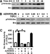
TGF-β1 induces Src activation, and AZD0530 inhibits TGF-β1–induced Src activation in a dose-dependent manner in human lung fibroblasts. (A) Serum-starved human lung fibroblasts were treated with TGF-β1 (10 ng/ml) in serum-free medium for the indicated time and were detergent lysed; whole-cell lysates were Western blotted with the indicated antibodies. (B) These lung fibroblasts were treated with the AZD0530 inhibitor at the indicated dose or with control vehicle, then treated with TGF-β1 (10 ng/ml) for 1 hour. Cells were detergent-lysed, and the lysates were analyzed by Western blot with the indicated antibodies. Src activation was determined by phosphorylation of tyrosine 416 of Src (pY416-Src). (C) Src kinase activities in these lung fibroblasts were examined by a luminescent kinase assay kit, and the data are presented as the percentage of relative luminescence to those fibroblasts cultured in serum-free medium (as 100%). The optimized AZD0530 dose (0.1 µM) was used in all tests. The data are presented as mean + S.E. *P < 0.01. RLU, relative light units.
Fig. 2.
Fig. 2.
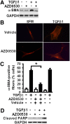
AZD0530 inhibits TGF-β1–induced α-SMA expression and formation of α-SMA–containing fibers in human lung fibroblasts. (A) Serum-starved human lung fibroblasts were treated with or without AZD0530 (0.1 µM) followed by TGF-β1 (10 ng/ml) for 48 hours. Fibroblasts were lysed and Western blotted with the indicated antibodies. (B) These fibroblasts were fixed and immunofluorescently stained with the Cy-3–labeled monoclonal antibody toward α-SMA. Fluorescent microscopic digital images were taken (original 200×), and representative pictures are shown. (C) Quantification of the percentage of cells with highly organized, thickened α-SMA–containing fibers as described in Materials and Methods. The data are presented as mean + S.E. *P < 0.01. (D) These fibroblasts were lysed, and an equivalent amount of whole cell lysates were Western blotted with the indicated antibodies. The antibody specifically recognizes the cleaved PARP and does not cross with the full-length form. The experiments were repeated three times, and representative pictures are shown. GAPDH, glyceraldehyde 3-phosphate dehydrogenase.
Fig. 3.
Fig. 3.
AZD0530 inhibits TGF-β1–induced three-dimensional collagen gel contraction and PDGF-induced migration in human lung fibroblasts. (A) Serum-starved human lung fibroblasts were treated with or without AZD0530 (0.1 µM) followed by TGF-β1 (10 ng/ml) treatment, and a collagen-gel contraction assay at 37°C, 5% CO2, for 60 hours. Representative digital images are shown. (B) Data are pooled from three individual experiments (each performed at least in duplicate) and are presented as the percentage of contracted collagen gel area relative to the area of culture wells (mean ± S.E.). The lower percentage represents a stronger gel contraction. (C) Serum-starved human lung fibroblasts were wounded, and treated with or without AZD0530 (0.1 µM) or vehicle followed by PDGF-BB (4 ng/ml) in serum-free medium (SFM) with 1% BSA for 24 hours. Monolayer wound closure assays were performed as described in Materials and Methods. Data obtained are pooled (n = 4 per group) and plotted as the percentage of wound area covered over 24 hours relative to the control (fibroblasts in vehicle-treated only). All data are presented as mean + S.E. *P < 0.01.
Fig. 4.
Fig. 4.
AZD0530 inhibits TGF-β1–induced procollagen and fibronectin expression, and reduces TGF-β1–induced FAK activation in human lung fibroblasts. Serum-starved human lung fibroblasts were treated with or without AZD0530 (0.1 µM) followed by TGF-β1 (10 ng/ml) for 24 hours. Fibroblasts were lysed and analyzed by Western blot with the indicated antibodies. (A) Expression of collagen and fibronectin (FN). Expression of collagen was examined through expression of procollagen 1A1 (Pro-Col). Representative images are shown. (B) FAK activation was examined through phosphorylation of the tyrosine 397 (pY397) of FAK. Representative images are shown. (C) Densitometry analysis of band intensity for pY397 of FAK. Results are normalized to the total FAK protein level, and basal pY397-FAK was used as 100%. Results are pooled from three individual experiments. *P < 0.01. GAPDH, glyceraldehyde 3-phosphate dehydrogenase.
Fig. 5.
Fig. 5.
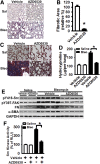
AZD0530 is protective against lung fibrosis. (A) Mice were intratracheally instilled with bleomycin (Bleo) or saline (Sal) control, and then were treated daily with AZD0530 compound (20 mg/kg body weight) or vehicle by oral gavage starting at day 7 after bleomycin treatment. Lung tissues were harvested at day 21 after bleomycin or saline treatment, sectioned, and H&E-stained. Representative lung sections are shown (magnification, 200×). (B) The lung fibrotic/lesion areas of bleomycin-challenged mice were examined morphometrically and reported by relative fibrotic areas (% of fibrotic area of mice treated with vehicle only). (C) Lung tissue sections were stained by a Masson’s trichrome staining kit to demonstrate the areas of collagen deposition (magnification, 400×). (D) Total lung collagen accumulation was determined by hydroxyproline assays. (E) Lungs were harvested at day 14 after bleomycin instillation. Whole-lung lysates were Western blotted for Src activation (pY416-Src), FAK activation (pY397-FAK), and expression of fibronectin (FN) and α-SMA. (F) Lungs were harvested at day 14 after bleomycin instillation, and Src kinase activities were examined (as in Fig. 1). These data are pooled and presented as mean + S.E. (n = 6–8 mice per group). *P < 0.01. GAPDH, glyceraldehyde 3-phosphate dehydrogenase; RLU, relative light units.

References

    1. Adamson IY, Bowden DH. (1974) The pathogenesis of bleomycin-induced pulmonary fibrosis in mice. Am J Pathol 77:185–197 - PMC - PubMed
    1. Ahluwalia MS, de Groot J, Liu WM, Gladson CL. (2010) Targeting SRC in glioblastoma tumors and brain metastases: rationale and preclinical studies. Cancer Lett 298:139–149 - PMC - PubMed
    1. Aleshin A, Finn RS. (2010) SRC: a century of science brought to the clinic. Neoplasia 12:599–607 - PMC - PubMed
    1. Boggon TJ, Eck MJ. (2004) Structure and regulation of Src family kinases. Oncogene 23:7918–7927 - PubMed
    1. Cai GQ, Chou CF, Hu M, Zheng A, Reichardt LF, Guan JL, Fang H, Luckhardt TR, Zhou Y, Thannickal VJ, et al. (2012) Neuronal Wiskott-Aldrich syndrome protein (N-WASP) is critical for formation of α-smooth muscle actin filaments during myofibroblast differentiation. Am J Physiol Lung Cell Mol Physiol 303:L692–L702 - PMC - PubMed

Publication types

MeSH terms