Review
. 2014 Sep 5;369(1650):20130457.
doi: 10.1098/rstb.2013.0457.

Centriole structure

Affiliations
Review

Centriole structure

Mark Winey et al. Philos Trans R Soc Lond B Biol Sci. .

Abstract

Centrioles are among the largest protein-based structures found in most cell types, measuring approximately 250 nm in diameter and approximately 500 nm long in vertebrate cells. Here, we briefly review ultrastructural observations about centrioles and associated structures. At the core of most centrioles is a microtubule scaffold formed from a radial array of nine triplet microtubules. Beyond the microtubule triplets of the centriole, we discuss the critically important cartwheel structure and the more enigmatic luminal density, both found on the inside of the centriole. Finally, we discuss the connectors between centrioles, and the distal and subdistal appendages outside of the microtubule scaffold that reflect centriole age and impart special functions to the centriole. Most of the work we review has been done with electron microscopy or electron tomography of resin-embedded samples, but we also highlight recent work performed with cryoelectron microscopy, cryotomography and subvolume averaging. Significant opportunities remain in the description of centriolar structure, both in mapping of component proteins within the structure and in determining the effect of mutations on components that contribute to the structure and function of the centriole.

Keywords: cartwheel; distal appendages; luminal density; pericentriolar material; subdistal appendages; triplet microtubules.


Figures

Figure 1.
Figure 1.
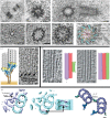
The microtubule scaffold and associated structures of the centriole. (a) Variations in centriole microtubule number. The top panels are Drosophila centrioles in longitudinal (left) and cross (right) section, showing doublet microtubules [6]. The lower panels are a cross section of a C. elegans centriole (left) showing singlet microtubules, and a human centriole (right) showing triplet microtubules. Bars, 100 nm. (b) Centrioles in mammalian cells. The top panel is a pair of orthogonally arranged centrioles in a centrosome. The bottom panels display cross sections of a centriole proximal region; the structural features are indicated on the right panel. Bars, 100 nm. (c) The left panel is a model of the yeast γ-tubulin complex (indicated in yellow and blues) anchoring a microtubule (grey) to a spindle pole body via a tether (brown, such as Spc110). See Kollman et al. [7] for details. The right panel is a tomographic slice of a budding yeast spindle pole body and attached spindle microtubules (as in O'Toole et al. [8]); arrows indicate the capped microtubule minus ends. Bar, 25 nm. (d). Capped minus ends are present on the A-tubule of assembling centrioles (left panel, asterisk; purple), B- and C-tubules have open ends (red, green). The A-tubule minus end cap is absent in mature centrioles (right panels). This figure is from Guichard et al. [9] with permission. Bars, 25 nm. (e). Models showing non-tubulin structures (purple) associated with the microtubule triplets (light blue) identified in cryoelectron tomograms of basal bodies in Chlamydomonas (as in Li et al. [10], left and centre panels). Left: the Y-shaped linker facing the basal body centre. Microtubule luminal structures such as the A- and B-tubule linker (arrowhead) and the A-tubule cone shaped structure (asterisk). The middle panels show microtubule luminal structures (arrows) present in the C-tubule in the distal region of the basal body. The right panel shows the location of the A-tubule to C-tubule linker (green) between microtubule triplets (purple) in Trychonympha ([11] with permission). Bar, 25 nm.
Figure 2.
Figure 2.
Detail of the cartwheel structure. (a) Cryoelectron tomographic slice (top) and models (lower) of the hub in Trichonympha reveal the stacking and dimensions of the Sas6 ring at the hub from [48] with permission. (bg) Selected slices from cryoelectron tomograms and models comparing the spoke and pinhead structures in Chlamydomonas and Trichonympha from [11] with permission. Cross-sectional (b) and longitudinal (c) views of the cartwheel (light blue), pinhead (medium blue), microtubule triplet (purple) and the A–C linker (green). Dimensions are shown, and C–SP is the ‘cartwheel spoke.’ Tomographic sections of the proximal region of Chlamydomonas basal bodies (d), were used for three-dimensional volume averaging (e), and higher magnification image (f) shows the structural similarity to the Trichonympha basal body (g). The black arrow indicates the pinhead–A-tubule connection. The red arrows and black arrowhead indicate the A-C linker connection to the A-tubule and the C-tubule, respectively. Bars, 25 nm. (h) A molecular model for the cartwheel hub, spoke and pinhead based on crystallographic data and known protein–protein interactions from [49] with permission.
Figure 3.
Figure 3.
Structures outside the microtubule triplets of the centriole. (a) Electron micrographs of centrioles in isolated centrosomes shown in longitudinal section and informative cross sections highlighting the distal and subdistal appendages. Reprinted from [69] based on original images in [58]. (b) Deuterosomes (arrows) organizing the formation of basal bodies in mouse tracheal epithelium cells as in [70]. Bar, 200 nm.

References

    1. Scheer U. 2014. Historical roots of centrosome research: discovery of Boveri's microscope slides in Würzburg. Phil. Trans. R. Soc. B 369, 20130469 (10.1098/rstb.2013.0469) - DOI - PMC - PubMed
    1. Sluder G. 2014. One to only two: a short history of the centrosome and its duplication. Phil. Trans. R. Soc. B 369, 20130455 (10.1098/rstb.2013.0455) - DOI - PMC - PubMed
    1. Gall JG. 2004. Early studies on centrioles and centrosomes. In Centrosomes in development and disease (ed. Nigg EA.), pp. 1–16. Weinheim, Germany: Wiley Blackwell.
    1. De Harven E, Bernhard W. 1956. Etude au microscope electronique de l'ultrastructure du centriole chez les vertebres. Z. Zellforschung. 45, 378–398. (10.1007/BF01106086) - DOI - PubMed
    1. Fawcett DW, Porter KR. 1954. A study of the fine structure of ciliated epithelia. J. Morphol. 94, 221–381. (10.1002/jmor.1050940202) - DOI

Publication types