Review
. 2014 Sep 5;369(1650):20130459.
doi: 10.1098/rstb.2013.0459.

Pericentriolar material structure and dynamics

Affiliations
Review

Pericentriolar material structure and dynamics

Jeffrey B Woodruff et al. Philos Trans R Soc Lond B Biol Sci. .

Abstract

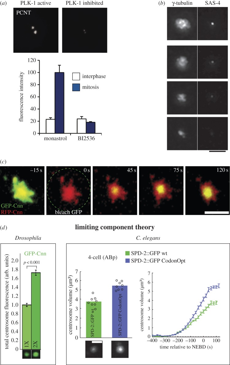
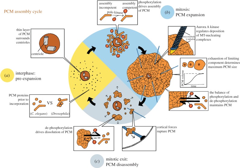
A centrosome consists of two barrel-shaped centrioles embedded in a matrix of proteins known as the pericentriolar material (PCM). The PCM serves as a platform for protein complexes that regulate organelle trafficking, protein degradation and spindle assembly. Perhaps most important for cell division, the PCM concentrates tubulin and serves as the primary organizing centre for microtubules in metazoan somatic cells. Thus, similar to other well-described organelles, such as the nucleus and mitochondria, the cell has compartmentalized a multitude of vital biochemical reactions in the PCM. However, unlike these other organelles, the PCM is not membrane bound, but rather a dynamic collection of protein complexes and nucleic acids that constitute the organelle's interior and determine its boundary. How is the complex biochemical machinery necessary for the myriad centrosome functions concentrated and maintained in the PCM? Recent advances in proteomics and RNAi screening have unveiled most of the key PCM components and hinted at their molecular interactions ( table 1). Now we must understand how the interactions between these molecules contribute to the mesoscale organization and the assembly of the centrosome. Among outstanding questions are the intrinsic mechanisms that determine PCM shape and size, and how it functions as a biochemical reaction hub.

Keywords: centrosome; microtubule-organizing centre; organelle scaling; pericentriolar material.


Figures

Figure 1.
Figure 1.
Structural organization of the PCM. (a) Negative stain electron micrograph of a centrosome isolated from Chinese hamster ovary cells. One hundred and twenty-five MTs emanate from the densely staining centre. (Adapted from [2].) (b) Structure of a purified Drosophila centrosome as revealed by electron tomography. A ninefold radially symmetric centriole can be seen at the centre surrounded by PCM. The inset shows a magnified view of a ring-like complex found within the PCM. These complexes measured 25–30 nm in diameter and were determined to contain γ-tubulin. (Adapted from [3].) (c) The γ-TuRC was later isolated from Drosophila cells and analysed by electron tomography. (Adapted from [4].) (d) Harsh treatment of Spisula centrosomes with potassium iodide revealed 12–15 nm wide filaments running throughout the PCM, leading to the hypothesis that a lattice-like network forms the structural foundation of the PCM. (Adapted from [5].) (e) The development of subdiffraction microscopy techniques allowed high precision localization of proteins within the PCM. The first picture depicts the localization of PCNT-like protein (D-PLP) in Drosophila cells determined with stochastic optical reconstruction microscopy (STORM). Comparison of the localization of numerous proteins revealed that the interphase PCM contains ordered subdomains of defined size. However, the expansive PCM that exists during mitosis is less ordered. Localization of PCNT and γ-tubulin in human cells using three-dimensional SIM is shown. (Adapted from [–8].)
Figure 2.
Figure 2.
Factors regulating PCM growth and final size. (a) Inhibition of PLK-1 kinase activity with the small molecule BI2436 reduces the incorporation of PCNT at centrosomes. Interestingly, inhibition of PLK-1 did not affect PCNT localization to the interphase centrosome. (Adapted from [42].) (b) γ-tubulin staining scales proportionally with the amount of SAS-4 localized to centrioles in C. elegans embryos, suggesting that centriole duplication and size determine PCM growth. Scale bar, 4 μm. (Adapted from [30].) (c) After photo-bleaching, GFP-Cnn first recovers at the centre of the centrosome, near the centrioles and then spreads outward to the periphery in Drosophila cells. This result indicates that Cnn is incorporated immediately around centrioles, suggesting that centrioles play a critical role in converting PCM proteins to an assembly-competent state. Scale bar, 3 μm. (Adapted from [35].) (d) Overexpression of Cnn and SPD-2 increases the final steady-state size of the PCM in Drosophila and C. elegans, respectively. SPD-2 expression levels were increased through a codon-optimization strategy for the C. elegans experiments (SPD-2::GFP CodonOpt, blue bar and line). These results suggest that Cnn and SPD-2 act as limiting components for the formation of PCM. Scale bar, 3 μm. (Adapted from [35,66].)
Figure 3.
Figure 3.
The PCM assembly cycle in embryonic systems. (a) During interphase, a thin layer of PCM surrounds the centrioles. In the cytoplasm are unincorporated PCM proteins, and there are species-specific differences in their assembly state. In C. elegans, the unincorporated core PCM components are separate entities, whereas in Drosophila, the core PCM proteins may already be pre-assembled into small complexes of defined stoichiometry. (b) As the cell progresses into mitosis, polo kinase phosphorylates the key scaffolding proteins, thereby inducing their incorporation around the established inner PCM layer. This new addition of protein causes the PCM to expand dramatically. As the mitotic PCM expands, Aurora A kinase phosphorylation promotes the deposition of protein complexes that aid in nucleating and stabilizing MTs (blue lines). The steady-state size of the PCM is determined by the total amount of a limiting component and the rates of phosphorylation and de-phosphorylation reactions. (c) Once mitosis is complete, MT-mediated cortical forces rupture the PCM, and protein phosphatases remove polo kinase-derived phosphorylations from the scaffold proteins. These two activities promote rapid disassembly of the mitotic PCM. (Illustration courtesy of Julia Eichhorn.)

References

    1. Robbins E, Jentzsch G, Micali A. 1968. The centriole cycle in synchronized HeLa cells. J. Cell Biol. 36, 329–339. (10.1083/jcb.36.2.329) - DOI - PMC - PubMed
    1. Gould RR, Borisy GG. 1977. The pericentriolar material in Chinese hamster ovary cells nucleates microtubule formation. J. Cell Biol. 73, 601–615. (10.1083/jcb.73.3.601) - DOI - PMC - PubMed
    1. Moritz M, Braunfeld MB, Sedat JW, Alberts B, Agard DA. 1995. Microtubule nucleation by gamma-tubulin-containing rings in the centrosome. Nature 378, 638–640. (10.1038/378638a0) - DOI - PubMed
    1. Moritz M, Braunfeld MB, Guénebaut V, Heuser J, Agard DA. 2000. Structure of the gamma-tubulin ring complex: a template for microtubule nucleation. Nat. Cell Biol. 2, 365–370. (10.1038/35014058) - DOI - PubMed
    1. Schnackenberg BJ, Khodjakov A, Rieder CL, Palazzo RE. 1998. The disassembly and reassembly of functional centrosomes in vitro. Proc. Natl Acad. Sci. USA 95, 9295–9300. (10.1073/pnas.95.16.9295) - DOI - PMC - PubMed

Substances

LinkOut - more resources