Review
. 2014 Sep 5;369(1650):20130460.
doi: 10.1098/rstb.2013.0460.

The centriole duplication cycle

Affiliations
Review

The centriole duplication cycle

Elif Nur Fırat-Karalar et al. Philos Trans R Soc Lond B Biol Sci. .

Abstract

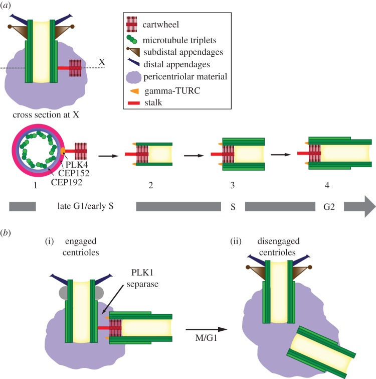
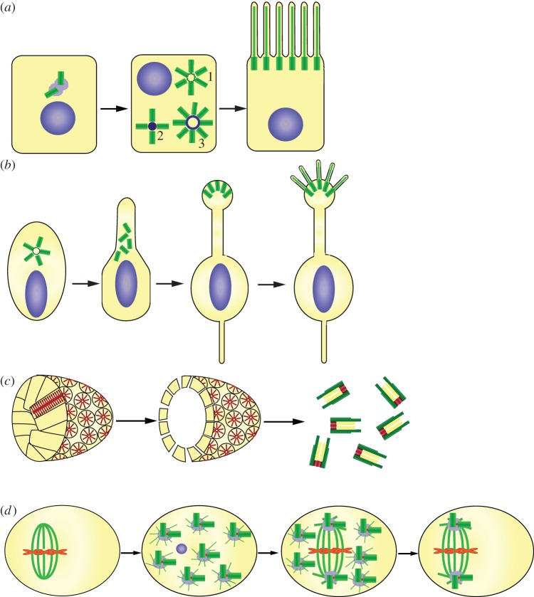
Centrosomes are the main microtubule-organizing centre of animal cells and are important for many critical cellular and developmental processes from cell polarization to cell division. At the core of the centrosome are centrioles, which recruit pericentriolar material to form the centrosome and act as basal bodies to nucleate formation of cilia and flagella. Defects in centriole structure, function and number are associated with a variety of human diseases, including cancer, brain diseases and ciliopathies. In this review, we discuss recent advances in our understanding of how new centrioles are assembled and how centriole number is controlled. We propose a general model for centriole duplication control in which cooperative binding of duplication factors defines a centriole 'origin of duplication' that initiates duplication, and passage through mitosis effects changes that license the centriole for a new round of duplication in the next cell cycle. We also focus on variations on the general theme in which many centrioles are created in a single cell cycle, including the specialized structures associated with these variations, the deuterosome in animal cells and the blepharoplast in lower plant cells.

Keywords: blepharoplast; centriole; centrosome; deuterosome.


Figures

Figure 1.
Figure 1.
Centriole assembly pathway in vertebrates. (a) Centrioles are cylindrical structures composed of nine microtubule triplets symmetrically arranged about a central core. The components important to the discussion here are indicated in the legend. Depicted is a longitudinal section of a mother centriole, which has two types of appendages, distal and subdistal, and lacks the internal cartwheel structure. The base of the mother centriole is embedded in the pericentriolar material. The formation of a procentriole has been initiated by assembly of the stalk and cartwheel from the side of the mother centriole. (a: 1–4) Stages of procentriole formation, depicted as viewed by cross section of centriole at X in longitudinal section. The mother centriole is not shown in (a: 2–4) for clarity, but is present and engaged to the procentriole throughout the process shown. (a-1) PLK4 accumulates at a single focus, in conjunction with CEP152 and CEP192, which are distributed in rings around the circumference of the centriole. PLK4 stimulates the assembly of a stalk and ninefold symmetric cartwheel that will provide structure to the procentriole and keep it engaged to the mother centriole. (a-2) Nine A-tubules are nucleated by the gamma-tubulin ring complex (gamma-TURC), in association with the cartwheel. These grow unidirectionally from the proximal to the distal end of the centriole. The A-tubules remain capped by the gamma-TURC throughout the assembly process, eventually being lost at the end of mitosis. (a-3) The B- and C-tubules form by a gamma-TURC-independent mechanism and grow until they reach the length of the A-tubule. (a-4) The distal end of the centriole is formed by elongation of the A- and B-tubules, creating a structurally distinct distal domain. (b) Centriole disengagement in the transition from M-G1. (i) A centrosome in metaphase of mitosis, with engaged mother centriole and procentriole. (ii) A centrosome in G1, after mitosis, with disengaged mother and daughter centrioles. The cartwheel has disassembled from the daughter centriole. Note that the subdistal appendages disassemble during mitosis, but the constituent proteins remain associated with the centriole. They are depicted as undifferentiated spheres in the mitotic centriole in place of the subdistal appendages. (Online version in colour.)
Figure 2.
Figure 2.
Centriole number control in specialized cell types. (a) Multiple centrioles in animal multi-ciliated epithelial cells. Some centrioles form by initiation in a rosette around the mother centrioles (1), but most form on a deuterosome (2 and 3). The deuterosome ranges in size from centriole size (2) to much larger rings (3); it is not clear whether these represent a pathway of maturation of the structure. The centrioles are freed from the surface they grew from and migrate to the cell surface to nucleate motile cilia. (b) Multiple centrioles in olfactory sensory cilia. A neuronal cell forms approximately 10 centrioles, which nucleate sensory cilia from the end of the dendrite process. In this case, it is unknown whether centriole formation occurs strictly on mother centrioles or whether the deuterosome pathway is invoked. (c) Multiple centrioles in ciliated land plants. Sperm cells of some primitive plants are multi-ciliated and from centrioles from a blepharoplast (shown without surrounding cell). The blepharoplast consists of many radially arranged ninefold symmetric cylinders which resemble the cartwheel. The blepharoplast enlarges and ultimately disintegrates into many procentrioles which elongate and ultimately nucleate cilia on the surface of the sperm cell. (d) De novo formation and segregation of multiple centrioles during parthenogenesis. In insects of the hymenoptera order, parthenogenetic development is accompanied by the formation of many centrosomes de novo. Although depicted as centriole pairs, it is unclear whether centrosomes have single or paired centrioles at this stage. Only two of the centrosomes associate with the mitotic spindle, whereas the remainder degenerate, resulting in restoration of the appropriate number of centrioles per nucleus. (Online version in colour.)

References

    1. Ishikawa H, Kubo A, Tsukita S. 2005. Odf2-deficient mother centrioles lack distal/subdistal appendages and the ability to generate primary cilia. Nat. Cell Biol. 7, 517–524. (10.1038/ncb1251) - DOI - PubMed
    1. Graser S, Stierhof YD, Lavoie SB, Gassner OS, Lamla S, Le Clech M, Nigg EA. 2007. Cep164, a novel centriole appendage protein required for primary cilium formation. J. Cell Biol. 179, 321–330. (10.1083/jcb.200707181) - DOI - PMC - PubMed
    1. Luders J, Stearns T. 2007. Microtubule-organizing centres: a re-evaluation. Nat. Rev. Mol. Cell Biol. 8, 161–167. (10.1038/nrm2100) - DOI - PubMed
    1. Carvalho-Santos Z, Azimzadeh J, Pereira-Leal JB, Bettencourt-Dias M. 2011. Evolution: tracing the origins of centrioles, cilia, and flagella. J. Cell Biol. 194, 165–175. (10.1083/jcb.201011152) - DOI - PMC - PubMed
    1. Andersen JS, Wilkinson CJ, Mayor T, Mortensen P, Nigg EA, Mann M. 2003. Proteomic characterization of the human centrosome by protein correlation profiling. Nature 426, 570–574. (10.1038/nature02166) - DOI - PubMed