Review
. 2014 Sep 5;369(1650):20130468.
doi: 10.1098/rstb.2013.0468.

Small organelle, big responsibility: the role of centrosomes in development and disease

Affiliations
Review

Small organelle, big responsibility: the role of centrosomes in development and disease

Pavithra L Chavali et al. Philos Trans R Soc Lond B Biol Sci. .

Abstract

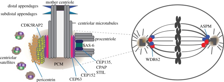
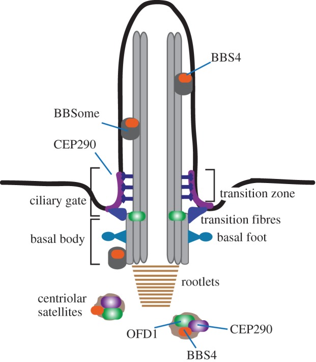
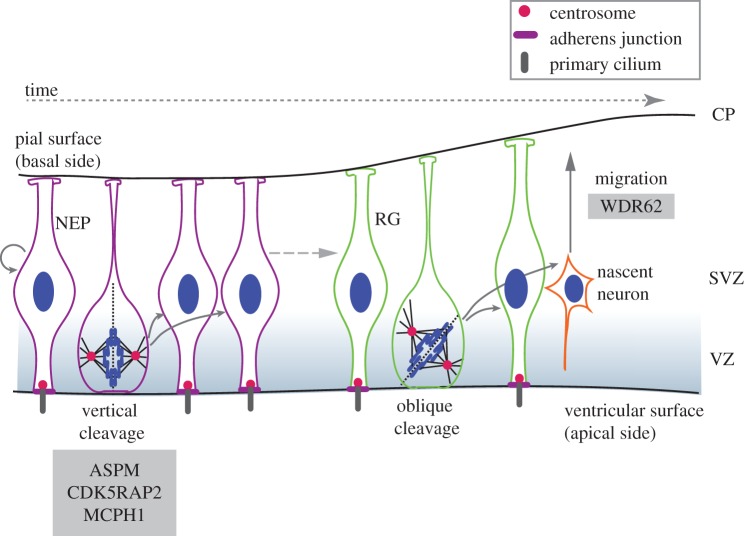
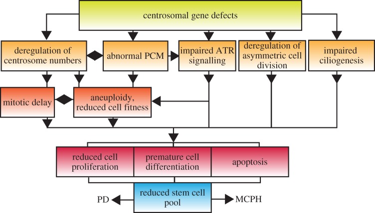
The centrosome, a key microtubule organizing centre, is composed of centrioles, embedded in a protein-rich matrix. Centrosomes control the internal spatial organization of somatic cells, and as such contribute to cell division, cell polarity and migration. Upon exiting the cell cycle, most cell types in the human body convert their centrioles into basal bodies, which drive the assembly of primary cilia, involved in sensing and signal transduction at the cell surface. Centrosomal genes are targeted by mutations in numerous human developmental disorders, ranging from diseases exclusively affecting brain development, through global growth failure syndromes to diverse pathologies associated with ciliary malfunction. Despite our much-improved understanding of centrosome function in cellular processes, we know remarkably little of its role in the organismal context, especially in mammals. In this review, we examine how centrosome dysfunction impacts on complex physiological processes and speculate on the challenges we face when applying knowledge generated from in vitro and in vivo model systems to human development.

Keywords: centriole; centrosome; cilia; ciliopathy; dwarfism; microcephaly.


Figures

Figure 1.
Figure 1.
Schematic of the vertebrate centrosome and the mitotic spindle. Key structural features of centrosomes are indicated. Locations of disease-associated centrosomal proteins are shown. Mother centrioles acquire two sets of appendages during centriole maturation. Procentriole formation requires assembly of SAS-6 homodimers into a cartwheel-like structure. STIL interacts with CPAP and possibly SAS-6 in the procentriole. By binding SAS-6, CPAP and centriolar microtubules, CEP135 could stabilize centrioles. CDK5RAP2 and pericentrin are scaffolding proteins located in the PCM, surrounded by centriolar satellites. Centrosomes are positioned at the mitotic spindle poles where spindle microtubules focus and nucleate astral microtubules. ASPM and WDR62 localize to the mitotic spindle poles. (Online version in colour.)
Figure 2.
Figure 2.
Schematic of the primary cilium. Key structural features of cilia are indicated. The basal body is attached to the plasma membrane via transition fibres derived from the distal appendages. Structural stability is provided by the ciliary rootlet. During ciliogenesis, centriolar satellites deliver proteins like CEP290, BBS4 and OFD1 to the cilium. CEP290 localizes to the transition zone and functions as the ciliary gate. BBS4 assembles into the BBSome at the ciliary base and is transported inside the cilium. OFD1 is localized to the distal end of the basal body. (Online version in colour.)
Figure 3.
Figure 3.
Neurogenesis in the rodent brain. NEPs and RG are collectively termed APs that reside in the ventricular zone (VZ). NEPs first expand through rapid symmetric divisions, but after the onset of neurogenesis they give rise to RG cells, which can divide symmetrically into two RGs or asymmetrically into an RG and a nascent neuron. APs undergoing symmetric proliferative divisions display a vertical cleavage plane relative to the apical surface, whereas those undergoing neurogenic divisions show oblique or horizontal cleavage planes. In the case of symmetric divisions, the cleavage furrow bisects the apical membrane and adjacent adherens junctions. In asymmetric divisions, the RG inherits the apical plasma membrane, whereas the newborn neuron migrates through the SVZ towards the cortical plate (CP) where it matures. Loss-of-function of CDK5RAP2 and ASPM in mouse models compromises symmetric AP divisions, similarly to CDK5RAP2-deficient human brain organoids. Individuals with WDR62 mutations exhibit cortical malformations and neuronal migration defects. (Online version in colour.)
Figure 4.
Figure 4.
Putative cellular mechanisms implicated in MCPH and PD. (Online version in colour.)

References

    1. Bornens M. 2012. The centrosome in cells and organisms. Science 335, 422–426. (10.1126/science.1209037) - DOI - PubMed
    1. Gonczy P. 2012. Towards a molecular architecture of centriole assembly. Nat. Rev. Mol. Cell Biol. 13, 425–435. (10.1038/nrm3373) - DOI - PubMed
    1. Ishikawa H, Marshall WF. 2011. Ciliogenesis: building the cell's antenna. Nat. Rev. Mol. Cell Biol. 12, 222–234. (10.1038/nrm3085) - DOI - PubMed
    1. Singla V, Reiter JF. 2006. The primary cilium as the cell's antenna: signaling at a sensory organelle. Science 313, 629–633. (10.1126/science.1124534) - DOI - PubMed
    1. Fliegauf M, Benzing T, Omran H. 2007. When cilia go bad: cilia defects and ciliopathies. Nat. Rev. Mol. Cell Biol. 8, 880–893. (10.1038/nrm2278) - DOI - PubMed

Publication types

Supplementary concepts

LinkOut - more resources