. 2014 Oct;35(31):8829-8839.
doi: 10.1016/j.biomaterials.2014.07.006. Epub 2014 Jul 18.

Dual growth factor delivery from bilayered, biodegradable hydrogel composites for spatially-guided osteochondral tissue repair

Affiliations

Dual growth factor delivery from bilayered, biodegradable hydrogel composites for spatially-guided osteochondral tissue repair

Steven Lu et al. Biomaterials. 2014 Oct.

Abstract

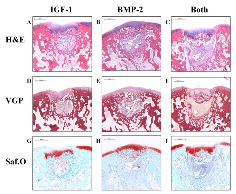
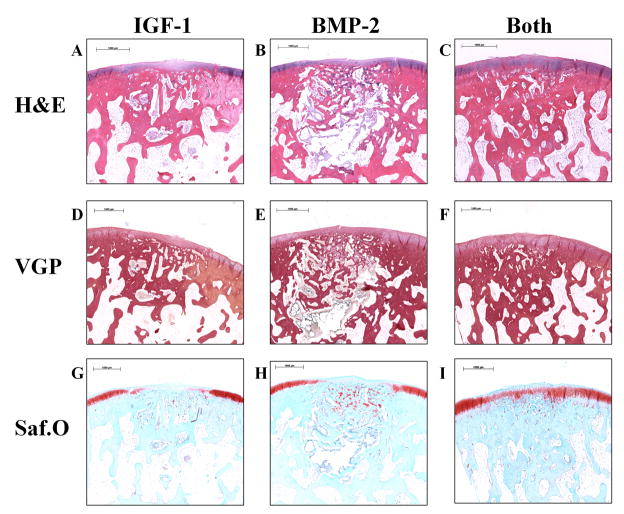
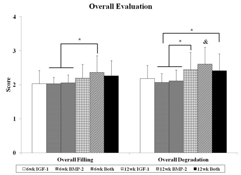
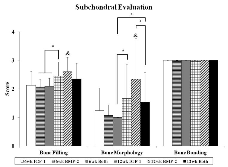
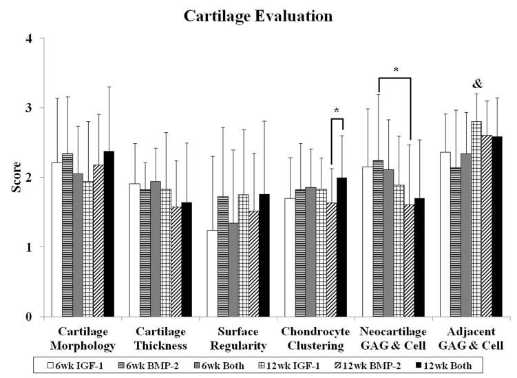
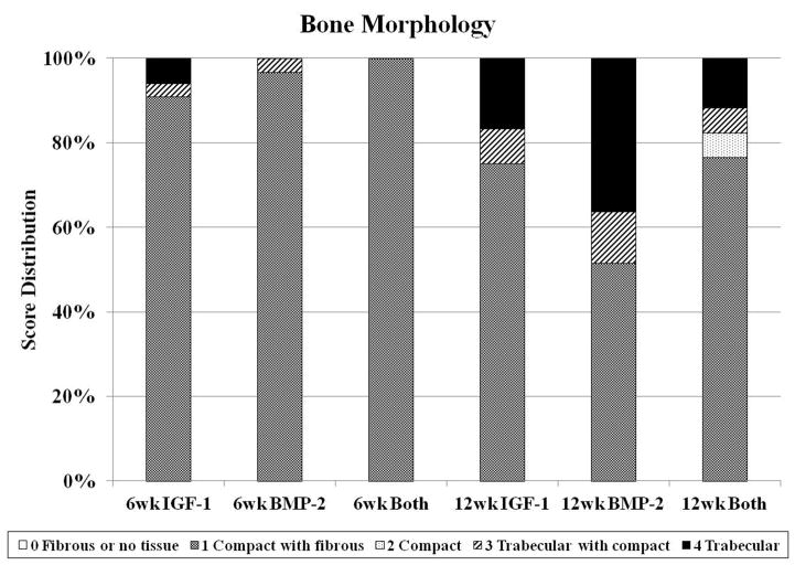
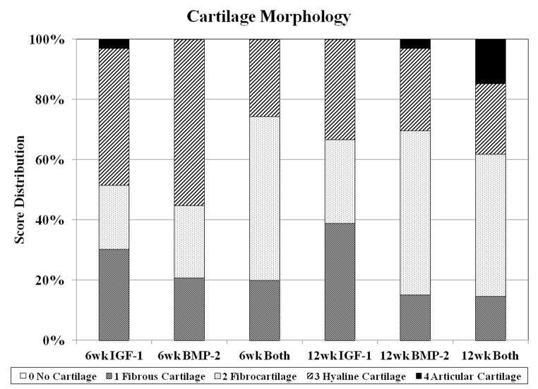
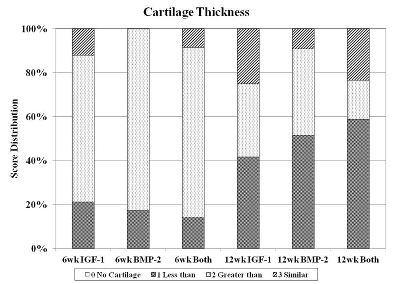
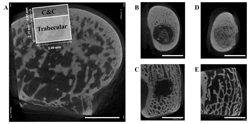
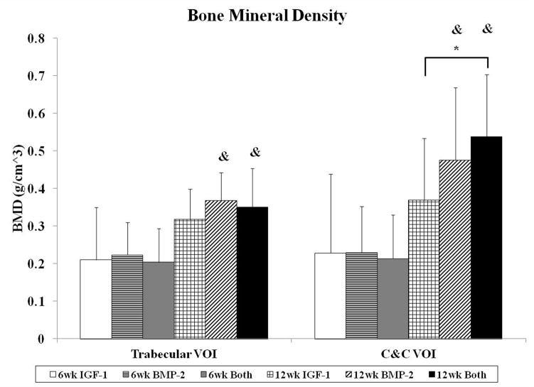
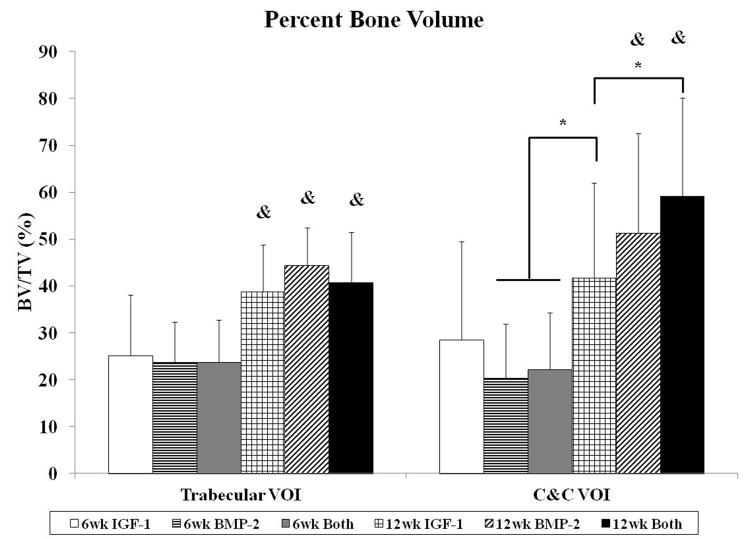
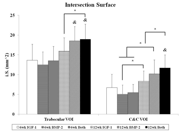
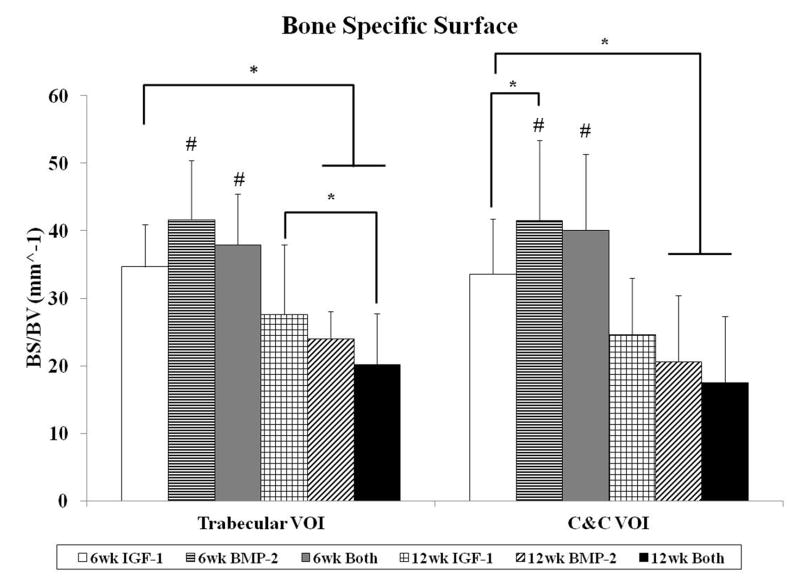
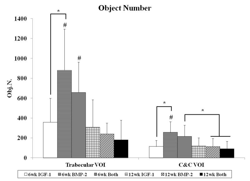
The present work investigated the use of biodegradable hydrogel composite scaffolds, based on the macromer oligo(poly(ethylene glycol) fumarate) (OPF), to deliver growth factors for the repair of osteochondral tissue in a rabbit model. In particular, bilayered OPF composites were used to mimic the structural layers of the osteochondral unit, and insulin-like growth factor-1 (IGF-1) and bone morphogenetic protein-2 (BMP-2) were loaded into gelatin microparticles and embedded within the OPF hydrogel matrix in a spatially controlled manner. Three different scaffold formulations were implanted in a medial femoral condyle osteochondral defect: 1) IGF-1 in the chondral layer, 2) BMP-2 in the subchondral layer, and 3) IGF-1 and BMP-2 in their respective separate layers. The quantity and quality of osteochondral repair was evaluated at 6 and 12 weeks with histological scoring and micro-computed tomography (micro-CT). While histological scoring results at 6 weeks showed no differences between experimental groups, micro-CT analysis revealed that the delivery of BMP-2 alone increased the number of bony trabecular islets formed, an indication of early bone formation, over that of IGF-1 delivery alone. At 12 weeks post-implantation, minimal differences were detected between the three groups for cartilage repair. However, the dual delivery of IGF-1 and BMP-2 had a higher proportion of subchondral bone repair, greater bone growth at the defect margins, and lower bone specific surface than the single delivery of IGF-1. These results suggest that the delivery of BMP-2 enhances subchondral bone formation and that, while the dual delivery of IGF-1 and BMP-2 in separate layers does not improve cartilage repair under the conditions studied, they may synergistically enhance the degree of subchondral bone formation. Overall, bilayered OPF hydrogel composites demonstrate potential as spatially-guided, multiple growth factor release vehicles for osteochondral tissue repair.

Keywords: Bone morphogenetic protein-2; Cartilage repair; Insulin-like growth factor-1; Rabbit model; Subchondral bone.


Figures

Fig. 1
Fig. 1
Representative histological sections of osteochondral tissue repair after 6 weeks of implantation for all three groups. Sections were stained with hematoxylin and eosin (H&E) (A–C), van Gieson’s Picrofuchsin (VGP) (D–F), or Safranin O/Fast Green (Saf.O) (G–I). All 6 week samples had incomplete degradation of the implant, evidenced by a light gray staining of the hydrogel surrounded by a yellow fibrous capsule in the VGP stains. The neo-cartilage formed was generally thicker than the surrounding cartilage with intense Saf.O staining and was present in the subchondral region. (scale bar: 1000 μm)
Fig. 2
Fig. 2
Representative histological sections of osteochondral tissue repair after 12 weeks of implantation for all three groups. Sections were stained with hematoxylin and eosin (H&E) (A–C), van Gieson’s Picrofuchsin (VGP) (D–F), or Safranin O/Fast Green (Saf.O) (G–I). 12 week samples generally had greater degradation than 6 week samples and a smaller fibrous capsule. Group IGF-1 had the most sections with fibrous tissue (D,G), group BMP-2 had the most sections with fibrocartilage (E,H), and group Both had the most sections with articular cartilage (F,I). (scale bar: 1000μm)
Fig. 3
Fig. 3
Histological scores for the overall defect evaluation (A), subchondral bone evaluation (B), and articulating cartilage evaluation (C). Data are shown as average scores with error bars representing standard deviation. (*) indicates a significant difference between groups regardless of time point and (&) indicates a significant difference between a 12 week group and all 6 week groups (p < 0.05).
Fig. 3
Fig. 3
Histological scores for the overall defect evaluation (A), subchondral bone evaluation (B), and articulating cartilage evaluation (C). Data are shown as average scores with error bars representing standard deviation. (*) indicates a significant difference between groups regardless of time point and (&) indicates a significant difference between a 12 week group and all 6 week groups (p < 0.05).
Fig. 4
Fig. 4
Distribution of histological scores for subchondral bone morphology (A), cartilage morphology (B), and cartilage thickness (C) for the three groups investigated at both 6 and 12 weeks.
Fig. 4
Fig. 4
Distribution of histological scores for subchondral bone morphology (A), cartilage morphology (B), and cartilage thickness (C) for the three groups investigated at both 6 and 12 weeks.
Fig. 4
Fig. 4
Distribution of histological scores for subchondral bone morphology (A), cartilage morphology (B), and cartilage thickness (C) for the three groups investigated at both 6 and 12 weeks.
Fig. 5
Fig. 5
Micro-CT images showing a transverse section of the osteochondral defect in a medial femoral condyle (A), sections of a 6 week Both sample in the cortical (B) and trabecular (C) region, and sections of a 12 week BMP-2 sample in the cortical (D) and trabecular (E) region. (A) also demonstrates the two volumes of interest used, cartilage and cortical (C&C) and trabecular, when analyzing the subchondral bone. (scale bar: 3 mm)
Fig. 6
Fig. 6
Morphological parameter analysis for both trabecular and cartilage and cortical (C&C) volumes of interest (VOI). Bone mineral density (A), percent bone volume (B), intersection surface (C), bone specific surface (D), and object number (E) were measured for all groups at both time points. Data are shown as average scores with error bars representing standard deviation. (*) indicates a significant difference between groups regardless of time point, (&) indicates a significant difference between a 12 week group and all 6 week groups, and (#) indicates a significant difference between a 6 week group and all 12 week groups (p < 0.05).
Fig. 6
Fig. 6
Morphological parameter analysis for both trabecular and cartilage and cortical (C&C) volumes of interest (VOI). Bone mineral density (A), percent bone volume (B), intersection surface (C), bone specific surface (D), and object number (E) were measured for all groups at both time points. Data are shown as average scores with error bars representing standard deviation. (*) indicates a significant difference between groups regardless of time point, (&) indicates a significant difference between a 12 week group and all 6 week groups, and (#) indicates a significant difference between a 6 week group and all 12 week groups (p < 0.05).

References

    1. Vinatier C, Mrugala D, Jorgensen C, Guicheux J, Noel D. Cartilage engineering: A crucial combination of cells, biomaterials and biofactors. Trends Biotechnol. 2009;27:307–14. - PubMed
    1. Vinatier C, Bouffi C, Merceron C, Gordeladze J, Brondello JM, Jorgensen C, et al. Cartilage tissue engineering: Towards a biomaterial-assisted mesenchymal stem cell therapy. Curr Stem Cell Res Ther. 2009;4:318–29. - PMC - PubMed
    1. Danisovic L, Varga I, Polak S. Growth factors and chondrogenic differentiation of mesenchymal stem cells. Tissue Cell. 2012;44:69–73. - PubMed
    1. Radin EL, Ehrlich MG, Chernack R, Abernethy P, Paul IL, Rose RM. Effect of repetitive impulsive loading on knee joints of rabbits. Clin Orthop Relat Res. 1978:288–93. - PubMed
    1. Serink MT, Nachemson A, Hansson G. Effect of impact loading on rabbit knee joints. Acta Orthop Scand. 1977;48:250–62. - PubMed

Publication types

MeSH terms

Substances