. 2014 Jul 21;9(7):e102627.
doi: 10.1371/journal.pone.0102627. eCollection 2014.

Reduced GABAergic inhibition in the basolateral amygdala and the development of anxiety-like behaviors after mild traumatic brain injury

Affiliations

Reduced GABAergic inhibition in the basolateral amygdala and the development of anxiety-like behaviors after mild traumatic brain injury

Camila P Almeida-Suhett et al. PLoS One. .

Abstract

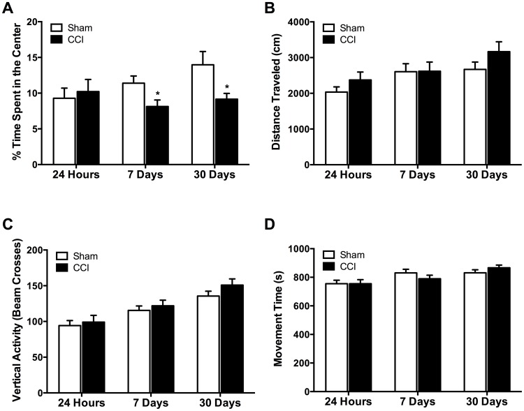
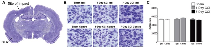
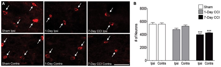
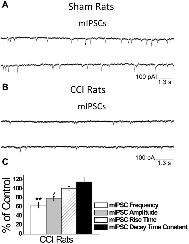
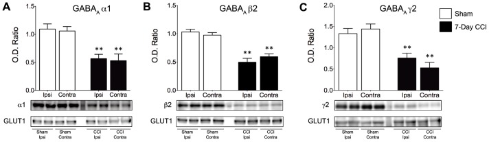
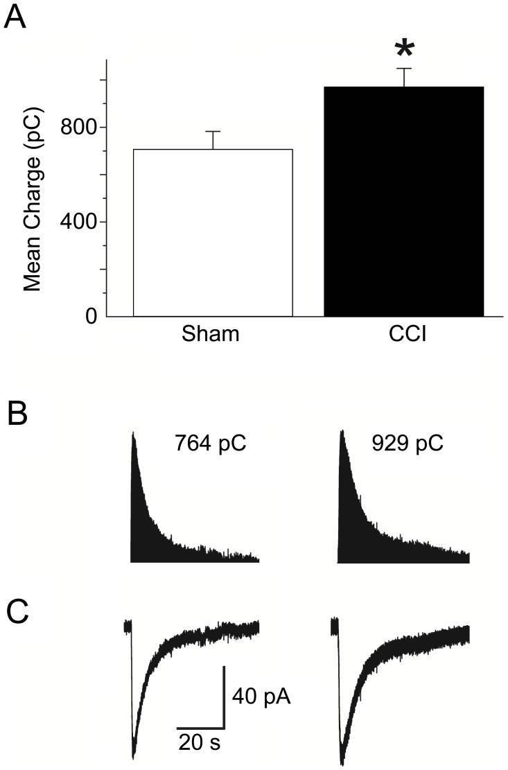
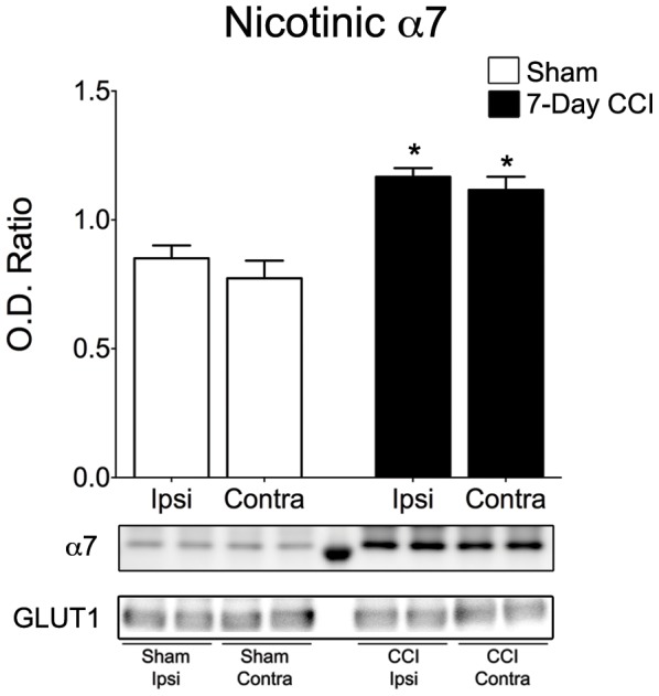
Traumatic brain injury (TBI) is a major public health concern affecting a large number of athletes and military personnel. Individuals suffering from a TBI risk developing anxiety disorders, yet the pathophysiological alterations that result in the development of anxiety disorders have not yet been identified. One region often damaged by a TBI is the basolateral amygdala (BLA); hyperactivity within the BLA is associated with increased expression of anxiety and fear, yet the functional alterations that lead to BLA hyperexcitability after TBI have not been identified. We assessed the functional alterations in inhibitory synaptic transmission in the BLA and one mechanism that modulates excitatory synaptic transmission, the α7 containing nicotinic acetylcholine receptor (α7-nAChR), after mTBI, to shed light on the mechanisms that contribute to increased anxiety-like behaviors. Seven and 30 days after a mild controlled cortical impact (CCI) injury, animals displayed significantly greater anxiety-like behavior. This was associated with a significant loss of GABAergic interneurons and significant reductions in the frequency and amplitude of spontaneous and miniature GABAA-receptor mediated inhibitory postsynaptic currents (IPSCs). Decreases in the mIPSC amplitude were associated with reduced surface expression of α1, β2, and γ2 GABAA receptor subunits. However, significant increases in the surface expression and current mediated by α7-nAChR, were observed, signifying increases in the excitability of principal neurons within the BLA. These results suggest that mTBI causes not only a significant reduction in inhibition in the BLA, but also an increase in neuronal excitability, which may contribute to hyperexcitability and the development of anxiety disorders.


Conflict of interest statement

Competing Interests: The authors have declared that no competing interests exist.

Figures

Figure 1
Figure 1. Mild TBI increases anxiety-like behaviors in the open field test.
(A) No differences in percent time spent in the center of the open field were found between CCI (n = 19) and sham (n = 19) animals 24 hours after injury. However, CCI rats spent significantly less time in the center of the open field 7- and 30-days after injury compared to sham animals. No significant differences were found between the sham and CCI animals in distance traveled (B), vertical activity (C), or movement time (D) at any of the time points. Bars show the mean ± SE of the percentage of time spent in the center (A), distance traveled (B), vertical activity (C), and movement time (D). *p<0.05.
Figure 2
Figure 2. Mild CCI does not cause a significant loss of neurons in the BLA 24 hours or 7 days after injury.
(A) Panoramic photomicrograph of Nissl-stained brain slice. Indicated are the sites of impact and the ipsilateral BLA. (B) Representative photomicrographs from Nissl-stained sections showing BLA cells from the ipsilateral (Top) and contralateral (Bottom) sides of sham, 1-day CCI, and 7-day CCI animals, respectively. Total magnification is 630X; scale bar, 50 µm. (C) Group data (mean ± SE; n = 8 for each group) of stereological estimation of the total number of Nissl-stained neurons in the BLA.
Figure 3
Figure 3. Delayed loss of GABAergic interneurons in the BLA within the first week after mild CCI.
(A) Representative photomicrographs of GAD-67 immunohistochemically stained GABAergic interneurons in the BLA of sham (left), 1-day CCI (middle), and 7-day CCI (right) animals. Total magnification is 630x; scale bar, 50 µm. (B) group data showing the mean and standard error of the stereologically estimated total number of GAD-67-positive cells in the BLA 1- and 7-days after CCI compared with sham. Only 7-days after CCI was there a significant bilateral reduction in GAD-67-positive cells indicating a delayed loss of GABAergic interneurons. ***p<0.001; n = 10 for each group.
Figure 4
Figure 4. Mild CCI causes a significant decrease in the frequency and amplitude of sIPSCs in the BLA 7-days after CCI.
sIPSCs were recorded from pyramidal-shaped neurons in the presence of CNQX, D-AP5, SCH50911, and LY 3414953, at a holding potential of −70 mV. Representative examples of recordings obtained in the BLA are shown in (A) and (B) for Sham and CCI 7-day animals, respectively. (C) Group data showing the change in the percentage frequency and amplitude of sIPSCs from CCI animals relative to Sham animals. The frequency and amplitude, but not the rise time and the decay time constant of the sIPSCs were significantly reduced in the CCI group compared to the sham controls. *p<0.05; **p<0.01; n = 18 for each group.
Figure 5
Figure 5. Mild CCI causes a significant decrease in the frequency and amplitude of mIPSCs in the BLA 7-days after CCI.
mIPSCs were recorded from pyramidal-shaped neurons in the presence of CNQX, D-AP5, SCH50911, LY 3414953, and TTX at a holding potential of –70 mV. Representative examples of recordings obtained in the BLA are shown in (A) and (B) for Sham and CCI 7 day animals, respectively. (C) Group data showing the change in the percentage frequency and amplitude of mIPSCs from CCI animals relative to Sham animals. The frequency and amplitude, but not the rise time and the decay time constant of the sIPSCs were significantly reduced in the CCI group compared to the sham controls. The recorded currents were blocked by the GABAA receptor antagonist bicuculline (data not shown). *p<0.05; **p<0.01; n = 17 for each group.
Figure 6
Figure 6. Surface expression of α1, β2, and γ2 GABAA receptor subunits is reduced in the BLA of CCI animals 7 days after mild CCI.
Western blot for subunits of (A) α1, (B) β2, and (C) γ2 subunits, respectively, was performed using biotinylated proteins isolated from the ipsilateral and contralateral sides of Sham and CCI 7-day animals. Group data showing the mean ± SE of the ratio between each subunit and GLUT1 optical densities. Top panel: representative Western blot for α1 (A), β2 (B), and γ2 (C) subunits of GABAA receptors, respectively. Bottom panel: representative Western blot for GLUT1, used as a loading control. Note that surface expression of GABAA α1, β2 and γ2 subunits are reduced in CCI animals when compared to Sham animals. *p<0.01; n = 4 for each group.
Figure 7
Figure 7. Activation of α7-nAChRs by fast application of the α7-nAChR agonist tricholine citrate, in the BLA, shows increased cholinergic conductance 7-days after CCI.
(A) Group data showing the mean ± SE charge transfer in pyramidal-shaped neurons in the BLA from CCI rats 7-days after injury (970±80 pC; n = 17) was significantly increased compared to sham rats (715±66 pC; n = 16). (B) Representative charge transfer from α7-nAChRs from sham (left), and 7-day CCI (right) animals. Note the increase in decay through α7-nAChRs in the BLA at day 7 post injury. (C) Representative α7-nAChR-mediated currents recorded from sham (left), and 7-day CCI (right) animals. The increase in the decay and amplitude of the α7-nAChR-mediated current 7-days post CCI led to increases in the charge transferred through by α7-nAChRs. Experiments were recorded in the presence of α-conotoxin Au1B, DHβE, atropine sulfate, D-AP5, CNQX, SCH50911, LY 3414953, and bicuculline. *p<0.05;
Figure 8
Figure 8. Surface expression of α7 subunit of neuronal nicotinic acetylcholine receptor is increased in the BLA of CCI animals 7-days after mild CCI.
Western blot was performed using biotinylated proteins isolated from the ipsilateral and contralateral sides of Sham and CCI 7-day animals. Group data showing the mean ± SE of the ratio between the α7 subunit and GLUT1 optical densities. Top panel: representative Western blot for α7-nAChRs. Bottom panel: representative Western blot for GLUT1, used as a loading control. *p<0.05; n = 4 for each group.

References

    1. Faul M, Xu L, Wald M, Coronado V (2010) Traumatic Brain Injury in the United States: Emergency Department Visits, Hospitalizations and Deaths 2002–2006. Atlanta (GA): Centers for Disease Control and Prevention, National Center for Injury Prevention and Control.
    1. Bigler ED, Maxwell WL (2012) Neuropathology of mild traumatic brain injury: relationship to neuroimaging findings. Brain Imaging Behav 6: 108–136. - PubMed
    1. Wagner AK, Postal BA, Darrah SD, Chen X, Khan AS (2007) Deficits in novelty exploration after controlled cortical impact. J Neurotrauma 24: 1308–1320. - PubMed
    1. Sosin DM, Sniezek JE, Thurman DJ (1996) Incidence of mild and moderate brain injury in the United States, 1991. Brain Inj 10: 47–54. - PubMed
    1. Kelly JP, Rosenberg JH (1997) Diagnosis and management of concussion in sports. Neurology 48: 575–580. - PubMed

Publication types