. 2014 Jul;16(7):595-605.
doi: 10.1016/j.neo.2014.06.005. Epub 2014 Jul 18.

SC-2001 overcomes STAT3-mediated sorafenib resistance through RFX-1/SHP-1 activation in hepatocellular carcinoma

Affiliations

SC-2001 overcomes STAT3-mediated sorafenib resistance through RFX-1/SHP-1 activation in hepatocellular carcinoma

Jung-Chen Su et al. Neoplasia. 2014 Jul.

Abstract

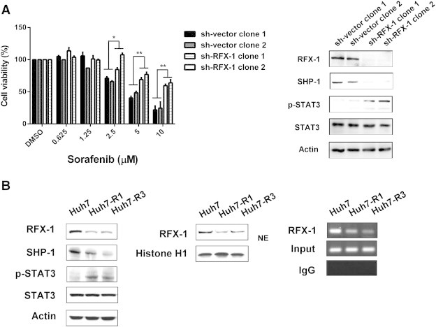
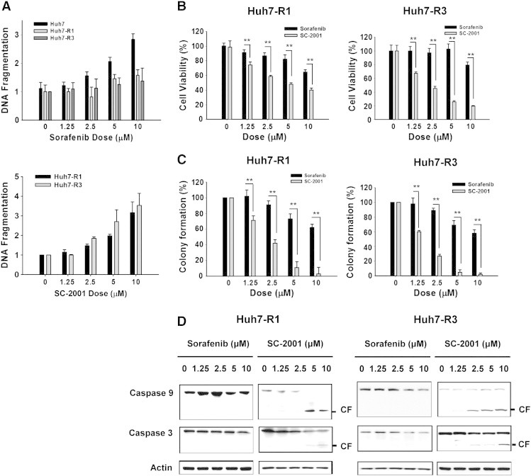
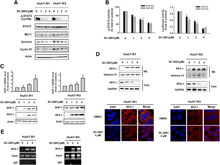
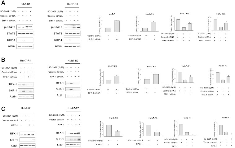
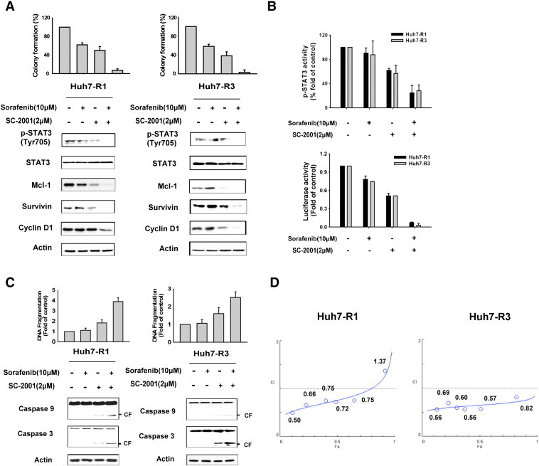
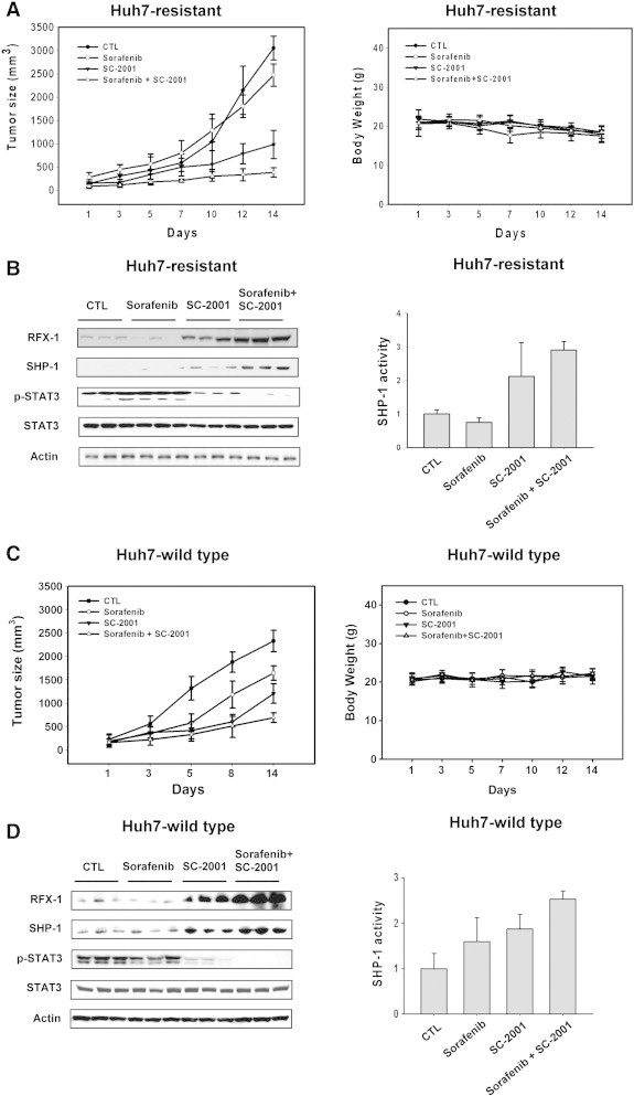
Hepatocellular carcinoma is the fifth most common solid cancer worldwide. Sorafenib, a small multikinase inhibitor, is the only approved therapy for advanced HCC. The clinical benefit of sorafenib is offset by the acquisition of sorafenib resistance. Understanding of the molecular mechanism of STAT3 overexpression in sorafenib resistance is critical if the clinical benefits of this drug are to be improved. In this study, we explored our hypothesis that loss of RFX-1/SHP-1 and further increase of p-STAT3 as a result of sorafenib treatment induces sorafenib resistance as a cytoprotective response effect, thereby, limiting sorafenib sensitivity and efficiency. We found that knockdown of RFX-1 protected HCC cells against sorafenib-induced cell apoptosis and SHP-1 activity was required for the process. SC-2001, a molecule with similar structure to obatoclax, synergistically suppressed tumor growth when used in combination with sorafenib in vitro and overcame sorafenib resistance through up-regulating RFX-1 and SHP-1 resulting in tumor suppression and mediation of dephosphorylation of STAT3. In addition, sustained sorafenib treatment in HCC led to increased p-STAT3 which was a key mediator of sorafenib sensitivity. The combination of SC-2001 and sorafenib strongly inhibited tumor growth in both wild-type and sorafenib-resistant HCC cell bearing xenograft models. These results demonstrate that inactivation of RFX/SHP-1 induced by sustained sorafenib treatment confers sorafenib resistance to HCC through p-STAT3 up-regulation. These effects can be overcome by SC-2001 through RFX-1/SHP-1 dependent p-STAT3 suppression. In conclusion, the use of SC-2001 in combination with sorafenib may constitute a new strategy for HCC therapy.


Figures

Figure 1
Figure 1
RFX-1 contributes to the growth inhibition effect of sorafenib in HCC cells. (A) Left, The cell proliferation of PLC5 sh-Vector (sh-vector clone 1 and 2) and sh-RFX-1 (sh-RFX-1 clone 1 and 2) cells were analyzed by MTT assay under sorafenib treatment. *P < 0.05, **P < 0.01. Right, The protein levels of RFX-1, SHP-1, and p-STAT3 in PLC5 sh-Vector (sh-vector clone 1 and 2) and sh-RFX-1 (sh-RFX-1 clone 1 and 2) cells were determined by western blot. Actin was used as a loading control. (B) Left, The protein levels of RFX-1, SHP-1, and p-STAT3 in Huh7 WT, R1, and R3 cells were determined by western blot. Actin was used as a loading control. Middle, The levels of nuclear extraction of RFX-1 in Huh7 WT, R1, and R3 cells were measured by western blot. Right, Chromatin immunoprecipitation assay was performed in Huh7 WT, R1, and R3 cells. The crude lysates from WT, R1, and R3 cells were immunoprecipitated, respectively, with RFX-1 antibody and rabbit IgG control and captured by protein A-agarose beads. The precipitated DNA fragments were amplified by PCR to detect fragments in SHP-1 promoter containing the RFX-1 binding site.
Figure 2
Figure 2
SC-2001 overcomes sorafenib resistance in HCC cells by enhancing cell viability, inhibition and apoptosis. (A) Top, DNA fragmentation assay was performed in Huh7 WT, R1, and R3 cells undergoing sorafenib treatment at different dosages. Bottom, DNA fragmentation assay was performed in Huh7 R1 and Huh7 R3 cells undergoing SC-2001 treatment at different dosages. (B) Huh7 R1 (left) and Huh7 R3 (right) cells were treated, respectively, with sorafenib and SC-2001 in dose dependent manner for 48 h. Cell viability was measured by MTT assay. **P < 0.01. (C) Huh7 R1 (left) and Huh7 R3 (right) cells were treated, respectively, with sorafenib and SC-2001 in dose dependent manner for 48 h. After treatment, the cells were seeded onto 6-well plates for colony formation assay. After 2 weeks, the cells were stained with crystal violet and the colonies were counted. **P < 0.01. (D) The protein levels of caspase-3 and caspase-9 were determined by western blot after Huh7 R1 and Huh7 R3 cells were exposed to sorafenib and SC-2001 for 48 h. Actin was used as a loading control.
Figure 3
Figure 3
Induction of RFX-1/SHP-1 by SC-2001 plays a role in overcoming STAT3 dependent-sorafenib-resistance in HCC cells. (A) Huh7 R1 and R3 cells were exposed to SC-2001 in dose dependent manner for 24 h. The cell lysates were subjected to western blot analysis with p-STAT3, Mcl-1, survivin, and cyclin D1 antibodies to analyze protein levels. Actin was used as a loading control. (B) Left, STAT3 activity ELISA was performed in Huh7 R1 and R3 cells after treatment with SC-2001 in dose dependent manner for 24 h. Right, Huh7 R1 and R3 cells were transfected with either vector or luciferase reporter driven by a promoter region containing STAT3-specific binding sites for 48 h. After transfection, the cells were treated with SC-2001 in dose dependent manner for 6 h. Luciferase activity was measured after the treatment. (C) The mRNA levels of SHP-1 and the protein levels of SHP-1 and RFX-1 in Huh7 R1 and R3 cells were determined. The mRNA level was analyzed by QPCR and the protein level was analyzed by western blot. (D) Top, Huh7 R1 and R3 cells were treated with SC-2001 in dose dependent manner. After treatment, the nuclear and cytosol extraction of RFX-1 was measured by western blot. Histone H1 was included as a loading control NE. Bottom, immunofluorescence staining, Huh7 R1 and R3 cells cells were treated with 4 μM SC-2001 for 24 h. After the end of treatment, RFX-1 was determined by immunofluorescence staining (red). Nuclei were counterstained with DAPI (blue). Representative confocal micrographs are shown. (E) Chromatin immunoprecipitation assay (CHIP) was conducted in Huh7 R1 and R3 cells after exposure to SC-2001 for 12 h. CHIP assay was performed as described above.
Figure 4
Figure 4
SC-2001-induced inhibition of colony formation in sorafenib-resistant HCC cells is RFX-1/SHP-1 dependent. (A) Huh7 R1 and R3 cells were transfected, respectively, with control siRNA or SHP-1 siRNA for 48 h. After transfection, the cells were treated with or without SC-2001 for 24 h and subjected to western blot assay to analyze the protein levels of p-STAT3 and SHP-1 or seeded on a 6-well plate for the colony forming assay. (B) Huh7 R1 and R3 cells were transfected, respectively, with control siRNA or RFX-1 siRNA for 48 h. After transfection, the cells were treated with or without SC-2001 for 24 h and subjected to western blot assay to analyze the protein levels of RFX-1 and SHP-1 or seeded on a 6-well plate for the colony forming assay. (C) Huh7 R1 and R3 cells were transfected, respectively, with control vector or RFX-1 overexpression plasmid for 48 h. After transfection, the cells were treated with or without SC-2001 for 24 h and the cells were subjected to western blot assay to analyze the protein levels of RFX-1 and SHP-1 or seeded on a 6-well plate for colony forming assay.
Figure 5
Figure 5
Use of SC-2001 in combination with sorafenib synergizes the anticancer effect. (A) Huh7 R1 and R3 cells were treated with a range of concentrations sorafenib and/or SC-2001 at a fixed ratio of 5:1 for 48 h. The colony formation (top) and western blot (bottom) were performed as described above after the treatment. (B) Top, Huh7 R1 and R3 cells were treated as described above and the STAT3 activity ELISA was performed after the treatment. Bottom, Huh7 R1 and R3 cells were transfected with either vector or luciferase reporter driven by a promoter region containing STAT3-specific binding sites for 48 h. After transfection, the cells were treated as described above and the luciferase activity was measured after the treatment. (C) The DNA fragmentation and protein levels of caspase-3 and caspase-9 were analyzed in Huh7 R1 and R3 cells after treatment with sorafenib and/or SC-2001 for 48 h as described above. (D) Huh7 R1 and R3 cells were treated with sorafenib and/or SC-2001 for 48 h administered over a range of concentrations at a fixed ratio of 5:1. After the cell viability was determined in each condition by MTT assay, the combination index (CI) was calculated by CompuSyn software. CI values of less than 1 were considered to respresent synergism.
Figure 6
Figure 6
SC-2001 potentiates the antitumor effect of sorafenib in both sorafenib-sensitive and sorafenib-resistant Huh7 xenograft models. (A) Left, Huh7-resistant cells were subcutaneously injected into nude mice. When tumors reached 100 mm3, the mice were then treated with sorafenib and/or SC-2001 orally for 2 weeks. Tumor volume was measured and calculated according to the formula described in Materials and Methods. Right, The body weight of nude mice bearing Huh7-resistant cells was measured. (B) Left, The protein levels of RFX-1, SHP-1, and p-STAT3 in Huh7-resistant cells in vivo were determined by western blot. Right, The in vivo SHP-1 activity in Huh7-resistant cells was determined. (C) Left, Huh7-wild type cells were subcutaneously injected into nude mice. When tumors reached 100 mm3, the mice were treated with sorafenib and/or SC-2001 orally for 2 weeks. The tumor volume was measured and calculated according to the formula described in Materials and Methods. Right, The body weight of nude mice bearing Huh7-wild type cells was measured. (D) Left, The in vivo protein levels of RFX-1, SHP-1, and p-STAT3 in Huh7 wild-type cells were determined by western blot. Right, The in vivo SHP-1 activity in Huh7 wild type cells was determined.

References

    1. Bruix J., Llovet J.M. HCC surveillance: who is the target population? Hepatology. 2003;37:507–509. - PubMed
    1. Llovet J.M., Di Bisceglie A.M., Bruix J., Kramer B.S., Lencioni R., Zhu A.X., Sherman M., Schwartz M., Lotze M., Talwalkar J. Design and endpoints of clinical trials in hepatocellular carcinoma. J Natl Cancer Inst. 2008;100:698–711. - PubMed
    1. Di Maio M., Daniele B., Perrone F. Targeted therapies: Role of sorafenib in HCC patients with compromised liver function. Nat Rev Clin Oncol. 2009;6:505–506. - PubMed
    1. Panka D.J., Wang W., Atkins M.B., Mier J.W. The Raf inhibitor BAY 43-9006 (Sorafenib) induces caspase-independent apoptosis in melanoma cells. Cancer Res. 2006;66:1611–1619. - PubMed
    1. Wilhelm S.M., Carter C., Tang L., Wilkie D., McNabola A., Rong H., Chen C., Zhang X., Vincent P., McHugh M. BAY 43-9006 exhibits broad spectrum oral antitumor activity and targets the RAF/MEK/ERK pathway and receptor tyrosine kinases involved in tumor progression and angiogenesis. Cancer Res. 2004;64:7099–7109. - PubMed

Publication types

MeSH terms