

RNAi-mediated TCR knockdown prevents autoimmunity in mice caused by mixed TCR dimers following TCR gene transfer
- PMID: 25048215
- PMCID: PMC4429734
- DOI: 10.1038/mt.2014.142
RNAi-mediated TCR knockdown prevents autoimmunity in mice caused by mixed TCR dimers following TCR gene transfer
Abstract
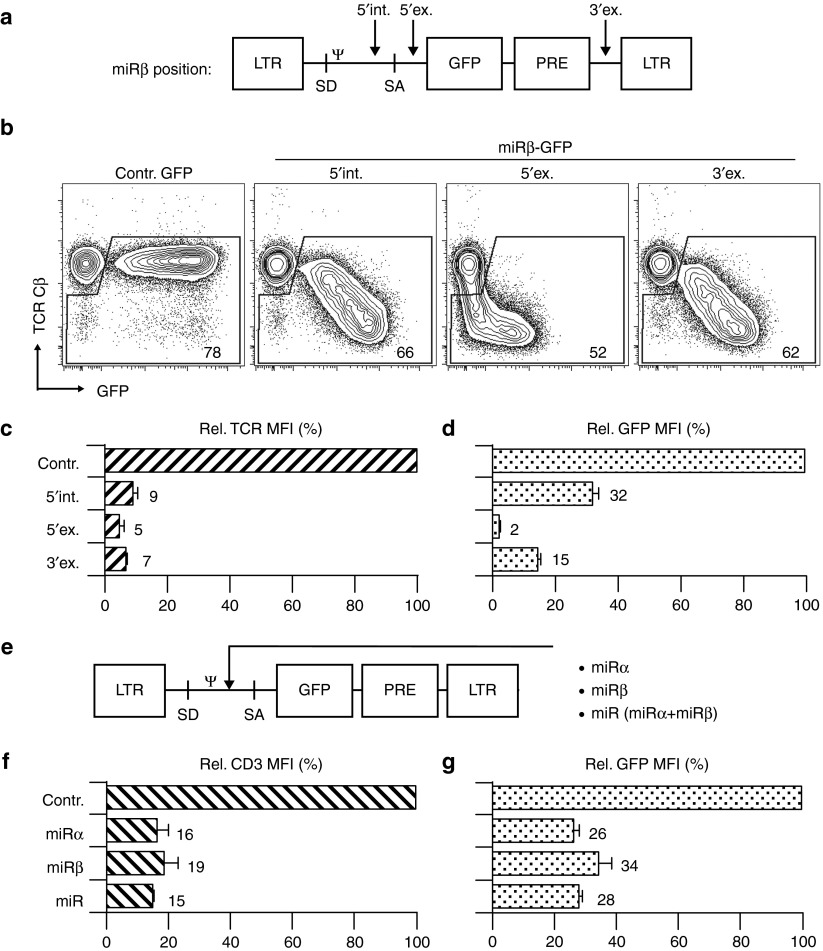
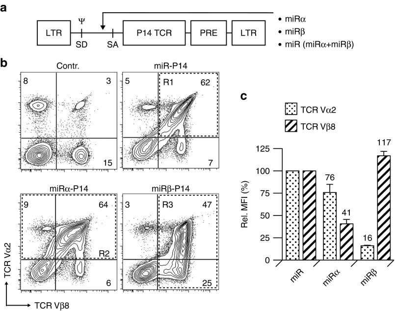
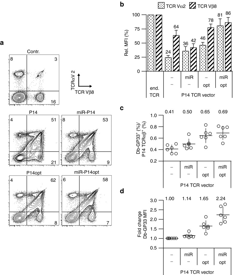
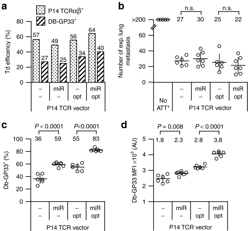
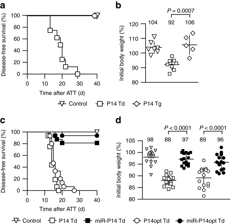
Genetically modified T cells that express a transduced T cell receptor (TCR) α/β heterodimer in addition to their endogenous TCR are used in clinical studies to treat cancer. These cells express two TCR-α and two TCR-β chains that do not only compete for CD3 proteins but also form potentially self-reactive mixed TCR dimers, composed of endogenous and transferred chains. To overcome these deficits, we developed an RNAi-TCR replacement vector that simultaneously silences the endogenous TCR and expresses an RNAi-resistant TCR. Transduction of the virus-specific P14 TCR without RNAi resulted in unequal P14 TCR-α and -β chain surface levels, indicating heterodimerization with endogenous TCR chains. Such unequal expression was also observed following TCR gene optimization. Equal surface levels of the introduced TCR chains were however achieved by silencing the endogenous TCR. Importantly, all mice that received cells transduced with the native or optimized P14 TCR developed lethal TCR gene transfer-induced graft-versus-host-disease (TI-GVHD) due to formation of mixed TCR dimers. In contrast, TI-GVHD was almost completely prevented when using the RNAi-TCR replacement vector. Our data demonstrate that RNAi-assisted TCR replacement reduces the formation of mixed TCR dimers, and thereby significantly reduces the risk of TI-GVHD in TCR gene therapy.
Figures
Similar articles
-
Immunotherapy with TCR-redirected T cells: comparison of TCR-transduced and TCR-engineered hematopoietic stem cell-derived T cells.J Immunol. 2014 Jan 1;192(1):206-13. doi: 10.4049/jimmunol.1202591. Epub 2013 Nov 29. J Immunol. 2014. PMID: 24293634
-
Retroviral transfer of a dominant TCR prevents surface expression of a large proportion of the endogenous TCR repertoire in human T cells.Gene Ther. 2008 Apr;15(8):625-31. doi: 10.1038/sj.gt.3303078. Epub 2008 Feb 28. Gene Ther. 2008. PMID: 18305579
-
Mixed T cell receptor dimers harbor potentially harmful neoreactivity.Proc Natl Acad Sci U S A. 2010 Jun 15;107(24):10972-7. doi: 10.1073/pnas.1005802107. Epub 2010 Jun 1. Proc Natl Acad Sci U S A. 2010. PMID: 20534461 Free PMC article.
-
Improved expression and reactivity of transduced tumor-specific TCRs in human lymphocytes by specific silencing of endogenous TCR.Cancer Res. 2009 Dec 1;69(23):9003-11. doi: 10.1158/0008-5472.CAN-09-1450. Epub 2009 Nov 10. Cancer Res. 2009. PMID: 19903853
-
The generation and selection of the T cell repertoire: insights from studies of the molecular basis of T cell recognition.Immunol Rev. 1988 Jan;101:81-113. doi: 10.1111/j.1600-065x.1988.tb00733.x. Immunol Rev. 1988. PMID: 2450828 Review.
Cited by
-
Breaking Bottlenecks for the TCR Therapy of Cancer.Cells. 2020 Sep 14;9(9):2095. doi: 10.3390/cells9092095. Cells. 2020. PMID: 32937956 Free PMC article. Review.
-
Toxicities Associated With Adoptive T-Cell Transfer for Cancer.Cancer J. 2015 Nov-Dec;21(6):506-9. doi: 10.1097/PPO.0000000000000157. Cancer J. 2015. PMID: 26588684 Free PMC article. Review.
-
Genome-edited allogeneic donor "universal" chimeric antigen receptor T cells.Blood. 2023 Feb 23;141(8):835-845. doi: 10.1182/blood.2022016204. Blood. 2023. PMID: 36223560 Free PMC article.
-
Donor T cells for CAR T cell therapy.Biomark Res. 2022 Apr 1;10(1):14. doi: 10.1186/s40364-022-00359-3. Biomark Res. 2022. PMID: 35365224 Free PMC article. Review.
-
Orthotopic T-Cell Receptor Replacement-An "Enabler" for TCR-Based Therapies.Cells. 2020 Jun 1;9(6):1367. doi: 10.3390/cells9061367. Cells. 2020. PMID: 32492858 Free PMC article. Review.
References
-
- Clay TM, Custer MC, Sachs J, Hwu P, Rosenberg SA, Nishimura MI. Efficient transfer of a tumor antigen-reactive TCR to human peripheral blood lymphocytes confers anti-tumor reactivity. J Immunol. 1999;163:507–513. - PubMed
-
- Fujio K, Misaki Y, Setoguchi K, Morita S, Kawahata K, Kato I, et al. Functional reconstitution of class II MHC-restricted T cell immunity mediated by retroviral transfer of the alpha beta TCR complex. J Immunol. 2000;165:528–532. - PubMed
-
- Kessels HW, Wolkers MC, van den Boom MD, van der Valk MA, Schumacher TN. Immunotherapy through TCR gene transfer. Nat Immunol. 2001;2:957–961. - PubMed
-
- de Witte MA, Coccoris M, Wolkers MC, van den Boom MD, Mesman EM, Song JY, et al. Targeting self-antigens through allogeneic TCR gene transfer. Blood. 2006;108:870–877. - PubMed
Publication types
MeSH terms
Substances
LinkOut - more resources
Full Text Sources
Other Literature Sources
Medical
Research Materials
