. 2014 Jul 21;9(7):e102164.
doi: 10.1371/journal.pone.0102164. eCollection 2014.

Real-time imaging of the epithelial-mesenchymal transition using microRNA-200a sequence-based molecular beacon-conjugated magnetic nanoparticles

Affiliations

Real-time imaging of the epithelial-mesenchymal transition using microRNA-200a sequence-based molecular beacon-conjugated magnetic nanoparticles

YoonSeok Choi et al. PLoS One. .

Abstract

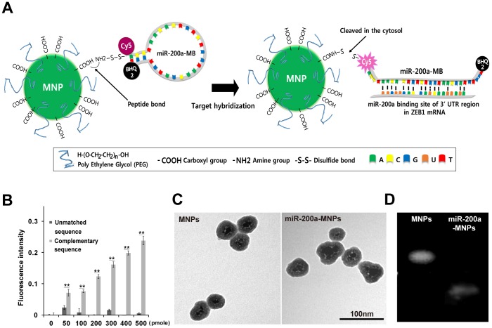
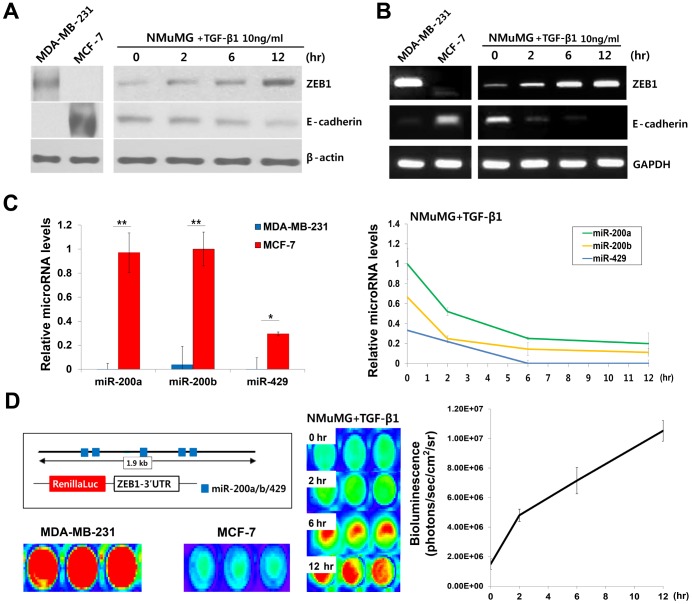
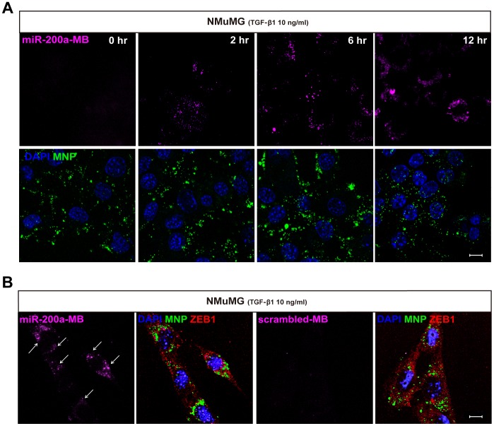
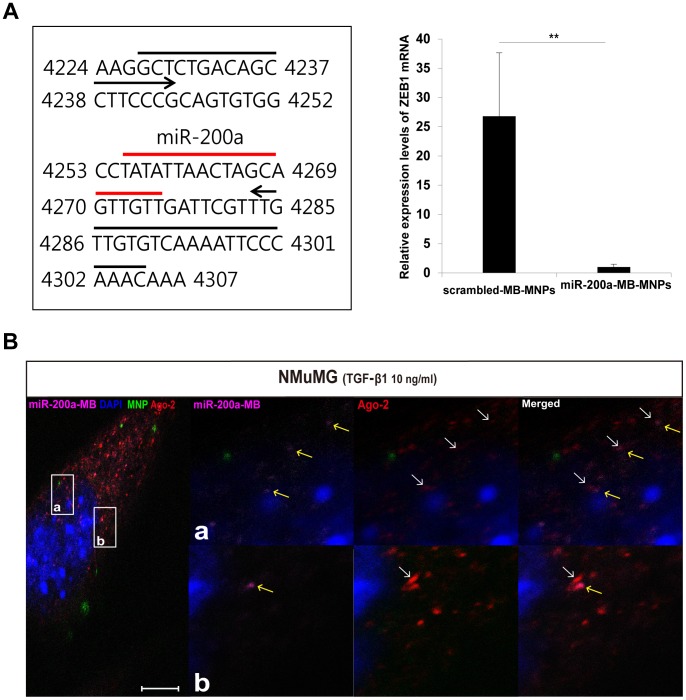
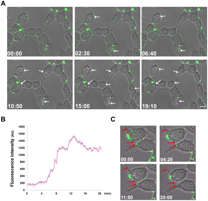
The epithelial-mesenchymal transition (EMT) plays important roles in tumor progression to metastasis. Thus, the development of an imaging probe that can monitor transient periods of the EMT process in live cells is required for a better understanding of metastatic process. Inspired by the fact that the mRNA expression levels of zinc finger E-box-binding homeobox 1 (ZEB1) increase when cells adopt mesenchyme characteristics and that microRNA-200a (miR-200a) can bind to ZEB1 mRNA, we conjugated molecular beacon (MB) mimicking mature miR-200a to magnetic nanoparticles (miR-200a-MB-MNPs) and devised an imaging method to observe transitional changes in the cells during EMT. Transforming growth factor-β1 treated epithelial cells and breast cancer cell lines representing both epithelial and mesenchymal phenotypes were used for the validation of miR-200a-MB-MNPs as an EMT imaging probe. The real-time imaging of live cells acquired with the induction of EMT revealed an increase in fluorescence signals by miR-200a-MB-MNPs, cell morphology alterations, and the loss of cell-cell adhesion. Our results suggest that miR-200a-MB-MNPs can be used as an imaging probe for the real-time monitoring of the EMT process in live cells.


Conflict of interest statement

Competing Interests: The authors have declared that no competing interests exist.

Figures

Figure 1
Figure 1. Characterization of miR-200a-MB-MNPs.
(A) A schematic representation of the miR-200a-MB-MNP structure and EMT imaging probe activation. (B) Cy5 fluorescence from miR-200a-MBs. Different concentrations of a synthetic oligonucleotide target either complementary to or unmatched to miR-200a were added to the miR-200a-MBs. ** indicates p<0.01. (C) TEM images of MNPs and miR-200a-MB-MNPs. The diameters of the MNPs and miR-200a-MB-MNPs were approximately 50 nm. (D) An agarose gel electrophoresis image; MNPs and miR-200a-MB-MNPs were loaded onto 0.5% agarose gels.
Figure 2
Figure 2. Reciprocal expression of E-cadherin, ZEB1, and the miR-200 family during EMT.
(A, B) Western blot and RT-PCR analyses of ZEB1 and E-cadherin in MCF-7, MDA-MB-231, and TGF-β1 treated NMuMG cells. (C) Real-time PCR analysis of the miR-200 family. The levels of miR-200a, b, and 429 in MCF-7, MDA-MB-231, and TGF-β1 treated NMuMG cells were evaluated. *, ** indicates p<0.05 or p<0.01, respectively. (D) A schematic image of the Renilla luciferase reporter and the analysis of luminescence signals for evaluating the binding availability of miR-200a-MBs. After transfection of pRL-ZEB1 plasmid, the luminescence signals in MCF-7, MDA-MB-231, and TGF-β1 treated NMuMG cells were analyzed. Data are from three independent experiments.
Figure 3
Figure 3. Validation of miR-200a-MB-MNPs as an EMT imaging probe.
(A) miR-200a-MB-MNPs were introduced into NMuMG cells at 0, 2, 6, and 12 hours post-TGF-β1 treatment. Cy5 fluorescence (pink) was observed at 2 hours after TGF-β1 treatment, and the Cy5 signals increased in a time-dependent manner. (B) Cy5 signals were analyzed in either miR-200a-MB-MNPs or scrambled-MB-MNPs delivered NMuMG cells after 6 hours of TGF-β1 treatment. Immunostaining of ZEB1 was performed to confirm the mesenchymal transformation of NMuMG into mesenchymal phenotype. Scale bar, 10 µm.
Figure 4
Figure 4. Analysis of the hybridization ability of miR-200a-MBs to ZEB1 mRNA using real-time PCR and immunostaining.
(A) The illustration represents the binding sites of the primers used in real-time PCR. Real-time PCR analysis showed reduced expression levels of ZEB1 mRNA in miR-200a-MB-MNPs delivered NMuMG cells compared to the cells with scrambled-MB-MNPs. ** indicates p<0.01. (B) The localization of miR-200a-MBs and the RISC subunit Ago-2 were observed within the several regions of TGF-β1 treated cell. The Cy5 signals (yellow arrows) of miR-200a-MBs were in close proximity to the Alexa-546 signals of Ago-2 (white arrows). Small boxes were marked with a & b for the indication of the magnified regions. Scale bar, 10 µm.
Figure 5
Figure 5. Real-time imaging of the 7EMT process in live cells.
(A) After the delivery of miR-200a-MB-MNPs (20 µg/ml) into NMuMG cells, real-time imaging was performed following TGF-β1 (10 ng/ml) treatment. From 2 minute 30 seconds after EMT induction, NMuMG cells containing MNPs (green) in their cytoplasm showed Cy5 fluorescence (white arrows) generated by the miR-200a-MBs. (B) The Cy5 fluorescence intensity was quantified with Image J, showing increased signals generated by miR-200a-MBs. (C) During the acquisition period of real-time imaging, the NMuMG cells exhibited morphological changes and the loss of cell-cell adhesions (red arrows) after TGF-β1 treatment. Scale bar, 10 µm.

References

    1. Acloque H, Adams MS, Fishwick K, Bronner-Fraser M, Nieto MA (2009) Epithelial-mesenchymal transitions: the importance of changing cell state in development and disease. J Clin Invest 119: 1438–1449. - PMC - PubMed
    1. Chaffer CL, Weinberg RA (2011) A perspective on cancer cell metastasis. Science 331: 1559–1564. - PubMed
    1. De Craene B, Berx G (2013) Regulatory networks defining EMT during cancer initiation and progression. Nat Rev Cancer 13: 97–110. - PubMed
    1. Eger A, Stockinger A, Schaffhauser B, Beug H, Foisner R (2000) Epithelial mesenchymal transition by c-Fos estrogen receptor activation involves nuclear translocation of beta-catenin and upregulation of beta-catenin/lymphoid enhancer binding factor-1 transcriptional activity. J Cell Biol 148: 173–188. - PMC - PubMed
    1. Eger A, Aigner K, Sonderegger S, Dampier B, Oehler S, et al. (2005) DeltaEF1 is a transcriptional repressor of E-cadherin and regulates epithelial plasticity in breast cancer cells. Oncogene 24: 2375–2385. - PubMed

Publication types