. 2014 Jul 22:5:4413.
doi: 10.1038/ncomms5413.

Protein painting reveals solvent-excluded drug targets hidden within native protein-protein interfaces

Affiliations

Protein painting reveals solvent-excluded drug targets hidden within native protein-protein interfaces

Alessandra Luchini et al. Nat Commun. .

Abstract

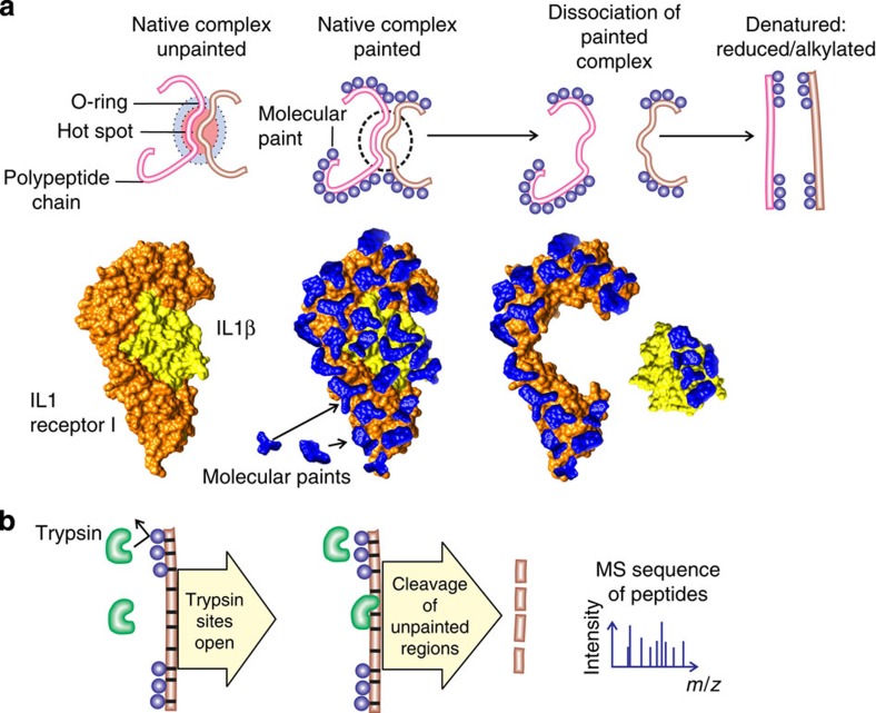
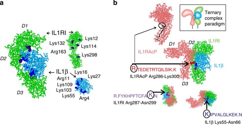
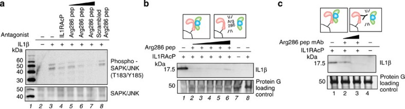
Identifying the contact regions between a protein and its binding partners is essential for creating therapies that block the interaction. Unfortunately, such contact regions are extremely difficult to characterize because they are hidden inside the binding interface. Here we introduce protein painting as a new tool that employs small molecules as molecular paints to tightly coat the surface of protein-protein complexes. The molecular paints, which block trypsin cleavage sites, are excluded from the binding interface. Following mass spectrometry, only peptides hidden in the interface emerge as positive hits, revealing the functional contact regions that are drug targets. We use protein painting to discover contact regions between the three-way interaction of IL1β ligand, the receptor IL1RI and the accessory protein IL1RAcP. We then use this information to create peptides and monoclonal antibodies that block the interaction and abolish IL1β cell signalling. The technology is broadly applicable to discover protein interaction drug targets.


Figures

Figure 1
Figure 1. Protein painting reveals hidden native hot spots of protein interactions.
(a) Paint molecules coat the surface of native protein complexes but cannot gain access to solvent-inaccessible protein–protein interface regions. Interleukin 1β receptor–ligand complex depicted with bound paint molecules to scale. (b) Trypsin cleavage is blocked by the presence of paint molecules that bind non-covalently near trypsin consensus sequences. Following dissociation of painted proteins the area of interaction remains unpainted and is susceptible to trypsin cleavage. Thus, trypsin cleavage peptides will be derived exclusively from unpainted interface areas.
Figure 2
Figure 2. Molecular paints rapidly coat native proteins in solution.
(ac) Association (blue) and dissociation (red) binding curves (moles of paint per mole of protein) for paint molecules (structures are shown inset) associating with CA II. Molecular paints have unusually high association rates and low dissociation rates: saturation is reached within 5 min and the off rate is <10% dissociation after 2 h. (d) Calibration data for RBB associated to CA (absorbance spectra for RBB, CA and the complex is reported in Supplementary Fig. 7). (e) Scatchard plot of CA and AO50 shows the number of binding sites to be five, confirming the saturation point of the binding kinetics in (b). (f) Scatchard plot of CA and ANSA shows the number of binding sites to be three.
Figure 3
Figure 3. Paint molecule coating withstands denaturation.
(a) CA was pulsed with RBB and the dissociation kinetics was studied both in native conditions (PBS, red plot) and in denaturing conditions (2 M urea, black plot). The amount of RBB bound to the protein is very similar in the two conditions (bound RBB in 2 M urea >95% of bound RBB in PBS). (b) The amount of RBB bound to CA was compared in native conditions (PBS=100%, column 1), reducing conditions (2 M urea, 10 mM dithiothreitol=86%, column 2) and alkylating conditions (2 M urea, 10 mM DTT, 50 mM iodoacetamide=86%, column 3) after 2 hours of incubation. The conditions tested for this experiment reproduce the experimental steps that are applied to proteins before trypsin digestion for MS and did not show a significant reduction in the amount of dye bound to the denatured reduced and alkylated protein.
Figure 4
Figure 4. Molecular paints block trypsin cleavage sites.
(a) Amino acids highlighted in red are consensus trypsin cleavage sites of CA II that were identified by reverse-phase liquid chromatography nanospray tandem MS in the absence of molecular paint (unpainted). Molecular paints (RBB, AO50, R49 and CR) blocked all (100%) consensus trypsin cleavage sites (painted, indicated by a blue X). CA was chosen because it contains 80% of the trypsin cleavage consensus sites that represent all the variations of the amino acids at the carboxy-side of the arginine and lysine. To further confirm that all the possible trypsin cleavage consensus domain were conserved as binding sites for the molecular paints, we conducted similar experiments for aprotinin and albumin in addition to the IL1β-IL1RI-IL1RAcP complex that documented full coverage of all known trypsin cleavage consensus sites for any permissible amino acid. Trypsin cleavage sites are the staple of mass spec sequencing because they mark the protein polypeptide chain at the highest frequency (resolution) compared with other protease cleavage sites, and they are preferentially distributed on protein surfaces, and in or near previously identified solvent-excluded hot spots. (b) 3D representation of crystallography structure of CA (PDB no. 1V9E). Surface trypsin cleavage sites are represented in magenta, (c) Yellow crosses indicate trypsin cleavage sites that were blocked by the four molecular paints used (RBB, AO50, R49 and CR). (d) Example representation of AO50 blocking Arg250 on the surface of CA as predicted by docking calculations (SwissDock, PDB no. 1V9E, ZINC25693528).
Figure 5
Figure 5. Protein painting reveals hot spots between IL1β, IL1RI and IL1RAcP.
(a) Identified opposing contact points revealed by the method for the ligand (blue) bound to its receptor (green) before and after dissociation (Fisher exact test P-value<0.0003). Fisher exact test was applied in order to determine whether there was an enrichment of trypsin cleavage sites belonging to protein–protein interface regions among all the trypsin cleavage sites identified by the protein painting method. The total number of trypsin cleavage sites of the two proteins was 49 (26 in the protein–protein interface region). The total number of trypsin cleavage sites identified by the protein painting method was 17, 15 of which belonged to the interface (Fisher exact test P-value<0.0003). (b) IL1RAcP (pink) bound to the receptor (green) ligand (blue) complex. Sequences identified for each protein were found to be opposing and juxtaposed, as noted. The sequence labelled Arg286 (represented in black in the protein model) was used to generate a synthetic peptide antagonist and was used as an antigen for a mouse IgG mAb to Arg286 peptide. Protein painting correctly revealed key contact points with closest proximity obtained by X-ray crystallography and PDBePISA structural analysis software. The two closest interaction points in the three-way complex are IL1RAcP:Arg286–IL1β:Asp54, distance 2.49 Å and IL1RI:Lys298–IL1β:Ser52, distance 2.51 Å (PDB no. 4DEP). These contact points were correctly identified by protein painting in both protein partners. D1, D2 and D3 indicate the domains of IL1RI and IL1RAcP.
Figure 6
Figure 6. IL1RAcP:Arg286 peptide and mAb abolish interleukin signalling.
(a) Synthetic antagonist peptide Arg286, identified with protein painting, inhibited SAPK/JNK signalling downstream from IL1RI as effectively as IL1RAcP recombinant protein. In lane 8, scrambled peptide obtained by randomly shuffling Arg286 sequence does not inhibit the signalling downstream from IL1RI. Data are representative of three independent experiments. (b) Arg286 peptide inhibition of ligand pull-down within the receptor complex by His-tagged IL1RAcP. Schematic representation of the complex is depicted in insert. IL1RAcP in the absence of IL1RI does not pull down IL1β, lane 8. (c) Arg286 peptide mAb specific for IL1RAcP peptide extinguishes complex formation depicted in insert.

Comment in

References

    1. Nero T. L., Morton C. J., Holien J. K., Wielens J. & Parker M. W. Oncogenic protein interfaces: small molecules, big challenges. Nat. Rev. Cancer 14, 248–262 (2014). - PubMed
    1. Johnson R. & Halder G. The two faces of Hippo: targeting the Hippo pathway for regenerative medicine and cancer treatment. Nat. Rev. Drug Discov. 13, 63–79 (2014). - PMC - PubMed
    1. Bogan A. A. & Thorn K. S. Anatomy of hot spots in protein interfaces. J. Mol. Biol. 280, 1–9 (1998). - PubMed
    1. Arkin M. R. & Wells J. A. Small-molecule inhibitors of protein-protein interactions: progressing towards the dream. Nat. Rev. Drug Discov. 3, 301–317 (2004). - PubMed
    1. Kortemme T. & Baker D. A simple physical model for binding energy hot spots in protein-protein complexes. Proc. Natl Acad. Sci. USA 99, 14116–14121 (2002). - PMC - PubMed

Publication types

MeSH terms