Review
. 2014 Aug;11(8):465-75.
doi: 10.1038/nrurol.2014.162. Epub 2014 Jul 22.

Molecular genetics and cellular features of TFE3 and TFEB fusion kidney cancers

Affiliations
Review

Molecular genetics and cellular features of TFE3 and TFEB fusion kidney cancers

Eric C Kauffman et al. Nat Rev Urol. 2014 Aug.

Abstract

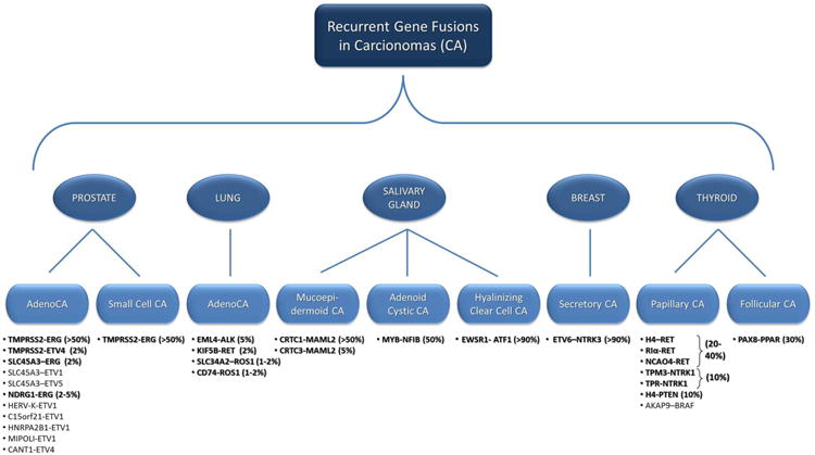
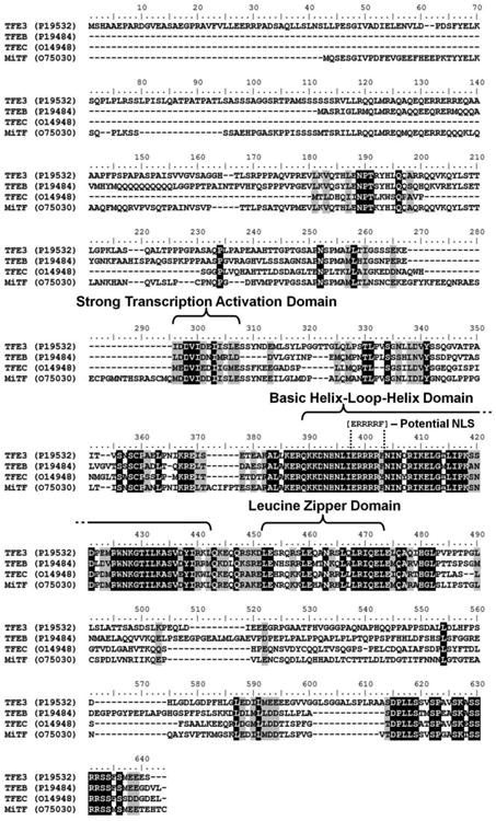
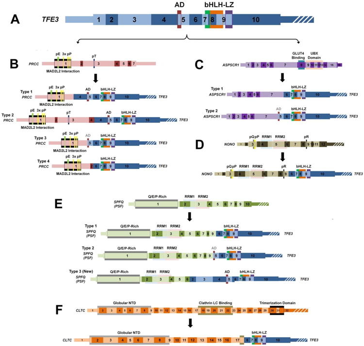
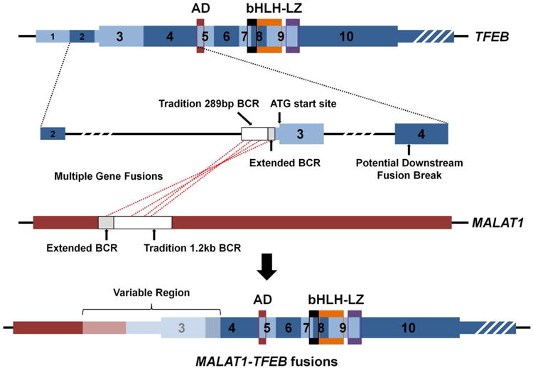
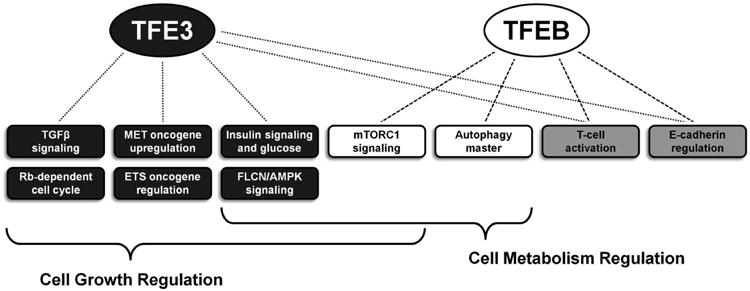
Despite nearly two decades passing since the discovery of gene fusions involving TFE3 or TFEB in sporadic renal cell carcinoma (RCC), the molecular mechanisms underlying the renal-specific tumorigenesis of these genes remain largely unclear. The recently published findings of The Cancer Genome Atlas Network reported that five of the 416 surveyed clear cell RCC tumours (1.2%) harboured SFPQ-TFE3 fusions, providing further evidence for the importance of gene fusions. A total of five TFE3 gene fusions (PRCC-TFE3, ASPSCR1-TFE3, SFPQ-TFE3, NONO-TFE3, and CLTC-TFE3) and one TFEB gene fusion (MALAT1-TFEB) have been identified in RCC tumours and characterized at the mRNA transcript level. A multitude of molecular pathways well-described in carcinogenesis are regulated in part by TFE3 or TFEB proteins, including activation of TGFβ and ETS transcription factors, E-cadherin expression, CD40L-dependent lymphocyte activation, mTORC1 signalling, insulin-dependent metabolism regulation, folliculin signalling, and retinoblastoma-dependent cell cycle arrest. Determining which pathways are most important to RCC oncogenesis will be critical in discovering the most promising therapeutic targets for this disease.


Figures

Figure 1
Figure 1. Recurrent Gene Fusions in Non-Renal Carcinomas
This demonstrates the currently known gene fusions that have been recurrently shown in Prostate, Lung, Salivary Gland, Breast and Thyroid carcinomas. The listed gene fusions appear in boldface if they occur with an incidence of greater that 1% with the reported percentage shown in parenthesis.
Figure 2
Figure 2. ClustalW Alignment of the TFE3, TFEB, TFEC, and MiTF gene protein sequences
Multiple sequence alignment using the protein sequence of the four genes of the micropthalmia transcription factor (MiT) gene family (TFE3, TFEB, TFEC, and MiTF) was performed using the ClustalW function of Bioedit Sequence Alignment Editor (http://www.mbio.ncsu.edu/bioedit/bioedit.html). The shared protein functional domains are highlighted as well as the potential nuclear localization signal (NLS) within the commonly retained region. Black shading = 100% homology, gray shading = 75% homology.
Figure 3
Figure 3. A schematic of the known TFE3 gene fusions
This scale schematic demonstrates the exons and functional domains of the TFE3 gene (A - blue), PRCC gene (B - pink), ASPSCR1 gene (C - purple), NONO gene (D - dark green), SPFQ (PSF) gene (E - light green), CLTC gene (F - orange), and highlights the region/type of fusion with the TFE3 gene. The known fusion genes are shown below each specific partner gene to demonstrate the retained exons/function domains for each fusion gene. The strong transcription activation domain (AD) crosses an exon boundary and is shaded black if all of the domain is retained and grey if only part of the domain is retained. Thin regions represent non-coding sequence, while thick regions represent the translated reading frame and white strips indicate the region is no longer to scale. bHLH = Basic Helix-Loop-Helix Domain, LZ = Leucine Zipper Domain, pE = Poly-Glutamate, pP = Poly-Proline, pT = Poly-Threonine, pQ = Poly-Glutamine, pR = Poly-Arginine, UBX = Ubiquitin Regulatory X, RMM = RNA-Recognition Motif, NTD = N-Terminal Domain.
Figure 4
Figure 4. A schematic of MALAT1-TFEB gene fusions
This scale schematic demonstrates the exons and functional domains with the TFEB gene and highlights the regions of fusion with the non-coding MALAT1 gene. The white box regions represent the traditional breakpoint cluster region (BCR) and the grey boxed regions represent the recently published extensions to BCR (Inamura et al). To date, all but one fusion has occurred before the initial ATG translational start of TFEB with a single downstream fusion breakpoint reported in exon 4 (Inamura et al). Thus, all MALAT1-TFEB gene fusions contain an upstream MALAT1 region (red) and the majority of the TFEB gene (blue) with differing amounts of the “variable region” (light red/blue). Thin regions represent non-coding sequence, while thick regions represent the translated reading frame and white strips indicate the region is no longer to scale. AD = Strong Transcription Activation Domain, bHLH = Basic Helix-Loop-Helix Domain, LZ = Leucine Zipper Domain.
Figure 5
Figure 5. TFE3 and TFEB functions that could potentially contribute to caricinogenesis
The functions and pathways associated solely with TFE3 are highlighted in dark grey boxes and those solely associated with TFEB in white boxes. The light grey boxes represent functions and pathways associated with both genes and the brackets highlight the multiple pathways involved in more broad global pathways such as growth and metabolism.

References

    1. Futreal PA, Coin L, Marshall M, Down T, Hubbard T, Wooster R, et al. A census of human cancer genes. Nat Rev Cancer. 2004;4:177. - PMC - PubMed
    1. Edwards PA. Fusion genes and chromosome translocations in the common epithelial cancers. J Pathol. 2010;220:244. - PubMed
    1. Stephens PJ, McBride DJ, Lin ML, Varela I, Pleasance ED, Simpson JT, et al. Complex landscapes of somatic rearrangement in human breast cancer genomes. Nature. 2009;462:1005. - PMC - PubMed
    1. Berger MF, Lawrence MS, Demichelis F, Drier Y, Cibulskis K, Sivachenko AY, et al. The genomic complexity of primary human prostate cancer. Nature. 2011;470:214. - PMC - PubMed
    1. Linehan WM, Bratslavsky G, Pinto PA, Schmidt LS, Neckers L, Bottaro DP, et al. Molecular diagnosis and therapy of kidney cancer. Annu Rev Med. 2010;61:329. - PMC - PubMed

MeSH terms

Substances