. 2014 Sep 5;289(36):24971-9.
doi: 10.1074/jbc.M114.577221. Epub 2014 Jul 21.

Homer2 protein regulates plasma membrane Ca²⁺-ATPase-mediated Ca²⁺ signaling in mouse parotid gland acinar cells

Affiliations

Homer2 protein regulates plasma membrane Ca²⁺-ATPase-mediated Ca²⁺ signaling in mouse parotid gland acinar cells

Yu-Mi Yang et al. J Biol Chem. .

Abstract

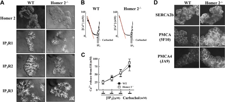
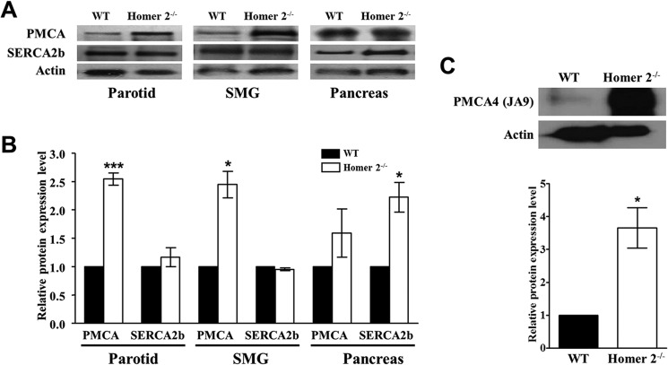
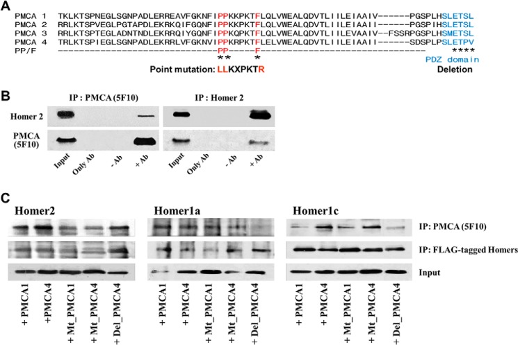
Homer proteins are scaffold molecules with a domain structure consisting of an N-terminal Ena/VASP homology 1 protein-binding domain and a C-terminal leucine zipper/coiled-coil domain. The Ena/VASP homology 1 domain recognizes proline-rich motifs and binds multiple Ca(2+)-signaling proteins, including G protein-coupled receptors, inositol 1,4,5-triphosphate receptors, ryanodine receptors, and transient receptor potential channels. However, their role in Ca(2+) signaling in nonexcitable cells is not well understood. In this study, we investigated the role of Homer2 on Ca(2+) signaling in parotid gland acinar cells using Homer2-deficient (Homer2(-/-)) mice. Homer2 is localized at the apical pole in acinar cells. Deletion of Homer2 did not affect inositol 1,4,5-triphosphate receptor localization or channel activity and did not affect the expression and activity of sarco/endoplasmic reticulum Ca(2+)-ATPase pumps. In contrast, Homer2 deletion markedly increased expression of plasma membrane Ca(2+)-ATPase (PMCA) pumps, in particular PMCA4, at the apical pole. Accordingly, Homer2 deficiency increased Ca(2+) extrusion by acinar cells. These findings were supported by co-immunoprecipitation of Homer2 and PMCA in wild-type parotid cells and transfected human embryonic kidney 293 (HEK293) cells. We identified a Homer-binding PPXXF-like motif in the N terminus of PMCA that is required for interaction with Homer2. Mutation of the PPXXF-like motif did not affect the interaction of PMCA with Homer1 but inhibited its interaction with Homer2 and increased Ca(2+) clearance by PMCA. These findings reveal an important regulation of PMCA by Homer2 that has a central role on PMCA-mediated Ca(2+) signaling in parotid acinar cells.

Keywords: Calcium ATPase; Calcium Transport; Cell Signaling; Homer Proteins; Parotid Gland; Plasma Membrane Ca2+-ATPase; Proline-rich Motif; Protein-Protein Interaction; Scaffold Protein.


Figures

FIGURE 1.
FIGURE 1.
Localization of Homer2, IP3Rs, Ca2+ pumps, and IP3-mediated Ca2+ release in WT and Homer2−/− cells. A, parotid acini from WT and Homer2−/− mice were stained for Homer2, IP3R1, IP3R2, and IP3R3. Deletion of Homer2 did not affect the expression and localization of the IP3Rs. B, cells from WT and Homer2−/− mice were permeabilized with SLO and allowed to reduce [Ca2+]i in the incubation media to approximately 75 nm. Next, Ca2+ release was measured by adding increasing concentrations of IP3 (solid arrows) and 1 mm carbachol (dotted arrows). C, quantitation of the results shown in B. D, parotid acini were stained for SERCA2b, total PMCA, and PMCA isoform 4 (PMCA4). Expression of PMCA increased in the apical membrane of Homer2−/− cells. Data are presented as the mean ± S.E.
FIGURE 2.
FIGURE 2.
Expression of Ca2+ pumps in WT and Homer2−/− cells. A, total PMCA and SERCA2b levels were analyzed by Western blot prepared from parotid, submandibular gland, and pancreatic acini from four WT and four Homer2−/− mice. Band intensity was analyzed by densitometry. B, quantitation of Western blot data in A. C, PMCA4 expression increased in the parotid of Homer2−/− mice. Quantitation is shown in the bar graph. Data were normalized to expression levels in WT cells and are presented as the mean ± S.E. *, p < 0.05; ***, p < 0.001 (compared with WT).
FIGURE 3.
FIGURE 3.
Characterization of Ca2+ signaling in WT and Homer2−/− cells. A, parotid acini from WT and Homer2−/− mice were stimulated with 1 mm carbachol while inhibiting SERCA with 100 μm CPA to elevate [Ca2+]i and prevent Ca2+ uptake by the ER. When [Ca2+]i stabilized at a steady state representing a balance of Ca2+ efflux and influx, Ca2+ clearance was initiated by removing external Ca2+ and adding of 10 μm atropine. B, average rate of Ca2+ clearance was determined from the slope of [Ca2+]i decline as a measure of PMCA activity. Results were normalized to the slope obtained from WT cells and are presented as the mean ± S.E. The rate of Ca2+ clearance increased significantly in Homer2−/− cells. C, WT and Homer2−/− parotid acinar cells in lightly Ca2+-buffered media were exposed to 1 mm carbachol while measuring [Ca2+]o. Homer2−/− cells exhibited an increased rate of [Ca2+]o, indicating a higher rate of PMCA in Homer2−/− than cells from WT mice. D, quantitation of multiple experiments similar to those shown in C. The average rate of increased [Ca2+]o was significantly higher in Homer2−/− cells. Data are depicted as the mean ± S.E. *, p < 0.05; **, p < 0.01 (compared with WT).
FIGURE 4.
FIGURE 4.
Interaction of Homer proteins with the PPXXF-like motif and PDZ-binding domain of PMCAs. A, amino acid sequences of mouse PMCA isoforms were aligned using BLAST. Alignments show the mutations in the PPXXF-like motif (red) and the PDZ-binding motif of PMCA (blue). B, PMCA and Homer2 co-immunoprecipitate from cell lysates prepared from parotid acini of WT mice. In immunoprecipitation (IP) experiments, Input denotes extract samples used for Western blot; Only Ab denotes control IP using antibodies without extract in the IP assay; −Ab denotes control IP using extract without antibodies in the IP assay; and +Ab denotes IP using extract and antibodies in the IP assay. C, co-IP of Homer proteins (FLAG-tagged) and PMCA (wild-type, PPXXF-like motif mutants (Mt_PMCA),or PMCA4 with deleted PDZ-binding motif (Del_PMCA4) co-transfected in HEK293 cells. Extracts prepared from the cells co-transfected with the respective FLAG-Homers and the indicated PMCA constructs were used to IP either PMCA with the 5F10 antibodies or the Homers with anti-FLAG antibodies, and the precipitates were probed with anti-FLAG antibodies to detect the Homers. Note that Mt_PMCAs reduced the interaction of PMCA with Homer2 but not the interaction of PMCA with Homer1a and Homer1c. However, the interaction of PMCA with Homer1a and Homer1c was decreased when the Del_PMCA4 mutant was used.
FIGURE 5.
FIGURE 5.
Effects of the Homers on Ca2+ clearance by PMCA in HEK293 cells. A, overexpression of Homer1a increases the rate of Ca2+ clearance, whereas expression of Homer1c and Homer2 do not (panel a). Panel b, quantitation of the results in (panel a). B, overexpression of WT PMCA (panel a) and Mt_PMCA (panel b) increase the rate of the Ca2+ clearance. C, overexpression of Homer2 significantly decreases the effect of WT PMCA (panel a) but not of Mt_PMCA (panel b) on Ca2+ efflux in co-transfected HEK293 cells. D, overexpression of Homer1a does not activate WT PMCA (panel a) or Mt_PMCA (panel b) in co-transfected cells. E, quantitation of PMCA activity in B–D. Results were normalized to GFP expression levels and are presented as the mean ± S.E. *, p < 0.05; **, p < 0.01; ***, p < 0.001 (compared with GFP).

References

    1. Berridge M. J., Lipp P., Bootman M. D. (2000) The versatility and universality of calcium signalling. Nat. Rev. Mol. Cell Biol. 1, 11–21 - PubMed
    1. Petersen O. H., Tepikin A. V. (2008) Polarized calcium signaling in exocrine gland cells. Annu. Rev. Physiol. 70, 273–299 - PubMed
    1. Berridge M. J., Bootman M. D., Roderick H. L. (2003) Calcium signalling: dynamics, homeostasis and remodelling. Nat. Rev. Mol. Cell Biol. 4, 517–529 - PubMed
    1. Fagni L., Chavis P., Ango F., Bockaert J. (2000) Complex interactions between mGluRs, intracellular Ca2+ stores and ion channels in neurons. Trends Neurosci. 23, 80–88 - PubMed
    1. Szumlinski K. K., Kalivas P. W., Worley P. F. (2006) Homer proteins: implications for neuropsychiatric disorders. Curr. Opin. Neurobiol. 16, 251–257 - PubMed

Publication types

MeSH terms

Substances

LinkOut - more resources