. 2014 Aug 5;111(31):E3177-86.
doi: 10.1073/pnas.1317022111. Epub 2014 Jul 21.

Phosphatidylinositol 4,5-bisphosphate triggers activation of focal adhesion kinase by inducing clustering and conformational changes

Affiliations

Phosphatidylinositol 4,5-bisphosphate triggers activation of focal adhesion kinase by inducing clustering and conformational changes

Guillermina M Goñi et al. Proc Natl Acad Sci U S A. .

Abstract

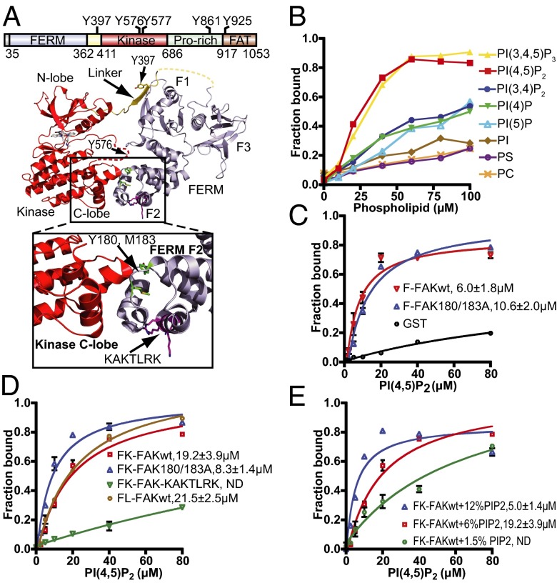
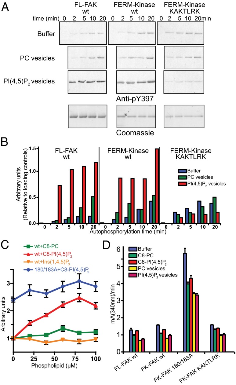
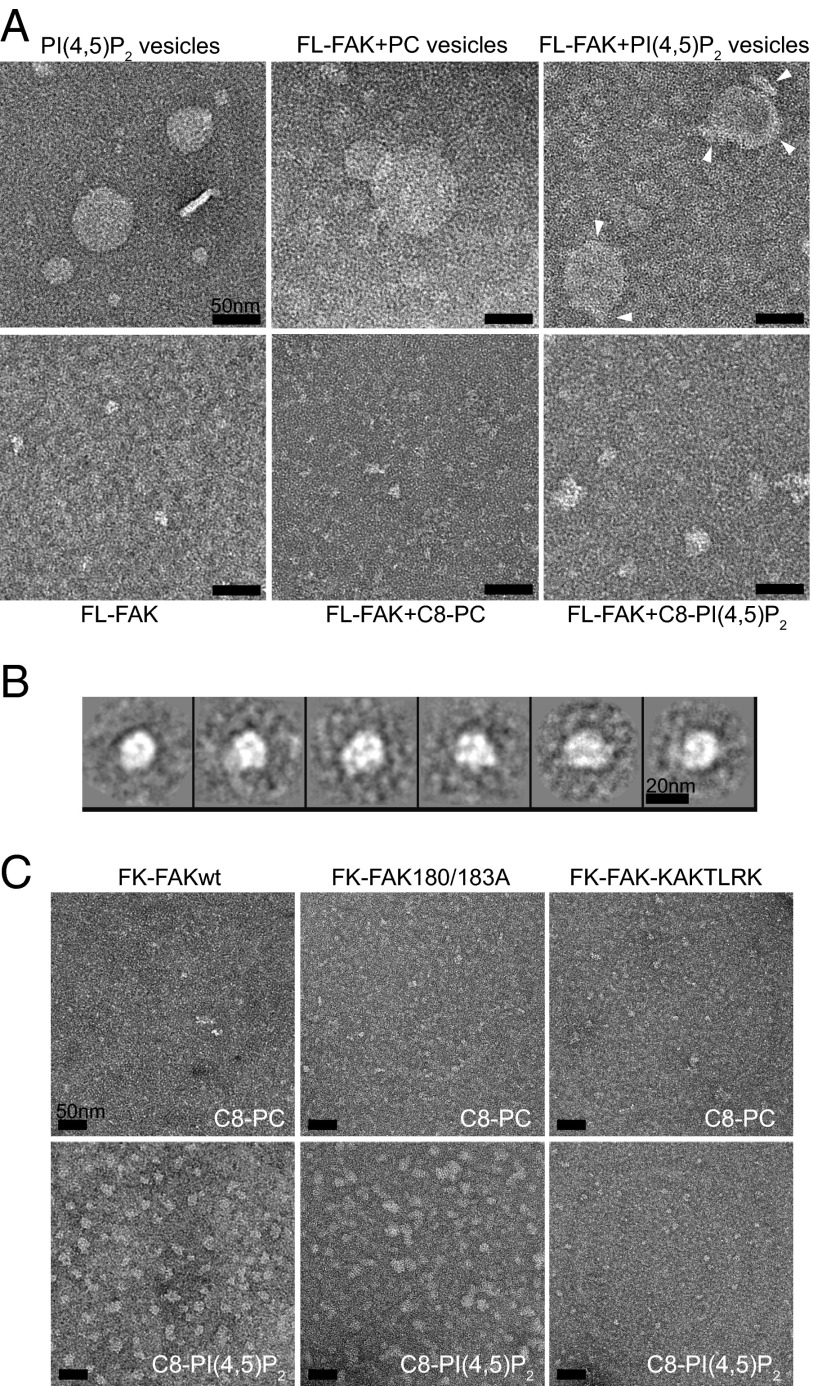
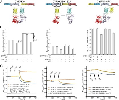
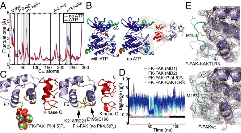
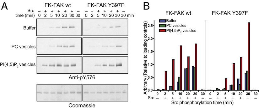
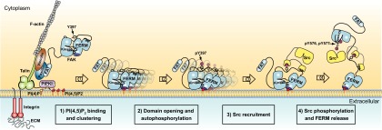
Focal adhesion kinase (FAK) is a nonreceptor tyrosine kinase (NRTK) with key roles in integrating growth and cell matrix adhesion signals, and FAK is a major driver of invasion and metastasis in cancer. Cell adhesion via integrin receptors is well known to trigger FAK signaling, and many of the players involved are known; however, mechanistically, FAK activation is not understood. Here, using a multidisciplinary approach, including biochemical, biophysical, structural, computational, and cell biology approaches, we provide a detailed view of a multistep activation mechanism of FAK initiated by phosphatidylinositol-4,5-bisphosphate [PI(4,5)P2]. Interestingly, the mechanism differs from canonical NRTK activation and is tailored to the dual catalytic and scaffolding function of FAK. We find PI(4,5)P2 induces clustering of FAK on the lipid bilayer by binding a basic region in the regulatory 4.1, ezrin, radixin, moesin homology (FERM) domain. In these clusters, PI(4,5)P2 induces a partially open FAK conformation where the autophosphorylation site is exposed, facilitating efficient autophosphorylation and subsequent Src recruitment. However, PI(4,5)P2 does not release autoinhibitory interactions; rather, Src phosphorylation of the activation loop in FAK results in release of the FERM/kinase tether and full catalytic activation. We propose that PI(4,5)P2 and its generation in focal adhesions by the enzyme phosphatidylinositol 4-phosphate 5-kinase type Iγ are important in linking integrin signaling to FAK activation.

Keywords: cell signaling; phosphoinositides.


Conflict of interest statement

The authors declare no conflict of interest.

Figures

Fig. 1.
Fig. 1.
FAK interacts with PI(4,5)P2 via the basic patch in the FERM domain. (A) Domain structure of FAK with the main phosphorylation sites indicated and a ribbon diagram of the FK-FAK crystal structure as reported by Lietha et al. (21) (PDB ID code 2J0J). In the zoom window, the interaction between the FERM F2 lobe and the kinase C-lobe is shown, with residues Y180 and M183 at the interface colored green and the basic KAKTLRK residues (K216, K218, R221, and K222) colored magenta. (B) Lipid binding specificity of FK-FAK was studied using vesicle pull-down assays with PC vesicles containing 6% (mol/mol) of the indicated phospholipids. Phosphorylation of the D4 and D5 positions of the inositol head group confers full binding affinity. PS, phosphatidyl serine. (C) Vesicle pull-downs with 6% (mol/mol) PI(4,5)P2 vesicles and GST-fused F-FAKwt (FERM = FAK31-405) or F-FAK180/183A (Y180 and M183 mutated to alanine). The mutations do not affect PI(4,5)P2 binding. GST fusions were used to obtain higher readouts. (D) PI(4,5)P2 vesicle pull-downs with FK-FAKwt, FK-FAK180/183A, FK-FAK-KAKTLRK (all KAKTLRK basic residues are mutated to alanine), or FL-FAKwt. Y180/M183A mutations result in ∼2.5-fold higher affinity to PI(4,5)P2, whereas the KAKTLRK mutations abolish binding. FL-FAK binds with similar affinity to FK-FAK. ND, not determined. (E) Vesicle pull-downs with 1.5% (mol/mol), 6% (mol/mol), or 12% (mol/mol) PI(4,5)P2 and FK-FAKwt. Increasing the PI(4,5)P2 density on vesicles results in higher affinity for FK-FAKwt, indicating an avidity effect. (CE) Error bars represent SD from three independent experiments and are shown if larger than the symbol. Kd values are determined by fitting a one-site binding model (cooperative fitting is shown in Fig. S1 B and C).
Fig. 2.
Fig. 2.
PI(4,5)P2 mediates FAK autophosphorylation but not catalytic turnover. (A) Autophosphorylation time course of FL-FAK, FK-FAKwt, and FK-FAK-KAKTLRK in the absence (buffer) or presence of PI(4,5)P2 or PC vesicles was monitored by immunoblotting using an anti-pY397 antibody. (Lower) Loading controls stained by Coomassie blue. Note that FL-FAK stains stronger because of its higher molecular weight. (B) Quantifications of blots from A relative to loading controls [using ImageJ (National Institutes of Health)]. For FL-FAK and FK-FAKwt, autophosphorylation is significantly faster in the presence of PI(4,5)P2 vesicles, whereas mutations in the basic patch of FK-FAK-KAKTLRK abrogate this effect. (C) Autophosphorylation efficiency of FK-FAK (wt or 180/183A mutant) was assessed using an ELISA method. The presence of C8-PI(4,5)P2, but not C8-PC or the head group Ins(1,4,5)P3, enhances autophosphorylation of FK-FAKwt to levels similar to FK-FAK180/183A, which was not affected by PI(4,5)P2. (D) Catalytic steady-state activity was assayed for the indicated FAK proteins using a kinase assay, which couples ADP production to NADH consumption (Methods). Whereas dissociation of FERM/kinase domains by mutation (180/183A) activates FAK, none of the tested lipids increase catalytic turnover. (C and D) Error bars represent SD from three experiments.
Fig. 3.
Fig. 3.
EM reveals PI(4,5)P2-induced FAK clustering. (A) Transmission electron micrographs of FL-FAK with lipid vesicles (Upper) or soluble lipids (Lower) imaged by negative staining. PI(4,5)P2 vesicles and soluble C8-PI(4,5)P2 mediate the formation of FAK clusters. Clusters on vesicles are indicated by arrowheads. (B) Reference-free 2D class averages of 574 FL-FAK/C8-PI(4,5)P2 clusters, representing a main cluster population, suggest a circular arrangement of FAK molecules in clusters. Volume calculations suggest that clusters consist of six to eight FL-FAK molecules per cluster. (C) FK-FAKwt and the 180/183A and KAKTLRK mutants were imaged in the presence of C8-PI(4,5)P2 or C8-PC. FK-FAKwt and FK-FAK180/183A, but not the KAKTLRK mutant, display clustering in the presence of PI(4,5)P2. (Scale bars: A and C, 50 nm.)
Fig. 4.
Fig. 4.
PI(4,5)P2 induces partial and Src induces full domain opening of the FAK FRET sensor in the presence of ATP. (A) Schematic illustration of the domain structure (Upper) and expected structural arrangement (Lower) of the conformational FRET sensors used. (B) Emission ratios of citrine (EmCitrine) and CFP (EmCFP) are plotted relative to CYFAKwt as a measure of FRET. The presence of C8-PI(4,5)P2 causes a reduction in FRET levels for all three sensors, suggesting that this effect is not conformational (labeled NC). The presence of ATP significantly increases FRET levels for CYFAKwt; however, this increase is reversed in the presence of PI(4,5)P2. This effect of ATP and PI(4,5)P2 is not seen for CYF-HFC, and is therefore likely conformational (labeled C). Error bars represent SD from a minimum of three experiments. (C) Relative FRET levels were monitored in real time, initially of the sensors alone and then following addition of (i) lipid [C8-PC (green plots) and C8-PI(4,5)P2 (blue plots)] or no lipid (brown and orange plots), (ii) Src (brown, green, and blue plots) or no Src (orange plots), and (iii) ATP/Mg2+ (all plots). As in B, PI(4,5)P2 reduces FRET levels of all sensors in all states (before/after phosphorylation), indicating a nonconformational effect. ATP results in a FRET spike only with CYFAKwt. If active Src is present, the spike is followed by a switch to the open conformation (lower FRET levels). In contrast, inactive SrcK298M induces only a modest FRET decrease (Fig. S5C). (Left) Gray control plot is without the CYFAK sensor to verify that PI(4,5)P2, Src, and ATP do not exhibit intrinsic fluorescence.
Fig. 5.
Fig. 5.
Effects of ATP and PI(4,5)P2 on FAK conformation. (A) Root mean square fluctuations (RMSFs) from unbiased MD simulations of the FAK kinase domain are shown for a window of 700 ns (discarding the first 200 ns of equilibration) for FAK alone (blue plot) and for FAK with ATP/Mg2+ (red plot). ATP binding stabilizes the αC- and αG-helices. (B) RMSF values from A are color-mapped onto the FAK structure. RMSFs range from blue (low values) to red (high values). Stabilizing ATP effects on αC- and αG-helices map autoinhibitory interaction sites as seen in the FK-FAK crystal structure (21). Neutralization of the basic patch by PI(4,5)P2 binding (C) or by mutation (D) interferes with a set of FERM F2 lobe-stabilizing salt bridges. MD simulations suggest that salt bridges between K218/R221 in the basic patch and E195/E198 are formed in the absence of PI(4,5)P2 (C, Right) but not in the presence of PI(4,5)P2 (C, Left), leading to a partial destabilization of the FERM F2 lobe and an altered force distribution, as shown in Fig. S7A. (D) Minimum distances for the residues K218/R221 and residue pair E195/E198 are shown during MD simulations with FK-FAK alone (MD1 and MD2), FK-FAK bound to PI(4,5)P2, or the basic patch mutant FK-FAK-KAKTLRK. In the two independent MD simulations of FK-FAKwt, these two pairs of oppositely charged residues strongly interact with each other. PI(4,5)P2 binding or KAKTLRK mutations to alanines significantly increase the minimum distances and fluctuations. (E) Crystal structure of the basic patch mutant FERM domain (F-FAK-KAKTLRK; PDB ID code 3ZDT; full structure is shown in Fig. S7C). In contrast to F-FAKwt (Lower, PDB ID code 4CYE), the structure of F-FAK-KAKTLRK (Upper) exhibits no electron density for the loop (cyan) containing the autoinhibitory residues Y180/M183 (green), indicating that this loop is disordered. The 2Fo-Fc electron density maps are shown as gray mesh countered at 1σ.
Fig. 6.
Fig. 6.
PI(4,5)P2 enhances Src phosphorylation of Y576 in FAK. (A) Time course of Src phosphorylation of FK-FAK in the absence or presence of PI(4,5)P2 or PC vesicles as monitored by Western blotting using an anti-pY576 antibody following a 2-min autophosphorylation reaction (Methods). Mutation of the autophosphorylation site (Y397F) significantly reduces the Y576 phosphorylation rate, whereas PI(4,5)P2 enhances Y576 phosphorylation for FK-FAKwt and FK-FAK-Y397F, indicating a combined effect of more efficient Src recruitment to the FAK autophosphorylation site and PI(4,5)P2-induced conformational changes. (Lower) Coomassie blue-stained loading controls are shown. (B) Quantifications of blots in A.
Fig. 7.
Fig. 7.
PIP5KIγ KD decreases cellular FAK activity and cell attachment. (A) Expression levels of PIP5KIγ668 (isoform 2) in HeLa cells after KD are shown for two different clones (KD1 and KD2) and scramble control (Ctr). (Upper) Representative immunoblot is shown. (Lower) Quantification of PIP5KIγ levels observed by immunoblotting is depicted in the histogram. Quantifications are from four blots (two independent experiments, each in duplicate). (B) Effect of PIP5KIγ reductions on the total level of FAK and on the total cellular levels of pFAK Y397, pFAK Y576, and pFAK Y577. A representative immunoblot and quantifications from four blots (two independent experiments, each in duplicate) are shown. (C) pFAKY397 levels in HeLa cells of Ctr or KD1 and KD2 cells transfected with WT (PIP5KIγ-WT) or a kinase-dead (PIP5Kγ-D316A) form of PIP5KIγ. Immunoblots and quantifications of two independent experiments are shown, with each performed twice. Note that PIP5KIγ-WT more than rescues but the kinase-dead mutant does not fully rescue FAK phosphorylation levels in KD cells compared with controls. The dashed line merges different lanes of the same immunoblot experiment. (D) Immunofluorescence staining of pFAK397 (red) and total FAK (green) in PIP5KIγ KD1 and KD2 clones and Ctr, and quantification of the immunofluorescence intensity of pFAK397 relative to total FAK signals specifically in FAs at 15 min and 2 h after stimulation with serum and fibronectin. (E) Functional cell adhesion assay to determine the ability of PIP5KIγ-deficient cells to attach to fibronectin (FN) compared with Ctr. Quantification of the number of attached cells for three independent experiments in triplicate is shown. Data represent the mean value ± SEM. *P < 0.05; **P < 0.01; ***P < 0.001; ****P < 0.0001 (unpaired Student t test).
Fig. 8.
Fig. 8.
Schematic model for integrin-mediated FAK activation by PI(4,5)P2. (Left) Cell adhesion via integrin receptors to the ECM results in integrin clustering and the recruitment of FA proteins (as illustrated here for talin, vinculin, paxillin, FAK, and PIP5KIγ) to form adhesion structures that link integrins to the actin cytoskeleton. Recruitment of PIP5KIγ results in a local increase of PI(4,5)P2 levels in FAs. PI(4,5)P2 in FAs binds FAK via the basic patch (dark blue) in the FERM domain of FAK, resulting in FAK clustering at the cell membrane (step 1). PI(4,5)P2-induced FAK clustering results in a relaxed FERM/kinase conformation, with the kinase N-lobe dissociated from the linker and FERM F1 lobe. PI(4,5)P2-induced clustering and conformational relaxation allow efficient autophosphorylation of Y397 (step 2) and Src recruitment via SH2 and SH3 domains (step 3). Recruited Src phosphorylates the activation loop residues Y576/Y577 of FAK, which results in full activation and release of the kinase from the membrane-clustered FERM domain (step 4).

References

    1. Schiller HB, Fässler R. Mechanosensitivity and compositional dynamics of cell-matrix adhesions. EMBO Rep. 2013;14(6):509–519. - PMC - PubMed
    1. Schaller MD. Cellular functions of FAK kinases: Insight into molecular mechanisms and novel functions. J Cell Sci. 2010;123(Pt 7):1007–1013. - PubMed
    1. Zhao X, Guan JL. Focal adhesion kinase and its signaling pathways in cell migration and angiogenesis. Adv Drug Deliv Rev. 2011;63(8):610–615. - PMC - PubMed
    1. Zamir E, Geiger B. Molecular complexity and dynamics of cell-matrix adhesions. J Cell Sci. 2001;114(Pt 20):3583–3590. - PubMed
    1. Ashton GH, et al. Focal adhesion kinase is required for intestinal regeneration and tumorigenesis downstream of Wnt/c-Myc signaling. Dev Cell. 2010;19(2):259–269. - PMC - PubMed

Publication types

MeSH terms

Substances

LinkOut - more resources