Review
. 2014 Jun;31(2):203-6.
doi: 10.1055/s-0034-1373794.

Multiple arteries supplying a single tumor vascular distribution: microsphere administration options for the interventional radiologist performing radioembolization

Affiliations
Review

Multiple arteries supplying a single tumor vascular distribution: microsphere administration options for the interventional radiologist performing radioembolization

Charles E Ray Jr et al. Semin Intervent Radiol. 2014 Jun.
No abstract available


Figures

Figure 1
Figure 1
Celiac angiogram demonstrating multiple hepatic artery anomalies. Open black arrow—undivided right hepatic artery. Solid black arrow—segment 4 (middle hepatic) artery. Solid white arrow—isolated segment 2/3 hepatic artery. Open white arrow—accessory segment 3 hepatic artery.
Figure 2
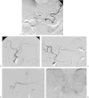
Figure 2
Flow redistribution performed due to left hepatic artery anomalies. (A) Celiac angiography demonstrating undivided right hepatic artery (open black arrow), segment 4 hepatic artery arising from the right hepatic artery (open white arrow), and separate origins of the segment 2 (solid white arrow) and segment 3 hepatic arteries (solid black arrow). The plan for this patient (who needed both right and left hepatic artery Y90 embolizations due to diffuse bilobar metastatic disease) was to treat the right hepatic and segment 4 hepatic artery distributions with one infusion, and the left hepatic artery distribution from a separate single infusion. To accomplish this single left hepatic artery infusion, it was decided to embolize the segment 2 branch and allow intrahepatic collaterals to develop from the segment 3 hepatic artery. (B) Angiography of the common hepatic artery following embolization of the gastroduodenal artery. The separate origins of the segments 2 and 3 hepatic arteries are visualized (circle). Open black arrow—right gastric artery, which also underwent protective embolization. (C) Proper hepatic angiography following embolization of the segment 2 hepatic artery demonstrating complete proximal occlusion. The most proximal coil overlays but is not in the segment 3 hepatic artery. (D) Diagnostic angiography performed on the day of Y90 administration into segment 2/3 (∼ 2 weeks after, A–C). Segment 3 hepatic angiography demonstrates reflux into the right hepatic artery (arrowhead). Also demonstrated is reconstitution of the intrahepatic segment 2 hepatic artery (arrow). (E) Delayed phase angiogram demonstrating near-complete coverage of the occluded segment 2 hepatic arterial distribution.

References

    1. Furuta T, Maeda E, Akai H. et al.Hepatic segments and vasculature: projecting CT anatomy onto angiograms. Radiographics. 2009;29(7):1–22. - PubMed
    1. Bilbao J I, Garrastachu P, Herráiz M J. et al.Safety and efficacy assessment of flow redistribution by occlusion of intrahepatic vessels prior to radioembolization in the treatment of liver tumors. Cardiovasc Intervent Radiol. 2010;33(3):523–531. - PubMed
    1. Uliel L, Royal H D, Darcy M D, Zuckerman D A, Sharma A, Saad N E. From the angio suite to the γ-camera: vascular mapping and 99mTc-MAA hepatic perfusion imaging before liver radioembolization—a comprehensive pictorial review. J Nucl Med. 2012;53(11):1736–1747. - PubMed
    1. Abdelmaksoud M H Louie J D Kothary N et al.Consolidation of hepatic arterial inflow by embolization of variant hepatic arteries in preparation for yttrium-90 radioembolization J Vasc Interv Radiol 201122101364–1371., e1 - PubMed
    1. Paprottka P M, Jakobs T F, Reiser M F, Hoffmann R T. Practical vascular anatomy in the preparation of radioembolization. Cardiovasc Intervent Radiol. 2012;35(3):454–462. - PubMed