. 2014 Apr;27(4):580-6.
doi: 10.5713/ajas.2013.13744.

Bacillus subtilis Protects Porcine Intestinal Barrier from Deoxynivalenol via Improved Zonula Occludens-1 Expression

Affiliations

Bacillus subtilis Protects Porcine Intestinal Barrier from Deoxynivalenol via Improved Zonula Occludens-1 Expression

Min Jeong Gu et al. Asian-Australas J Anim Sci. 2014 Apr.

Abstract

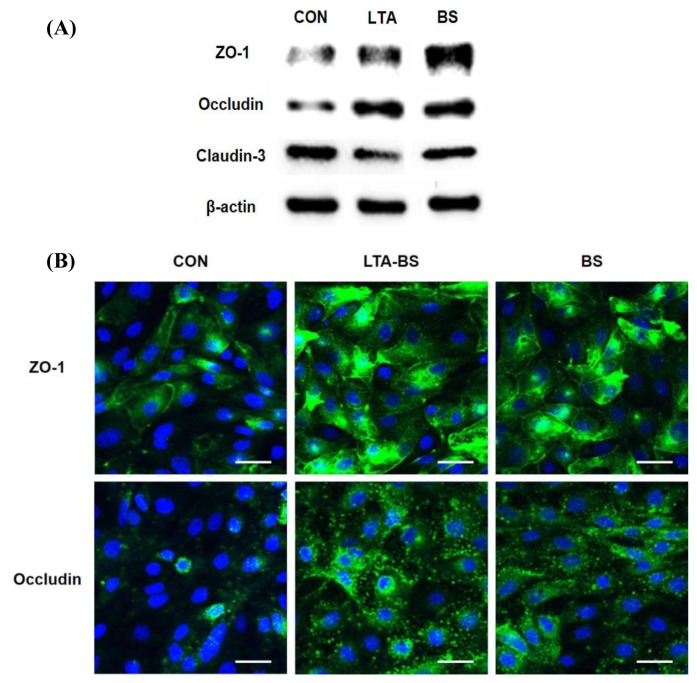
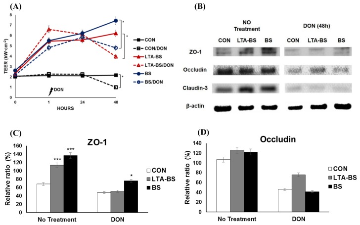
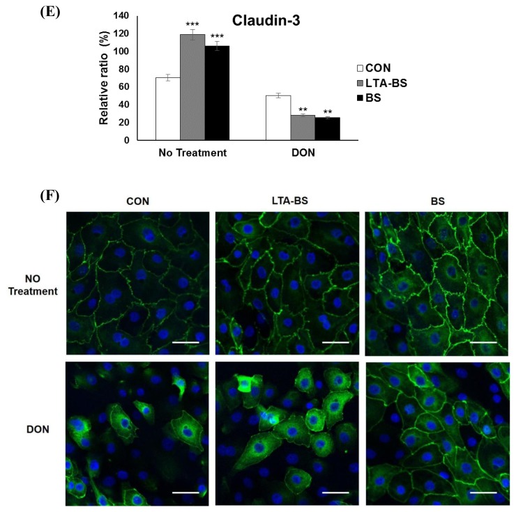
Intestinal epithelial cells (IECs) forming the barrier for the first-line of protection are interconnected by tight junction (TJ) proteins. TJ alteration results in impaired barrier function, which causes potentially excessive inflammation leading to intestinal disorders. It has been suggested that toll-like receptor (TLR) 2 ligands and some bacteria enhance epithelial barrier function in humans and mice. However, no such study has yet to be claimed in swine. The aim of the present study was to examine whether Bacillus subtilis could improve barrier integrity and protection against deoxynivalenol (DON)-induced barrier disruption in porcine intestinal epithelial cell line (IPEC-J2). We found that B. subtilis decreased permeability of TJ and improved the expression of zonula occludens (ZO)-1 and occludin during the process of forming TJ. In addition, ZO-1 expression of IPEC-J2 cells treated with B. subtilis was up-regulated against DON-induced damage. In conclusion, B. subtilis may have potential to enhance epithelial barrier function and to prevent the cells from DON-induced barrier dysfunction.

Keywords: Bacillus subtilis; Deoxynivalenol; Porcine Intestinal Epithelial Cell Line (IPEC-J2); Tight Junction; Zonula Occludens-1.


Figures

Figure 1
Figure 1
Increased expression of TJ proteins of IPEC-J2 cells treated short-term with B. subtilis. (A) IPEC-J2 cells were treated with 1×10⁹ cfu/mL of B. subtilis and LTA-BS (as a control) for 1 h. Tight junction proteins were analyzed by western blotting. Lysed cells were loaded on gel, electrophoresed and transferred to PVDF membrane. Then, the membrane was stained with primary and secondary antibodies, visualized with ECL kit and analyzed by ChemiDoc XRS. (B) IPEC-J2 cells were treated with 1×10⁹ cfu/mL of B. subtilis and stained with anti-occludin or -ZO-1. Nuclei were stained with DAPI. The expression of target proteins was visualized by confocal laser scanning microscope. Scale bar denotes 20 μm. BS, Bacillus subtilis.
Figure 2
Figure 2
i) Maintained ZO-1 expression in IPEC-J2 treated with B. subtilis and then exposed to DON. (A) IPEC-J2 cells, grown on 0.4-μm polyester membrane trans-well, were treated with B. subtilis or LTA-BS for 1 h, and then the cells were treated with 2 μg/mL of DON for 48 h. TEER was determined at 0, 1, 24, and 48 h post-treatment. TEER values are expressed as means±SEM (n = 3). * p<0.05. (B) IPEC-J2 cells were pretreated with B. subtilis for 1 h, and then treated with DON for 48 h. Then, the cells were analyzed for the expression of ZO-1, occludin, and claudin-3 by western blotting (C, D, E). These panels show the relative intensity of the TJ proteins, (C) ZO-1, (D) occludin and (E) claudin-3, those quantified after the normalization with β-actin. Value was compared to each control within the treatment group. * p<0.05, ** p<0.01, *** p<0.001. (F) IPEC-J2 cells were treated with 1×10⁹ cfu/mL of B. subtilis for 1 h prior to DON treatment for 48 h and stained with anti-ZO-1 antibody. Nuclei were stained with DAPI. The expression of target proteins was visualized by confocal laser scanning microscope. Scale bar denotes 20 μm. Figure 2. ii) Maintained ZO-1 expression in IPEC-J2 treated with B. subtilis and then exposed to DON. (A) IPEC-J2 cells, grown on 0.4-μm polyester membrane trans-well, were treated with B. subtilis or LTA-BS for 1 h, and then the cells were treated with 2 μg/mL of DON for 48 h. TEER was determined at 0, 1, 24, and 48 h post-treatment. TEER values are expressed as means±SEM (n = 3). * p<0.05. (B) IPEC-J2 cells were pretreated with B. subtilis for 1 h, and then treated with DON for 48 h. Then, the cells were analyzed for the expression of ZO-1, occludin, and claudin-3 by western blotting (C, D, E). These panels show the relative intensity of the TJ proteins, (C) ZO-1, (D) occludin and (E) claudin-3, those quantified after the normalization with β-actin. Value was compared to each control within the treatment group. * p<0.05, ** p<0.01, *** p<0.001. (F) IPEC-J2 cells were treated with 1×10⁹ cfu/mL of B. subtilis for 1 h prior to DON treatment for 48 h and stained with anti-ZO-1 antibody. Nuclei were stained with DAPI. The expression of target proteins was visualized by confocal laser scanning microscope. Scale bar denotes 20 μm.
Figure 2
Figure 2
i) Maintained ZO-1 expression in IPEC-J2 treated with B. subtilis and then exposed to DON. (A) IPEC-J2 cells, grown on 0.4-μm polyester membrane trans-well, were treated with B. subtilis or LTA-BS for 1 h, and then the cells were treated with 2 μg/mL of DON for 48 h. TEER was determined at 0, 1, 24, and 48 h post-treatment. TEER values are expressed as means±SEM (n = 3). * p<0.05. (B) IPEC-J2 cells were pretreated with B. subtilis for 1 h, and then treated with DON for 48 h. Then, the cells were analyzed for the expression of ZO-1, occludin, and claudin-3 by western blotting (C, D, E). These panels show the relative intensity of the TJ proteins, (C) ZO-1, (D) occludin and (E) claudin-3, those quantified after the normalization with β-actin. Value was compared to each control within the treatment group. * p<0.05, ** p<0.01, *** p<0.001. (F) IPEC-J2 cells were treated with 1×10⁹ cfu/mL of B. subtilis for 1 h prior to DON treatment for 48 h and stained with anti-ZO-1 antibody. Nuclei were stained with DAPI. The expression of target proteins was visualized by confocal laser scanning microscope. Scale bar denotes 20 μm. Figure 2. ii) Maintained ZO-1 expression in IPEC-J2 treated with B. subtilis and then exposed to DON. (A) IPEC-J2 cells, grown on 0.4-μm polyester membrane trans-well, were treated with B. subtilis or LTA-BS for 1 h, and then the cells were treated with 2 μg/mL of DON for 48 h. TEER was determined at 0, 1, 24, and 48 h post-treatment. TEER values are expressed as means±SEM (n = 3). * p<0.05. (B) IPEC-J2 cells were pretreated with B. subtilis for 1 h, and then treated with DON for 48 h. Then, the cells were analyzed for the expression of ZO-1, occludin, and claudin-3 by western blotting (C, D, E). These panels show the relative intensity of the TJ proteins, (C) ZO-1, (D) occludin and (E) claudin-3, those quantified after the normalization with β-actin. Value was compared to each control within the treatment group. * p<0.05, ** p<0.01, *** p<0.001. (F) IPEC-J2 cells were treated with 1×10⁹ cfu/mL of B. subtilis for 1 h prior to DON treatment for 48 h and stained with anti-ZO-1 antibody. Nuclei were stained with DAPI. The expression of target proteins was visualized by confocal laser scanning microscope. Scale bar denotes 20 μm.

References

    1. Abreu MT. Toll-like receptor signalling in the intestinal epithelium: How bacterial recognition shapes intestinal function. Nat Rev Immunol. 2010;10:131–144. - PubMed
    1. Aperce CC, Burkey TE, KuKanich B, Crozier-Dodson BA, Dritz SS, Minton JE. Interaction of bacillus species and salmonella enterica serovar typhimurium in immune or inflammatory signaling from swine intestinal epithelial cells. J Anim Sci. 2010;88:1649–1656. - PubMed
    1. Cario E. Barrier-protective function of intestinal epithelial toll-like receptor 2. Mucosal Immunol. 2008;1(Suppl 1):S62–66. - PubMed
    1. Cario E, Gerken G, Podolsky DK. Toll-like receptor 2 enhances zo-1-associated intestinal epithelial barrier integrity via protein kinase C. Gastroenterology. 2004;127:224–238. - PubMed
    1. Cario E, Gerken G, Podolsky DK. Toll-like receptor 2 controls mucosal inflammation by regulating epithelial barrier function. Gastroenterology. 2007;132:1359–1374. - PubMed