. 2014 Jul;35(7):3517-28.
doi: 10.1002/hbm.22418. Epub 2013 Nov 25.

Neuronal oscillations and functional interactions between resting state networks

Neuronal oscillations and functional interactions between resting state networks

Xu Lei et al. Hum Brain Mapp. 2014 Jul.

Abstract

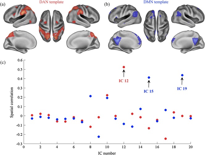
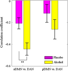
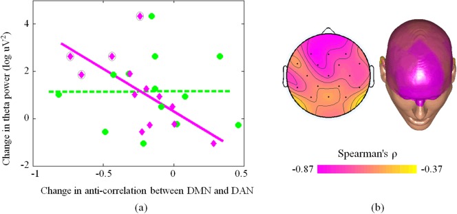
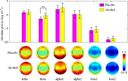
Functional magnetic imaging (fMRI) studies showed that resting state activity in the healthy brain is organized into multiple large-scale networks encompassing distant regions. A key finding of resting state fMRI studies is the anti-correlation typically observed between the dorsal attention network (DAN) and the default mode network (DMN), which - during task performance - are activated and deactivated, respectively. Previous studies have suggested that alcohol administration modulates the balance of activation/deactivation in brain networks, as well as it induces significant changes in oscillatory activity measured by electroencephalography (EEG). However, our knowledge of alcohol-induced changes in band-limited EEG power and their potential link with the functional interactions between DAN and DMN is still very limited. Here we address this issue, examining the neuronal effects of alcohol administration during resting state by using simultaneous EEG-fMRI. Our findings show increased EEG power in the theta frequency band (4-8 Hz) after administration of alcohol compared to placebo, which was prominent over the frontal cortex. More interestingly, increased frontal tonic EEG activity in this band was associated with greater anti-correlation between the DAN and the frontal component of the DMN. Furthermore, EEG theta power and DAN-DMN anti-correlation were relatively greater in subjects who reported a feeling of euphoria after alcohol administration, which may result from a diminished inhibition exerted by the prefrontal cortex. Overall, our findings suggest that slow brain rhythms are responsible for dynamic functional interactions between brain networks. They also confirm the applicability and potential usefulness of EEG-fMRI for central nervous system drug research.


Figures

Figure 1
Figure 1
Selection of independent components (ICs) using DAN and DMN templates. The DAN (a) and DMN (b) templates are colored red and blue respectively, and are overlaid onto a standard cortical model (left and right lateral, left and right medial, and dorsal views). (c) The scatter plot shows the spatial correlations between each IC map and the templates maps (red dots for DAN, blue dots for DMN). The selected ICs are indicated with a black arrow. [Color figure can be viewed in the online issue, which is available at http://wileyonlinelibrary.com.]
Figure 2
Figure 2
Group‐level maps for the anterior and posterior DMN (aDMN and pDMN, respectively) and the DAN in (a) placebo and (b) alcohol sessions. Brain areas with intensities of two standard deviations greater than the mean are shown. The aDMN, pDMN, and DAN are colored in blue, red, and green, respectively. [Color figure can be viewed in the online issue, which is available at http://wileyonlinelibrary.com.]
Figure 3
Figure 3
Anterior (blue) and posterior (red) DMN and the DAN (green) in a single subject (placebo session). Brain areas with intensities of two standard deviations above the mean are shown in the spatial maps. The time courses of the networks were first regressed out global signal and then displayed with the same colors of the corresponding maps. Note that the anterior part of DMN (aDMN) positively correlates with the posterior part of DMN (pDMN) and negatively correlates with the DAN. [Color figure can be viewed in the online issue, which is available at http://wileyonlinelibrary.com.]
Figure 4
Figure 4
Increased anti‐correlation between DMN and DAN. The bar plots show the correlation coefficients (mean ± standard error) between DAN and the anterior and posterior part of DMN in alcohol and placebo sessions. Significant changes between conditions were assessed by means of paired t‐tests. Double asterisks indicate significant differences (P < 0.01). [Color figure can be viewed in the online issue, which is available at http://wileyonlinelibrary.com.]
Figure 5
Figure 5
Power spectra of all channels in (a) placebo and alcohol (b) sessions, and their difference (c). For the difference between alcohol and placebo, the topographies of EEG are illustrated in 3, 6, 10, 12, 18, 25 Hz. [Color figure can be viewed in the online issue, which is available at http://wileyonlinelibrary.com.]
Figure 6
Figure 6
Relationship between changes in DMN‐DAN anti‐correlation and theta power EEG activity across subjects. (a) The pink line indicates the correlation between change in anti‐correlation between DAN and aDMN, whereas each pink diamond represents a single subject. The green dotted line indicates the correlation between change in anti‐correlation between DAN and pDMN, whereas each green circle represents a given subject. A pink diamond outlined in a black circle represents an individual reporting intense euphoria. (b) The correlation between theta rhythm power in each EEG channel and anti‐correlation between DAN and DMN is represented in a topographical EEG map. [Color figure can be viewed in the online issue, which is available at http://wileyonlinelibrary.com.]
Figure 7
Figure 7
The grand‐average absolute powers (mean ± standard error) and topographies of EEG bands (delta, theta, alpha 1, alpha 2, beta 1, and beta 2) in placebo and alcohol sessions. [Color figure can be viewed in the online issue, which is available at http://wileyonlinelibrary.com.]

References

    1. Allen PJ, Josephs O, Turner R (2000): A method for removing imaging artifact from continuous EEG recorded during functional MRI. Neuroimage 12:230–239. - PubMed
    1. Asada H, Fukuda Y, Tsunoda S, Yamaguchi M, Tonoike M (1999): Frontal midline theta rhythms reflect alternative activation of prefrontal cortex and anterior cingulate cortex in humans. Neurosci Lett 274:29–32. - PubMed
    1. Bressler SL, Menon V (2010): Large‐scale brain networks in cognition: Emerging methods and principles. Trends Cogn Sci 14:277–290. - PubMed
    1. Buckner RL, Andrews‐Hanna JR, Schacter DL (2008): The brain's default network: Anatomy, function, and relevance to disease. Ann NY Acad Sci 1124:1–38. - PubMed
    1. Calhoun VD, Adali T, Pearlson GD, Pekar JJ (2001a): A method for making group inferences from functional MRI data using independent component analysis. Hum Brain Mapp 14:140–151. - PMC - PubMed

Publication types

MeSH terms