. 2015 Jan;135(1):170-180.
doi: 10.1038/jid.2014.310. Epub 2014 Aug 22.

Suppressing AP1 factor signaling in the suprabasal epidermis produces a keratoderma phenotype

Affiliations

Suppressing AP1 factor signaling in the suprabasal epidermis produces a keratoderma phenotype

Ellen A Rorke et al. J Invest Dermatol. 2015 Jan.

Erratum in

  • Erratum.
    [No authors listed] [No authors listed] J Invest Dermatol. 2021 Jul;141(7):1862. doi: 10.1016/j.jid.2021.05.008. J Invest Dermatol. 2021. PMID: 34167723 No abstract available.

Abstract

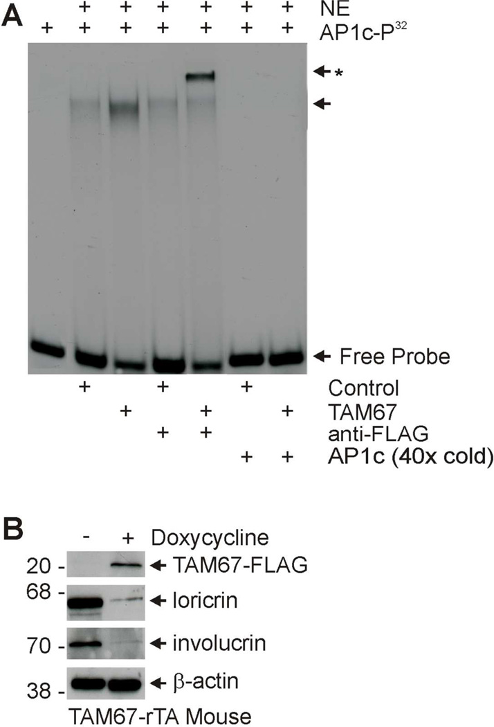
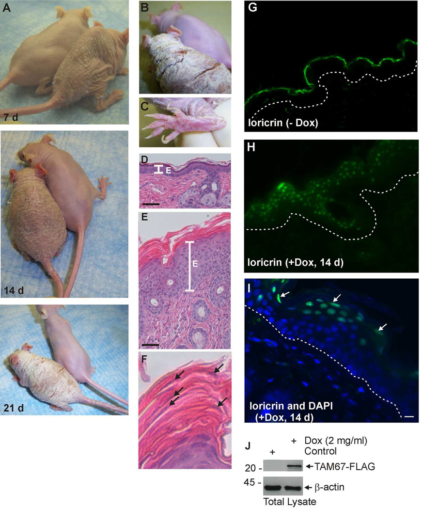
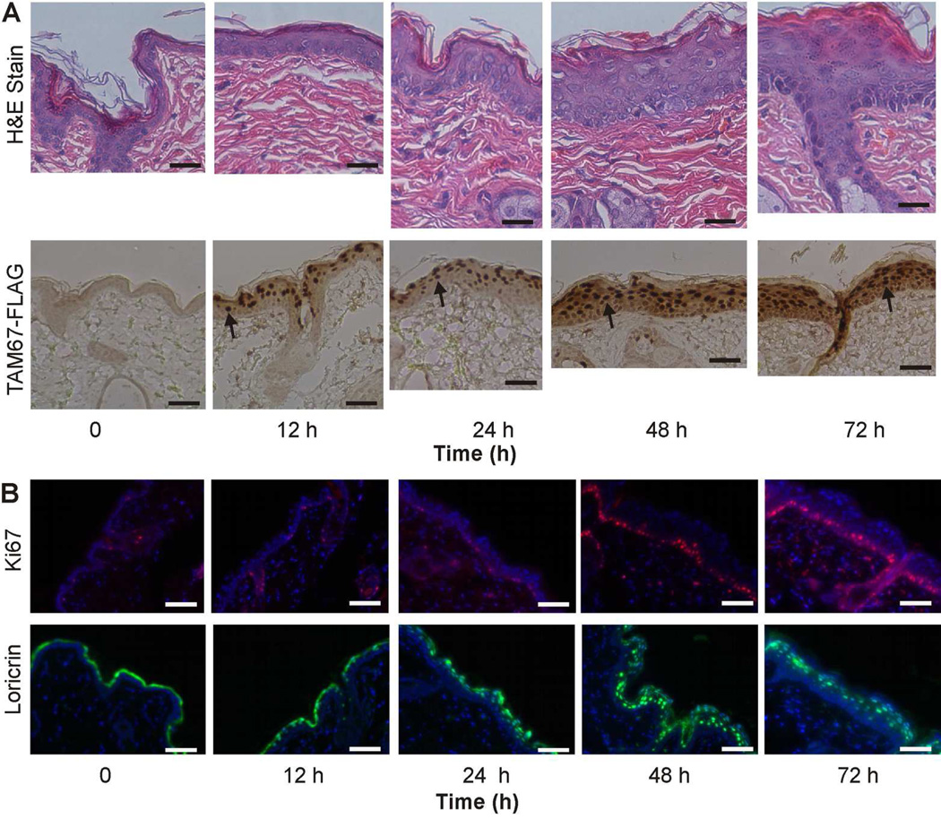
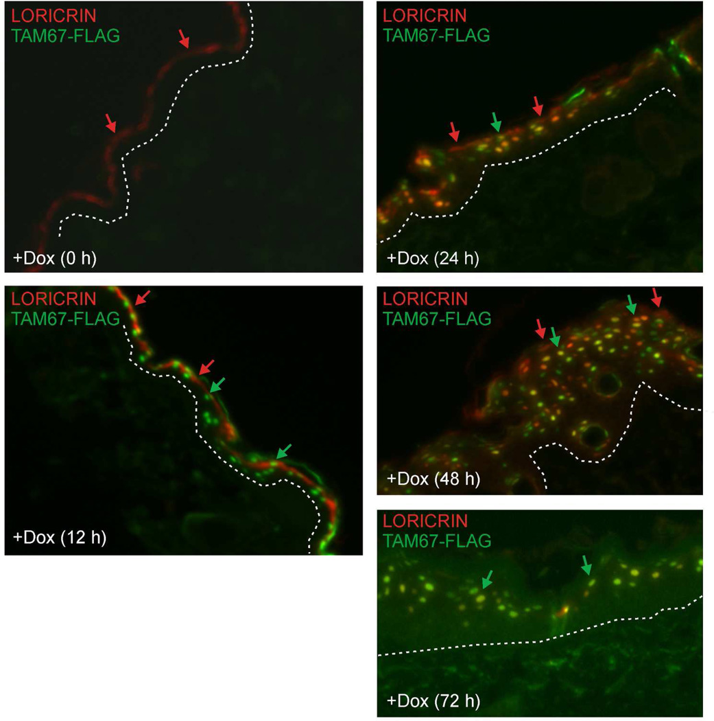
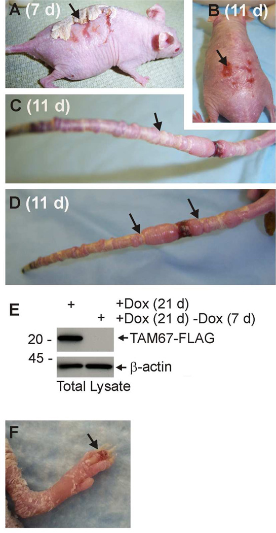
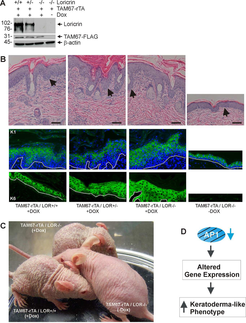
Keratodermas comprise a heterogeneous group of highly debilitating and painful disorders characterized by thickening of the skin with marked hyperkeratosis. Some of these diseases are caused by genetic mutation, whereas other forms are acquired in response to environmental factors. Our understanding of signaling changes that underlie these diseases is limited. In the present study, we describe a keratoderma phenotype in mice in response to suprabasal epidermis-specific inhibition of activator protein 1 transcription factor signaling. These mice develop a severe phenotype characterized by hyperplasia, hyperkeratosis, parakeratosis, and impaired epidermal barrier function. The skin is scaled, constricting bands encircle the tail and digits, the footpads are thickened and scaled, and loricrin staining is markedly reduced in the cornified layers and increased in the nucleus. Features of this phenotype, including nuclear loricrin localization and pseudoainhum (autoamputation), are characteristic of the Vohwinkel syndrome. We confirm that the phenotype develops in a loricrin-null genetic background, indicating that suppressed suprabasal AP1 factor function is sufficient to drive this disease. We also show that the phenotype regresses when suprabasal AP1 factor signaling is restored. Our findings suggest that suppression of AP1 factor signaling in the suprabasal epidermis is a key event in the pathogenesis of keratoderma.


Conflict of interest statement

Conflict of Interest

The authors have no conflict of interest financial or otherwise.

Figures

Fig. 1
Fig. 1
TAM67-FLAG interacts with AP1 response element and suppresses AP1-responsive genes. TAM67-rTA mice were treated with (+) or without (−) 2 mg/ml doxycycline in drinking water for 3 days. A Interaction of TAM67 with AP1 site consensus element. Epidermis from control and TAM67-expressing epidermis was collected by high temperature separation (Rorke et al., 2010) and nuclear extracts were prepared and incubated with AP1c-P32 as indicated. FP indicates free probe and NE indicates nuclear extract. The arrow indicates migration of the mobility shifted bands, and the arrow/asterisk indicates migration of the supershifted band (anti-FLAG treated sample). A 40-fold excess of AP1c was added as indicated to demonstrate binding specificity. B Murine epidermis was collected free of the dermis by high temperature separation as previously described (Rorke et al., 2010). Total extract was prepared for immunoblot to detect the indicated proteins. TAM67-FLAG was detected with anti-FLAG. Similar results were observed in each of three experiments.
Fig. 2
Fig. 2. Characterization of SKH1-TAM67-rTA mice
SKH1-TAM67-rTA mice were administered drinking water supplemented with or without 2 mg/ml doxycycline. A Doxycycline-treated and untreated littermates were photographed at 7, 14 and 21 d. B/C Enlarged images showing hyperkeratinization of the dorsal epidermis and foot in mice treated for 21 d with doxycycline. Panel B includes an image of an untreated littermate. D Histological appearance of epidermis from untreated littermate. E Histological appearance of epidermis from 14 d doxycycline-treated littermate. The line labeled “E” marks the extent of the living epidermis. F Histological appearance of the cornified layer from panel E shows incomplete nuclear destruction (parakeratosis) in the cornified layer. The arrows indicate nuclei. G/H Loricrin distribution in untreated and 14 d doxycycline-treated SKH1-TAM67-rTA mice. The dotted lines indicate the dermal/epidermal junction. I Co-staining with DAPI (nuclear) and anti-loricrin confirms loricrin nuclear distribution in epidermis from 14 d doxycycline-treated mouse. J Immunoblot shows accumulation of TAM67-FLAG in 14 d doxycycline-treated mice and the absence of expression in untreated littermate.
Fig. 3
Fig. 3. Time course of impact of TAM67-FLAG on epidermal endpoints
SKH1-TAM67-rTA mice were administered 2 mg/ml doxycycline in drinking water and at the indicated times tissue was harvested for characterization. A Epidermal morphology (H&E stain) and immunodetection of TAM67-FLAG using a peroxidase-based secondary antibody. The arrows indicate nuclear TAM67-FLAG accumulation in cells of the suprabasal epidermis. B Immunodetection of Ki67 and loricrin. Ki67 is detected in the basal and immediately suprabasal cell layers in TAM67-FLAG expressing mice. Loricrin shifts from staining the margin of the cornified layer 0 and 12 h, to staining of nuclei at 24, 48 and 72 h.
Fig. 4
Fig. 4
Staining of loricrin (red) and TAM67-FLAG (green) showing that TAM67-FLAG appears in the nucleus at 12 h and loricrin appears in the nucleus beginning at 24 h. Red arrows indicate loricrin localization, green arrows indicate TAM67-FLAG localization, and yellow signal indicates co-localization. TAM67-FLAG and loricrin co-localize in many but not all suprabasal nuclei.
Fig. 5
Fig. 5. Pseudoainhum (autoamputation) in TAM67-expressing mice
SKH1-TAM67-rTA mice were treated for 21 d with 2 mg/ml doxycycline and the doxycycline was then removed for 7 or 11 d. A/B Appearance of mice at 7 and 11 d after discontinuation of doxycycline treatment. The accumulated cornified material is progressively shed from the skin surface. The arrows indicate the remaining cornified material and a residual scar. C/D Appearance of the tails of two mice after 21 d doxycycline treatment followed by 11 d without doxycycline. The arrows show representative pseudoainhum bands on each tail. In some mice the tail is lost due to autoamputation. E An immunoblot showing TAM67- FLAG level in 21 d doxycycline treatment mice versus mice treated for 21 d followed by 7 d of discontinued treatment. F Early stage pseudoainhum of digits in mouse treated for 21 d with doxycycline. The arrow indicats pseudoainhum-associated edema.
Fig. 6
Fig. 6. Suppression of suprabasal AP1 factor signaling is sufficient for phenotype development - loricrin is not required
A Immunoblot confirmation of mouse genotype. SKH-1 mice of the indicated TAM67-FLAG and loricrin genotypes were treated for 21 d with (+) or without (−) 2 mg/ml doxycycline. At 21 d epidermal extracts were prepared for detection of the indicated proteins. B H&E, K1 and K6 staining of epidermis from 21 d doxycycline-treated mice. Arrows or dotted lines indicate that epidermal/dermal junction. C Appearance of mice of the indicated genotype after 21 d of doxycycline treatment. Absence of loricrin did not prevent phenotype development. D Hypothesis: Inhibition of suprabasal AP1 factor signaling produces a keratoderma-like phenotype that closely resembles human keratoderma by altering gene expression.

Comment in

  • Findings of Research Misconduct.
    [No authors listed] [No authors listed] Fed Regist. 2024 Aug 15;89(158):66420-66422. Fed Regist. 2024. PMID: 39161428 Free PMC article. No abstract available.

References

    1. Adhikary G, Crish J, Lass J, et al. Regulation of involucrin expression in normal human corneal epithelial cells: a role for activator protein one. Invest Ophthalmol Vis Sci. 2004;45:1080–1087. - PubMed
    1. Adhikary G, Crish JF, Gopalakrishnan R, et al. Involucrin expression in the corneal epithelium: an essential role for Sp1 transcription factors. Invest Ophthalmol Vis Sci. 2005;46:3109–3120. - PubMed
    1. Amagai M, Stanley JR. Desmoglein as a target in skin disease and beyond. J Invest Dermatol. 2012;132:776–784. - PMC - PubMed
    1. Armstrong DK, McKenna KE, Hughes AE. A novel insertional mutation in loricrin in Vohwinkel's Keratoderma. J Invest Dermatol. 1998;111:702–704. - PubMed
    1. Brown PH, Chen TK, Birrer MJ. Mechanism of action of a dominant-negative mutant of c-Jun. Oncogene. 1994;9:791–799. - PubMed

Publication types

MeSH terms

Supplementary concepts

LinkOut - more resources