

Insular and hippocampal gray matter volume reductions in patients with major depressive disorder
- PMID: 25051163
- PMCID: PMC4106847
- DOI: 10.1371/journal.pone.0102692
Insular and hippocampal gray matter volume reductions in patients with major depressive disorder
Abstract
Background: Major depressive disorder is a serious psychiatric illness with a highly variable and heterogeneous clinical course. Due to the lack of consistent data from previous studies, the study of morphometric changes in major depressive disorder is still a major point of research requiring additional studies. The aim of the study presented here was to characterize and quantify regional gray matter abnormalities in a large sample of clinically well-characterized patients with major depressive disorder.
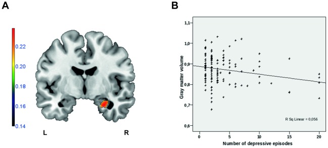
Methods: For this study one-hundred thirty two patients with major depressive disorder and 132 age- and gender-matched healthy control participants were included, 35 with their first episode and 97 with recurrent depression. To analyse gray matter abnormalities, voxel-based morphometry (VBM8) was employed on T1 weighted MRI data. We performed whole-brain analyses as well as a region-of-interest approach on the hippocampal formation, anterior cingulate cortex and amygdala, correlating the number of depressive episodes.
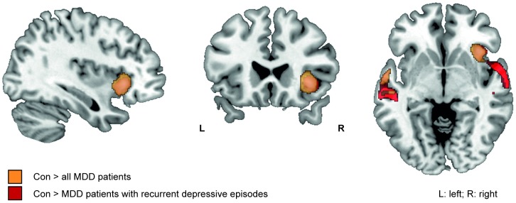
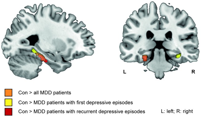
Results: Compared to healthy control persons, patients showed a strong gray-matter reduction in the right anterior insula. In addition, region-of-interest analyses revealed significant gray-matter reductions in the hippocampal formation. The observed alterations were more severe in patients with recurrent depressive episodes than in patients with a first episode. The number of depressive episodes was negatively correlated with gray-matter volume in the right hippocampus and right amygdala.
Conclusions: The anterior insula gray matter structure appears to be strongly affected in major depressive disorder and might play an important role in the neurobiology of depression. The hippocampal and amygdala volume loss cumulating with the number of episodes might be explained either by repeated neurotoxic stress or alternatively by higher relapse rates in patients showing hippocampal atrophy.
Conflict of interest statement
Figures
Similar articles
-
Longitudinal brain volume changes in major depressive disorder.J Neural Transm (Vienna). 2018 Oct;125(10):1433-1447. doi: 10.1007/s00702-018-1919-8. Epub 2018 Aug 27. J Neural Transm (Vienna). 2018. PMID: 30167933
-
Comparison of regional gray matter volume abnormalities in Alzheimer׳s disease and late life depression with hippocampal atrophy using VSRAD analysis: a voxel-based morphometry study.Psychiatry Res. 2015 Apr 30;232(1):71-5. doi: 10.1016/j.pscychresns.2015.01.018. Epub 2015 Jan 31. Psychiatry Res. 2015. PMID: 25773003
-
Grey matter volume abnormalities in the first depressive episode of medication-naïve adult individuals: a systematic review of voxel based morphometric studies.Int J Psychiatry Clin Pract. 2021 Nov;25(4):407-420. doi: 10.1080/13651501.2020.1861632. Epub 2020 Dec 22. Int J Psychiatry Clin Pract. 2021. PMID: 33351672
-
Effects of cumulative illness severity on hippocampal gray matter volume in major depression: a voxel-based morphometry study.Psychol Med. 2018 Oct;48(14):2391-2398. doi: 10.1017/S0033291718000016. Epub 2018 Feb 8. Psychol Med. 2018. PMID: 29415775
-
Cortical and Subcortical Gray Matter Volume in Youths With Conduct Problems: A Meta-analysis.JAMA Psychiatry. 2016 Jan;73(1):64-72. doi: 10.1001/jamapsychiatry.2015.2423. JAMA Psychiatry. 2016. PMID: 26650724 Review.
Cited by
-
Neuroanatomical Differences Among Sexual Offenders: A Targeted Review with Limitations and Implications for Future Directions.Violence Gend. 2020 Sep 1;7(3):86-97. doi: 10.1089/vio.2019.0051. Epub 2020 Sep 11. Violence Gend. 2020. PMID: 32939353 Free PMC article. Review.
-
Morphological Biomarkers in the Amygdala and Hippocampus of Children and Adults at High Familial Risk for Depression.Diagnostics (Basel). 2022 May 12;12(5):1218. doi: 10.3390/diagnostics12051218. Diagnostics (Basel). 2022. PMID: 35626374 Free PMC article.
-
Role of inflammation in depression relapse.J Neuroinflammation. 2019 Apr 17;16(1):90. doi: 10.1186/s12974-019-1475-7. J Neuroinflammation. 2019. PMID: 30995920 Free PMC article. Review.
-
Brain structure abnormalities in young women who presented conduct disorder in childhood/adolescence.Cogn Affect Behav Neurosci. 2017 Aug;17(4):869-885. doi: 10.3758/s13415-017-0519-7. Cogn Affect Behav Neurosci. 2017. PMID: 28695488 Free PMC article.
-
Different spatial patterns of brain atrophy and global functional connectivity impairments in major depressive disorder.Brain Imaging Behav. 2017 Dec;11(6):1678-1689. doi: 10.1007/s11682-016-9645-z. Brain Imaging Behav. 2017. PMID: 27766588 Free PMC article.
References
-
- Kessler RC, Berglund P, Demler O, Jin R, Koretz D, et al. (2003) The epidemiology of major depressive disorder: results from the National Comorbidity Survey Replication (NCS-R). JAMA 289: 3095–3105. - PubMed
-
- WHO (2008) The global burden of disease. Update 2004: World Health Organization, Geneva, Switzerland.
-
- Bora E, Fornito A, Pantelis C, Yücel M (2011) Gray matter abnormalities in Major Depressive Disorder: A meta-analysis of voxel based morphometry studies. J Affect Disord. - PubMed
-
- Arnone D, McIntosh AM, Ebmeier KP, Munafò MR, Anderson IM (2012) Magnetic resonance imaging studies in unipolar depression: systematic review and meta-regression analyses. Eur Neuropsychopharmacol 22: 1–16. - PubMed
Publication types
MeSH terms
LinkOut - more resources
Full Text Sources
Other Literature Sources
