. 2015 Feb;21(3-4):417-25.
doi: 10.1089/ten.TEA.2013.0615. Epub 2014 Oct 15.

In vitro characterization of patches of human mesenchymal stromal cells

Affiliations

In vitro characterization of patches of human mesenchymal stromal cells

Stephan Roux et al. Tissue Eng Part A. 2015 Feb.

Abstract

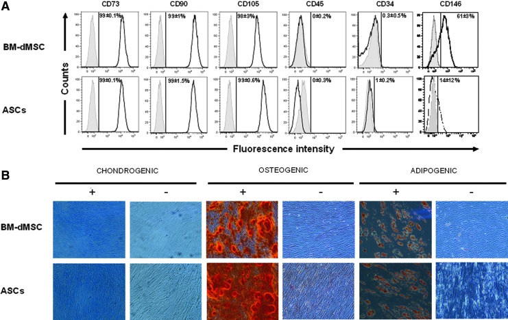
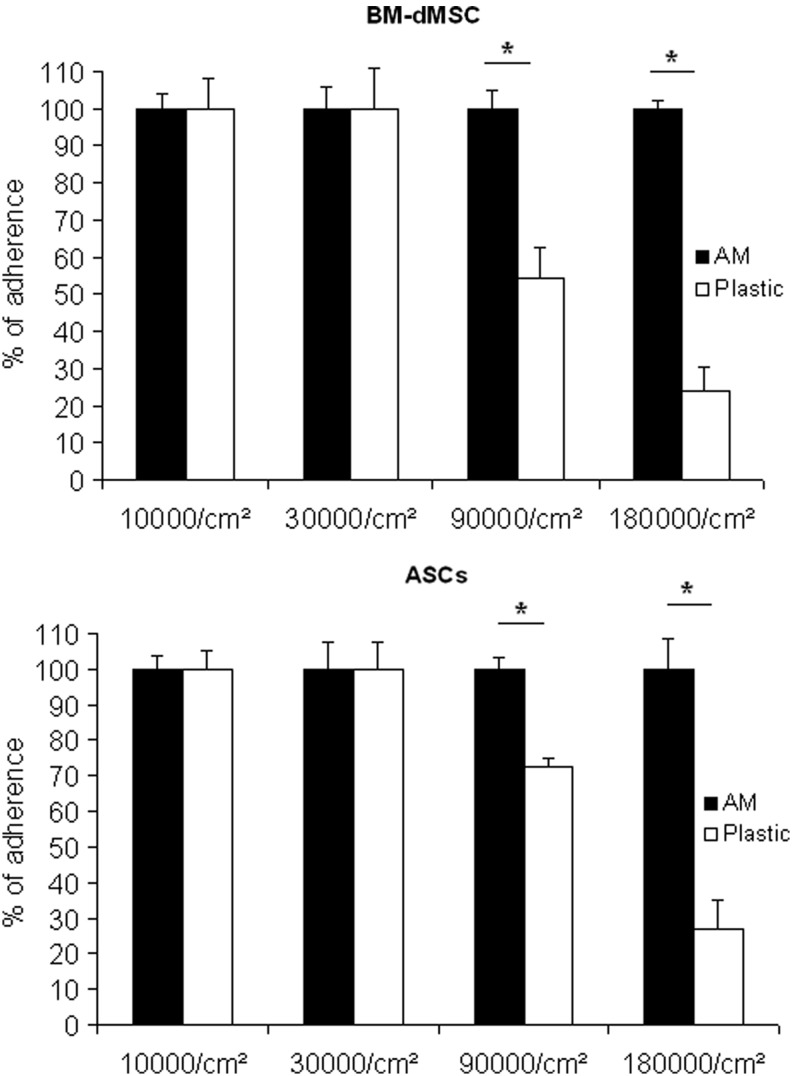
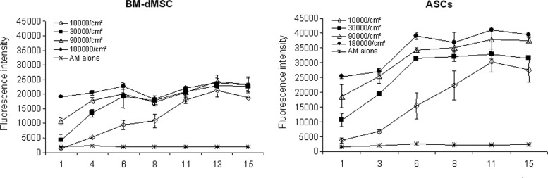
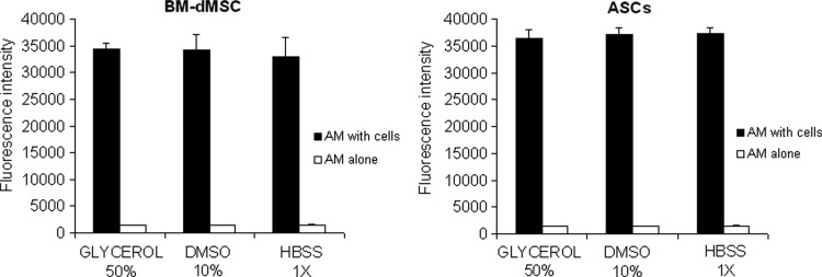
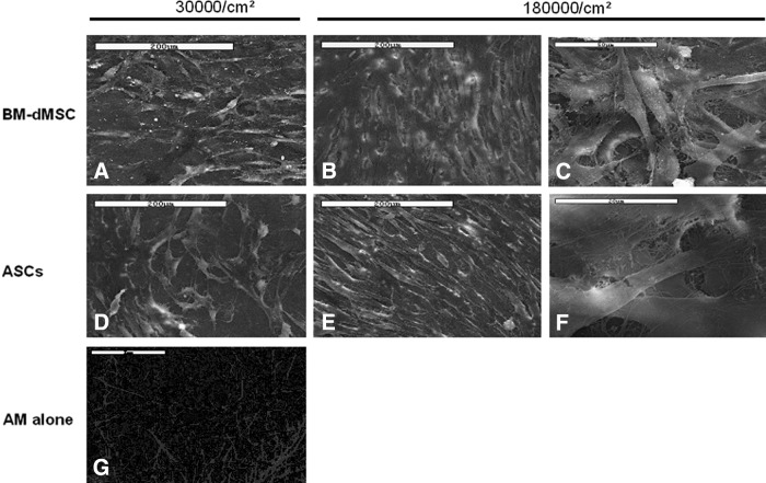
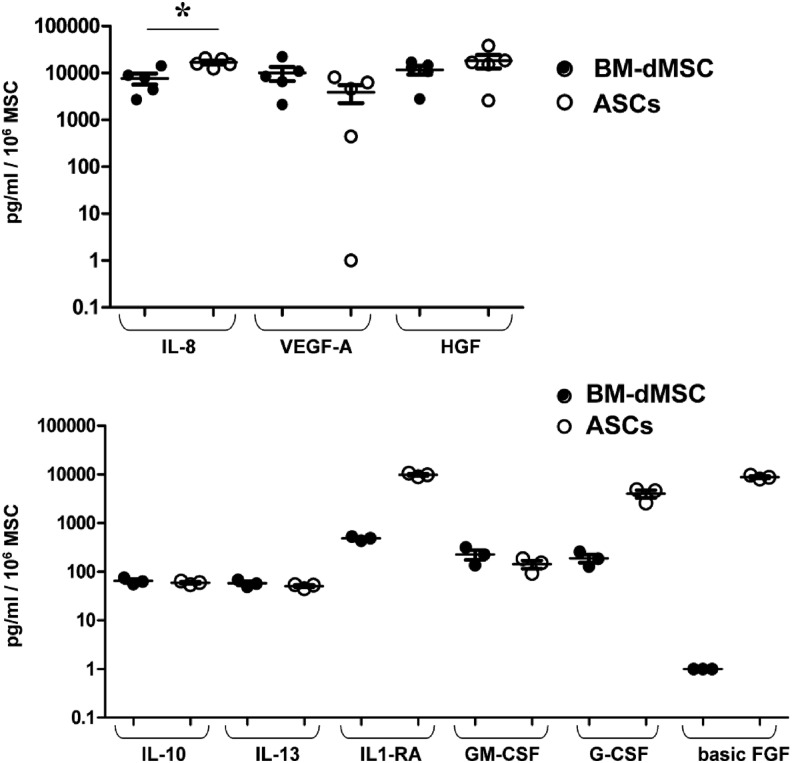
Stem cells may represent an excellent strategy to improve the healing of skin ulcers. Today the administration mode of stem cells to skin defects remains unsatisfactory. Delivering stem cells with topical treatments represents a new strategy and answering the patients' need. Mesenchymal stromal cells (MSC) have been shown to improve wound healing of cutaneous lesions and amniotic membrane (AM) is known to represent a natural scaffold for cells. The aim of this study is to develop a tissue-engineered product combining MSC and AM for clinical use. In this work we investigated whether the stromal matrix of intact human AM could constitute a scaffold for human MSC derived from either bone marrow (BM) or adipose tissue (AT). For this purpose, clinical-grade AM, MSC, and culture medium were used. We performed experiments of short-term adherence and proliferation for 15 days after the seeding of the cells. Morphological aspects and secretion profiles of MSC onto AM were studied, respectively, by scanning electron microscopy and Luminex analysis. Results demonstrated that the stromal matrix allow the adherence in much greater amount of MSC from BM or AT compared to 2D material. Experiments of proliferation showed that both kinds of MSC could proliferate on the stromal matrix and remain viable 15 days after the seeding of the cells. The 3D analysis of MSC culture demonstrated that both types of MSC invaded the stromal matrix and grew in multiple layers while retaining their fibroblastic morphology. By studying the secretion profile of MSC onto the stromal matrix, we found that both kinds of MSC secrete important cytokines and growth factors for wound healing of cutaneous lesions, such as vascular endothelial growth factor, hepatocyte growth factor, and basic fibroblast growth factor. In conclusion, these results suggest that the stromal matrix of AM seeded with MSC represents a bioactive scaffold that should be evaluated in patients with a nonhealing cutaneous wound.


Figures

<b>FIG. 1.</b>
FIG. 1.
Characterization of mesenchymal stromal cells (MSC). (A) Phenotype of MSC. MSC derived from bone marrow (BM-dMSC) (passage 2) and adipose tissue (ASC) (passage 3) were analyzed by fluorescence-activated cell sorting after staining with conjugated monoclonal antibodies against surface markers (black line) or IgG control isotypes (gray line). Results were representative of three independent experiments. The mean percentage of fluorescence above the threshold for isotype controls is indicated. (B) Differentiation potential of MSC for mesodermic lineage: MSC were cultured in a medium with (+) or without (−) differentiation cocktails for 3 weeks. Chondrogenic, osteogenic, and adipogenic differentiation were observed, respectively by coloration with Alcian Blue, which detects proteoglycans of chondroblasts, Alizarin Red, which detects calcium mineralization of osteoblasts and Oil red O, which detects lipid vesicles in adipocytes. Results depicted the differentiation of MSC derived from the bone marrow (BM-dMSC) or ASC from one donor out of three. Color images available online at www.liebertpub.com/tea
<b>FIG. 2.</b>
FIG. 2.
MSC adherence on amniotic membrane (AM). MSC were allowed to adhere on the stromal side of AM or on culture plastic at doses from 10,000 cells/cm2 to 180,000 cells/cm2. An Alamar Blue assay was performed 6 h after loading. Results are representative of two independent experiments performed in triplicate and mean data±SEM are presented (*p<0.05).
<b>FIG. 3.</b>
FIG. 3.
MSC proliferation on AM. MSC were loaded at various doses from 10,000 cells/cm2 to 180,000 cells/cm2 on the stromal side of AM and cultured for a period of 15 days. Cell viability was assessed using an Alamar Blue assay performed in triplicate. Results are representative of two independent experiments performed in duplicate and mean data±SEM are presented.
<b>FIG. 4.</b>
FIG. 4.
Effect of AM preparation on MSC adherence. MSC seeded at 180,000 cells/cm2 were allowed to adhere on the stromal side of AM cryopreserved in glycerol 50%, DMSO 10% or HBSS 1×. An Alamar Blue assay was performed 48 h after the loading. Results are representative of two independent experiments performed in triplicate and mean data±SEM are presented. The ANOVA test was used to compare the three preparations of AM: pBM-dMSC=0.71; pASC=0.56. The Mann–Whitney U-test was performed to compare MSC for both sources of cells: pGlycerol 50%=0.20; pDMSO 10%=0.40; pHBSS 1×=0.20.
<b>FIG. 5.</b>
FIG. 5.
MSC morphology on AM. BM-dMSC were cultured at 30,000 cells/cm2 (A) or 180,000 cells/cm2 (B and C) and ASC were cultured at 30,000 cells/cm2 (D) or 180,000 cells/cm2 (E and F) on the stromal side of AM in duplicate. MSC morphology was observed at 48 h of culture by scanning electron microscopy. The stromal side of AM without MSC is presented on (G). Scale bars: (A–F) 200 μm; (G) 2 μm.
<b>FIG. 6.</b>
FIG. 6.
Study of MSC secretion profile on AM. MSC derived from one BM and one adipose tissue were incubated at 180,000 cells/cm2 on the stromal side of AM. Supernatants were harvested at 48 h to study the secretion of proteins related to wound healing. Protein concentration was normalized with the quantity of cells determined using an Alamar Blue assay. Values represent the mean±SEM of triplicate (for the first experiment) and duplicate (for the second experiment) of two independent experiments for interleukin (IL)-8, vascular endothelial growth factor (VEGF)-A, and hepatocyte growth factor (HGF) or triplicate of one experiment for IL-10, IL-13, IL-1-RA, platelet-derived growth factor (PDGF-BB), granulocyte-macrophage colony-stimulating factor (GM-CSF), granulocyte-colony-stimulating factor (G-CSF), and basic fibroblast growth factor (FGF) (*p<0.05 between BM-dMSC and ASC).

References

    1. Simon D.A., Dix F.P., and McCollum C.N.Management of venous leg ulcers. BMJ 328,1358, 2004 - PMC - PubMed
    1. Pittenger M.F., Mackay A.M., Beck S.C., Jaiswal R.K., Douglas R., Mosca J.D., et al. . Multilineage potential of adult human mesenchymal stem cells. Science 284,143, 1999 - PubMed
    1. Delorme B., Ringe J., Gallay N., Le Vern Y., Kerboeuf D., Jorgensen C., et al. . Specific plasma membrane protein phenotype of culture-amplified and native human bone marrow mesenchymal stem cells. Blood 111,2631, 2008 - PubMed
    1. Mitchell J.B., McIntosh K., Zvonic S., Garrett S., Floyd Z.E., Kloster A., et al. . Immunophenotype of human adipose-derived cells: temporal changes in stromal-associated and stem cell-associated markers. Stem Cells 24,376, 2006 - PubMed
    1. Strioga M., Viswanathan S., Darinskas A., Slaby O., and Michalek J.Same or not the same? Comparison of adipose tissue-derived versus bone marrow-derived mesenchymal stem and stromal cells. Stem Cells Dev 21,2724, 2012 - PubMed

Publication types

LinkOut - more resources