. 2014 Dec;171(23):5417-30.
doi: 10.1111/bph.12857.

δ-Opioid receptors up-regulate excitatory amino acid transporters in mouse astrocytes

Affiliations

δ-Opioid receptors up-regulate excitatory amino acid transporters in mouse astrocytes

Jianfeng Liang et al. Br J Pharmacol. 2014 Dec.

Abstract

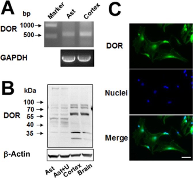
Background and purpose: Astrocytic excitatory amino acid transporters (EAATs) regulate extracellular glutamate concentrations and play a role in preventing neuroexcitotoxicity. As the δ-opioid receptor (DOP receptor) is neuroprotective against excitotoxic injury, we determined whether DOP receptor activation up-regulates EAAT expression and function.

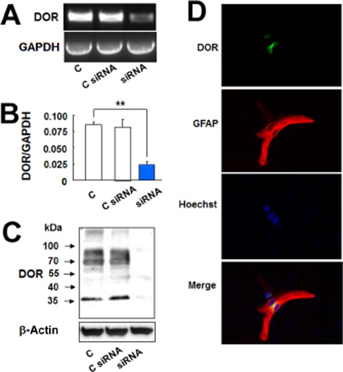
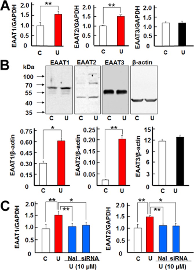
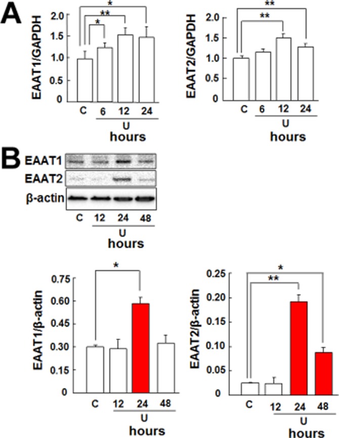
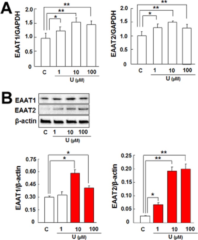
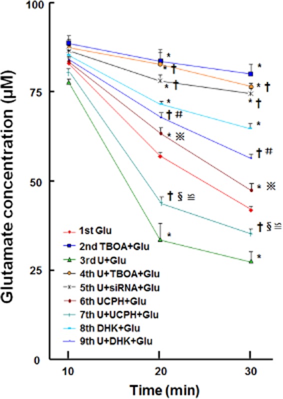
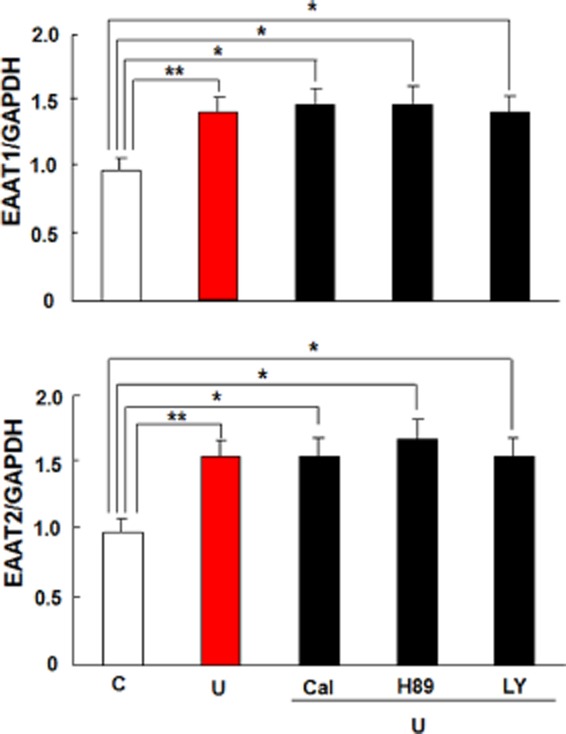
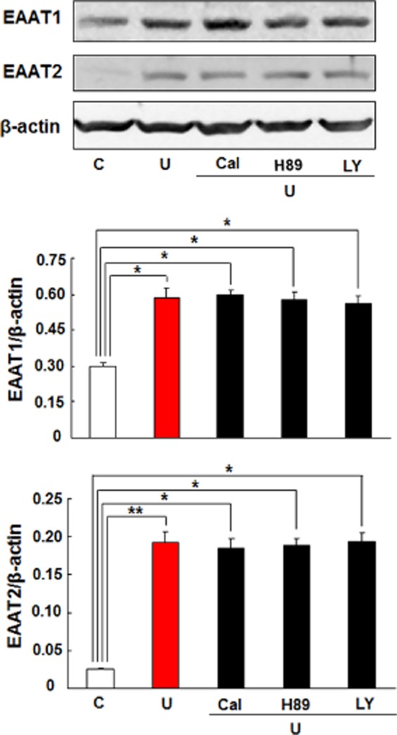
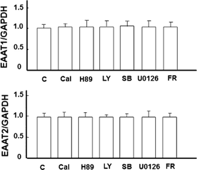
Experimental approach: We measured mRNA and protein expression of EAAT1, EAAT2 and EAAT3 in cultured mouse astrocytes exposed to a specific DOP receptor agonist (UFP-512) with or without a DOP receptor antagonist, DOP receptor siRNA or inhibitors of PKC, PKA, PI3K, p38, MAPK, MEK and ERK, and evaluated the function of EAATs by measuring glutamate uptake.

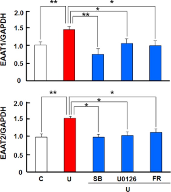
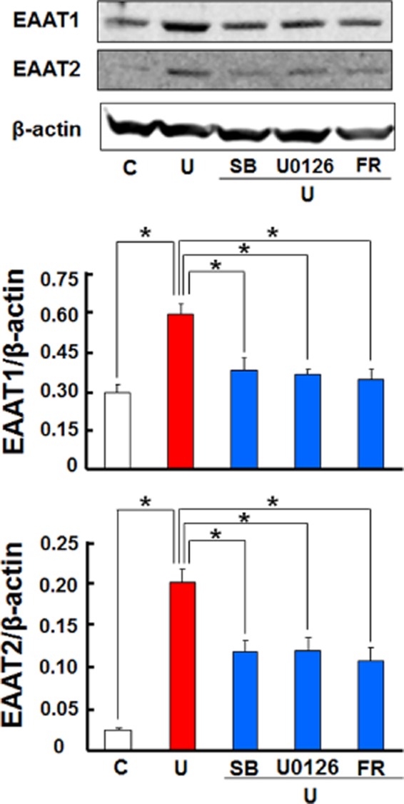
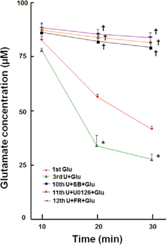
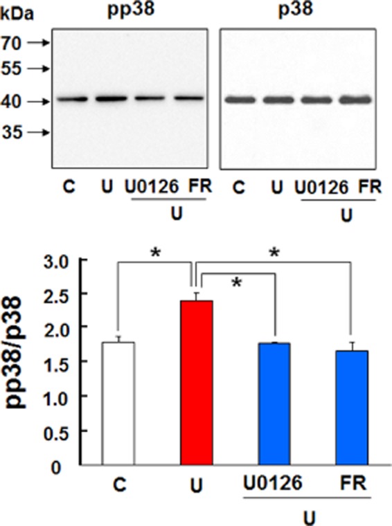
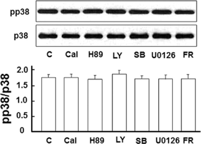
Key results: Astrocytic DOP receptor mRNA and protein were suppressed by DOP receptor siRNA knockdown. DOP receptor activation increased mRNA and protein expression of EAAT1 and EAAT2, but not EAAT3, thereby enhancing glutamate uptake of astrocytes. DOP receptor-induced EAAT1 and EAAT2 expression was largely reversed by DOP receptor antagonist naltrindole or by DOP receptor siRNA knockdown, and suppressed by inhibitors of MEK, ERK and p38. DOP receptor-accelerated glutamate uptake was inhibited by EAAT blockers, DOP receptor siRNA knockdown or inhibitors of MEK, ERK or p38. In contrast, inhibitors of PKA, PKC or PI3K had no significant effect on DOP receptor-induced EAAT expression.

Conclusions and implications: DOP receptor activation up-regulates astrocytic EAATs via MEK-ERK-p38 signalling, suggesting a critical role for DOP receptors in the regulation of astrocytic EAATs and protection against neuroexcitotoxicity. As decreased EAAT expression contributes to pathophysiology in many neurological diseases, including amyotrophic lateral sclerosis, our findings present a new platform for potential treatments of these diseases.


Figures

Figure 1
Figure 1
DOP receptor (DOR) expression in the astrocytes. (A) DOP receptor mRNA analysis by RT-PCR. Total mRNA was extracted from the astrocytes and cortical tissues. (B) DOP receptor protein detection by Western blot analysis. Total proteins were extracted from the astrocytes, UFP-512 treated astrocytes, cortex and whole brain. (C) Immunocytochemistry results showing that DOP receptor protein existed in the membrane, cytoplasma and nuclei of the astrocytes. Scale bar, 50 μm. Ast, astrocytes; U, astrocytes treated with 10 μM UFP-512 for 24 h. Note that the astrocytes expressed DOP receptor mRNA and 36 and 72 kDa DOP receptor protein.
Figure 2
Figure 2
DOP receptor siRNA reduced DOP receptor (DOR) expression in the astrocytes. (A) DOP receptor mRNA analysis by RT-PCR. Total mRNA was extracted from the astrocytes and those treated with control siRNA or DOP receptor siRNA. (B) DOP receptor mRNA analysis by quantitative PCR. Total mRNA was extracted from the control astrocytes and those treated with control siRNA or DOP receptor siRNA. (C) DOP receptor protein detection by Western blot analysis. Total proteins were extracted from the control astrocytes and those treated with control siRNA or DOP receptor siRNA. (D) Astrocytes were treated with DOP receptor siRNA for 24 h and then were stained with DOP receptor and GFAP antibodies. Immunocytochemistry results showing DOP receptor siRNA markedly reduced astrocytic DOP receptor expression. C, non-treated astrocytes; C siRNA, astrocytes treated with control siRNA for 24 h; siRNA, astrocytes treated with DOP receptor siRNA for 24 h. **P < 0.01. Note that there was a significant reduction in astrocytic DOP receptor mRNA and protein expression after addition of DOP receptor siRNA.
Figure 3
Figure 3
DOP receptors induced EAAT1 and EAAT2 expression in the astrocytes. (A) Quantitative PCR analysis showing that UFP-512 induced EAAT1 and EAAT2 mRNA expression, but did not change EAAT3 mRNA expression. Astrocytes were treated with 10 μM UFP-512 for 12 h and total mRNA was extracted for detection. (B) Western blot analysis showing that UFP-512 induced EAAT1 (65 kDa) and EAAT2 (70 kDa) protein expression, whereas it did not alter EAAT3 (57 kDa) protein expression. The astrocytes were treated with 10 μM UFP-512 for 24 h and total protein was extracted for measurements. (C) Effect of DOP receptor antagonism and siRNA knockdown on astrocytic EAATs. For DOP receptor antagonism, the astrocytes were pretreated with naltrindole for 1 h, and then treated with 10 μM UFP-512 and 10 μM naltrindole for 12 h. For DOP receptor siRNA knockdown, the astrocytes were pretreated with DOP receptor siRNA for 24 h and then treated with 10 μM UFP-512 for 12 h. C, non-treatment; Nal, 10 μM naltrindole, a DOP receptor antagonist; U, UFP-512, a specific DOP receptor agonist. *P < 0.05; **P < 0.01. Note that UFP-512 elevated EAAT1 and EAAT2 mRNA expression, while both naltrindole and DOP receptor siRNA abolished the UFP-512-induced increase in EAAT1 and EAAT2 expression.
Figure 4
Figure 4
Time course of DOP receptor-induced EAAT1 and EAAT2 expression in the astrocytes. (A) Quantitative PCR analysis showing that UFP-512 induced EAAT1 and EAAT2 mRNA expression in a time-dependent manner with expression reaching peak at 12 h. (B) Western blot showing that UFP-512 induced EAAT1 and EAAT2 protein production in a time-dependent manner with production reaching peak at 24 h. C, non-treatment; U, 10 μM UFP-512. *P < 0.05; **P < 0.01. Note that UFP-512 induced both EAAT1 and EAAT2 mRNA as well as protein expression in a time-dependent manner.
Figure 5
Figure 5
DOP receptor induced astrocytic EAAT1 and EAAT2 expression in a dose-dependent manner. (A) Astrocytes were treated with UFP-512 for 12 h. Then total mRNA was extracted for mRNA analysis. Quantitative PCR analysis showing that UFP-512 induced EAAT1 and EAAT2 mRNA expression in a dose-dependent manner with expression reaching peak at 10 μM. (B) Astrocytes were treated with UFP-512 for 24 h. Then total protein was extracted for protein analysis. Western blot showing that UFP-512 induced EAAT1 protein production in a dose-dependent manner with production reaching a peak at 10 μM and also up-regulated EAAT2 production, which reached a plateau at 10 μM and 100 μM. C, non-treatment; U, UFP-512. *P < 0.05; **P < 0.01. Note that UFP-512 induced EAAT1 and EAAT2 mRNA as well as protein expression in a time-dependent manner.
Figure 6
Figure 6
DOP receptor induced increase in astrocytic glutamate uptake. Nine groups of astrocytic culture were used for this set of experiments. Each group of cultured astrocytes was treated as detailed in the Methods section. Two microlitres of the treated astrocytic-conditioned media were collected from each group at 10, 20 and 30 min time points for extracellular glutamate analysis with the L-glutamate assay. *P < 0.05 versus Glu. †P < 0.05 versus U + Glu. §P < 0.05 U + UCPH + Glu versus UCPH + Glu. #P < 0.05 U + DHK + Glu versus DHK + Glu. ≌ P < 0.05 U + UCPH + Glu versus U + DHK + Glu. P < 0.05 UCPH + Glu versus DHK + Glu. Glu, 100 μM glutamate; DHK, 300 μM dihydrokainic acid, a potent EAAT2 inhibitor; siRNA, DOP receptor siRNA; TBOA, 100 μM threo-ß-benzyloxyaspartate, a potent blocker of all subtypes of the excitatory amino acid transporters; U, 10 μM UFP-512; UCPH, 10 μM UCPH-101, a specific EAAT1 inhibitor. Note that UFP-512 enhanced astrocytic glutamate uptake and this enhancement was completely blocked by the glutamate inhibitor TBOA as well as DOP receptor siRNA. Both DHK and UCPH-101 partially suppressed glutamate uptake in astrocyte culture in the presence or absence of UFP-512 stimulation. DHK had a greater effect than UCPH-101, suggesting that EAAT2 has a greater effect than EAAT1.
Figure 7
Figure 7
Effects of PKC, PKA or PI3K inhibitors on DOP receptor-induced EAAT1 and EAAT2 mRNA expression in the astrocytes. Astrocytes were pretreated with the indicated inhibitor for 1 h, followed by 10 μM UFP-512 and the inhibitor for 12 h. Total mRNA was extracted for quantitative PCR analysis. C, non-treatment; Cal, 500 nM calphostin C, a PKC inhibitor; H89, a PKA inhibitor, 10 μM; LY, 10 μM LY294002, a PI3K inhibitor; U: 10 μM UFP-512. *P < 0.05; **P < 0.01. The values are the means ± SD. At least three independent trials were performed for each assay. Note that the inhibitors of PKC, PKA or PI3K did not suppress astrocytic EAAT1 and EAAT2 mRNA expression induced by DOP receptor activation.
Figure 8
Figure 8
Effects of PKC, PKA or PI3K inhibitors on DOP receptor-induced EAAT1 and EAAT2 protein expression in the astrocytes. The astrocytes were pretreated with the indicated inhibitor for 1 h, followed by 10 μM UFP-512 and the inhibitor for 24 h. C, non-treatment; Cal, 500 nM calphostin C, PKC inhibitor; H89, a PKA inhibitor, 10 μM; LY, 10 μM LY294002, a PI3K inhibitor; U, 10 μM UFP-512. *P < 0.05; **P < 0.01. Note that the inhibitors of PKC, PKA or PI3K did not alter astrocytic EAAT1 and EAAT2 protein expression induced by DOP receptor activation.
Figure 9
Figure 9
No appreciable effect of signal pathway inhibitors on EAAT1 and EAAT2 expression in the absence of UFP-512 stimulation. Astrocytes were treated with indicated inhibitor for 12 h. Then total mRNA was extracted for mRNA analysis. Quantitative PCR analysis showing that EAAT1 and EAAT2 expression was not affected. C, non-treatment; Cal: 500 nM calphostin C, a PKC inhibitor; FR: 500 nM FR180204, ERK inhibitor; H89, a PKA inhibitor, 10 μM; LY, 10 μM LY294002, a PI3K inhibitor; SB, 10 μM SB203580, p38 inhibitor; U0126, an MEK inhibitor, 5 μM. Note that the inhibitors had no effect on astrocytic EAAT1 and EAAT2 expression.
Figure 10
Figure 10
DOP receptor-induced expression of EAAT1 and EAAT2 mRNAs via MEK-ERK-p38 signalling in the astrocytes. The astrocytes were pretreated with the indicated inhibitor for 1 h, followed by the inhibitor plus 10 μM UFP-512 for 12 h. C, non-treatment; FR: 500 nM FR180204, ERK inhibitor; SB, 10 μM SB203580, p38 inhibitor; U, 10 μM UFP-512; U0126, an MEK inhibitor, 5 μM. *P < 0.05; **P < 0.01. Note that the inhibitors of MEK, ERK and p38 MAPKs blocked astrocytic EAAT1 and EAAT2 mRNA expression induced by DOP receptor activation with UFP-512.
Figure 11
Figure 11
DOP receptor induced expression of EAAT1 and EAAT2 proteins through MEK-ERK-p38 signalling in the astrocytes. The astrocytes were pretreated with the indicated inhibitor for 1 h, followed by the inhibitor plus 10 μM UFP-512 for 24 h. C, non-treatment; FR: 500 nM FR180204, an ERK inhibitor; SB, 10 μM SB203580, p38 inhibitor; U, 10 μM UFP-512; U0126, an MEK inhibitor, 5 μM. *P < 0.05; **P < 0.01. Note that the inhibitors of MEK, ERK and p38 blocked astrocytic EAAT1 and EAAT2 protein expression induced by DOP receptor activation with UFP-512.
Figure 12
Figure 12
Effects of MEK, ERK and p38 inhibitors on DOP receptor-accelerated glutamate uptake in the astrocyte. Each group of cultured astrocytes was treated as detailed in the Methods section. After these treatments, 2 μL of astrocytic condition medium were collected from each group at 10, 20 and 30 min time points for extracellular glutamate analysis with the L-glutamate assay. FR, 500 nM FR180204, an ERK inhibitor; SB, 10 μM SB203580, p38 inhibitor; U0126, an MEK inhibitor, 5 μM. *P < 0.05 versus Glu. †P < 0.05 versus U + Glu. Note that UFP-512 enhanced astrocytic glutamate uptake was completely blocked by inhibitors for MEK, ERK and p38.
Figure 13
Figure 13
Effects of MEK and ERK inhibitors on DOP receptor-induced p38 phosphorylation in the astrocytes. The astrocytes were pretreated with the indicated inhibitor for 1 h, followed by the inhibitor plus 10 μM UFP-512 for 24 h. C, non-treatment; FR, 500 nM FR180204, an ERK inhibitor; pp38: phosphorylated p38; U, 10 μM UFP-512; U0126, an MEK inhibitor, 5 μM. *P < 0.05. Note that the inhibitors of MEK and ERK suppressed UFP-512-induced p38 phosphorylation.
Figure 14
Figure 14
No appreciable change in p38 activation after the treatment with PK and MAPK inhibitors in the absence of UFP-512 stimulation. Astrocytes were treated with indicated inhibitor for 24 h. Then total protein was extracted for Western blot. C, non-treatment; Cal, 500 nM calphostin C, a PKC inhibitor; FR, 500 nM FR180204, ERK inhibitor; H89, a PKA inhibitor, 10 μM; LY, 10 μM LY294002, a PI3K inhibitor; pp38, phosphorylated p38; SB, 10 μM SB203580, p38 inhibitor; U0126, an MEK inhibitor, 5 μM. Note that the inhibitors had no effect on p38 activation.

References

    1. Abe K, Saito H. Possible linkage between glutamate transporter and mitogen-activated protein kinase cascade in cultured rat cortical astrocytes. J Neurochem. 2001;76:217–223. - PubMed
    1. Alexander SPH, Benson HE, Faccenda E, Pawson AJ, Sharman JL, Spedding M, et al. The Concise Guide to PHARMACOLOGY 2013/14: G protein-coupled receptors. Br J Pharmacol. 2013a;170:1459–1581. - PMC - PubMed
    1. Alexander SPH, Benson HE, Faccenda E, Pawson AJ, Sharman JL, Spedding M, et al. The Concise Guide to PHARMACOLOGY 2013/14: Transporters. Br J Pharmacol. 2013b;170:1706–1796. - PMC - PubMed
    1. Alexander SPH, Benson HE, Faccenda E, Pawson AJ, Sharman JL, Spedding M, et al. The Concise Guide to PHARMACOLOGY 2013/14: Enzymes. Br J Pharmacol. 2013c;170:1797–1867. - PMC - PubMed
    1. Arriza JL, Fairman WA, Wadiche JI, Murdoch GH, Kavanaugh MP, Amara SG. Functional comparisons of three glutamate transporter subtypes cloned from human motor cortex. J Neurosci. 1994;14:5559–5569. - PMC - PubMed

Publication types

MeSH terms

Substances