. 2014 Jul;26(7):3090-100.
doi: 10.1105/tpc.114.124008. Epub 2014 Jul 22.

Uric acid accumulation in an Arabidopsis urate oxidase mutant impairs seedling establishment by blocking peroxisome maintenance

Affiliations

Uric acid accumulation in an Arabidopsis urate oxidase mutant impairs seedling establishment by blocking peroxisome maintenance

Oliver K Hauck et al. Plant Cell. 2014 Jul.

Abstract

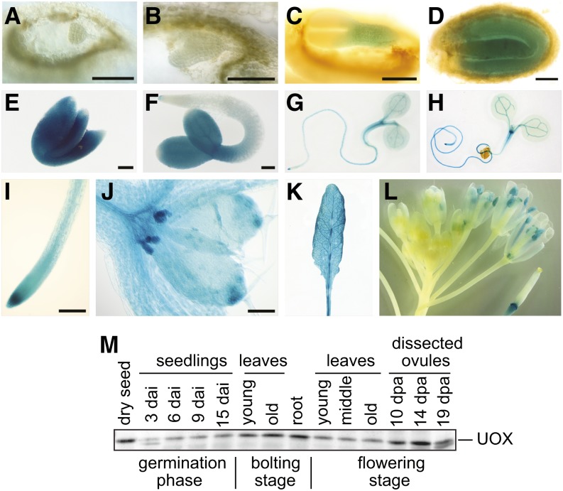
Purine nucleotides can be fully catabolized by plants to recycle nutrients. We have isolated a urate oxidase (uox) mutant of Arabidopsis thaliana that accumulates uric acid in all tissues, especially in the developing embryo. The mutant displays a reduced germination rate and is unable to establish autotrophic growth due to severe inhibition of cotyledon development and nutrient mobilization from the lipid reserves in the cotyledons. The uox mutant phenotype is suppressed in a xanthine dehydrogenase (xdh) uox double mutant, demonstrating that the underlying cause is not the defective purine base catabolism, or the lack of UOX per se, but the elevated uric acid concentration in the embryo. Remarkably, xanthine accumulates to similar levels in the xdh mutant without toxicity. This is paralleled in humans, where hyperuricemia is associated with many diseases whereas xanthinuria is asymptomatic. Searching for the molecular cause of uric acid toxicity, we discovered a local defect of peroxisomes (glyoxysomes) mostly confined to the cotyledons of the mature embryos, which resulted in the accumulation of free fatty acids in dry seeds. The peroxisomal defect explains the developmental phenotypes of the uox mutant, drawing a novel link between uric acid and peroxisome function, which may be relevant beyond plants.


Figures

Figure 1.
Figure 1.
Overview of Purine Nucleotide Catabolism. The scheme focuses on the XDH and UOX reactions. HIU, 5-hydroxyisourate; Pi, phosphate.
Figure 2.
Figure 2.
UOX and XDH Amount and Activity in the Wild Type and Mutants. Leaf samples were from 4-week-old plants of the wild type, uox and xdh mutants, the uox xdh double mutant, and the uox mutant transformed with a 35S promoter–driven UOX cDNA construct for complementation (35S:UOX). Panels from top to bottom are as follows: quantification of UOX activity per milligram of total protein (error bars are sd; n = 3); immunoblot developed with anti-UOX antiserum; visualization of XDH activity in gel; and immunoblot developed with anti-XDH antiserum. mU, milliunits.
Figure 3.
Figure 3.
Seedling Establishment of the Wild Type, the uox and xdh Mutants, the uox Complementation Line (35S:UOX), and the Double Mutant. Seeds were sown on half-strength Murashige and Skoog agar medium, stratified in the dark for 2 d, and grown under long-day conditions (16 h of light). The medium contained 2% (w/v) Suc where indicated. Bars = 1 mm.
Figure 4.
Figure 4.
Promoter Activity and Expression of UOX. (A) to (E) Promoter-GUS activity during embryonic development. (A) Globular stage. (B) Late heart stage. (C) Torpedo stage. (D) Bent cotyledon stage. (E) Mature embryo dissected from the dry seed. (F) Germinated seedling, 3 DAI. (G) Young seedling, 5 DAI. (H) Young seedling, 7 DAI. (I) Root tip. (J) Shoot apical tip of seedling, 9 DAI. (K) Rosette leaf from bolting plant. (L) Inflorescence. Bars = 100 μm. (M) Immunoblot of proteins extracted from different tissues (10 μg of protein per lane), developed with UOX antiserum. dpa, days after anthesis.
Figure 5.
Figure 5.
Uric Acid and Xanthine Metabolite Profiles. (A) Tissue uric acid content in the uox mutant grown under long-day conditions (16 h of light). Top row, uric acid concentration in reference to the dry weight (dw) of young leaves (yl; leaves 13 to the top leaf), middle-aged leaves (ml; leaves 7 to 12), old leaves (ol; leaves 1 to 6), roots (rt), whole siliques at different DAA (dpa), developing seeds (ds) and silique walls (sw) at 19 DAA, and seedlings during germination at 0 to 15 DAI. Bottom row, as in the top row but with reference to the fresh weight (fw). (B) Uric acid and xanthine content of seeds of the wild type, the uox and xdh mutants, the xdh uox double mutant, and the complementation line (35S:UOX) grown under long-day conditions (16 h of light). Mean values ± sd are shown (n = 3 or as indicated).
Figure 6.
Figure 6.
Ultrastructural Alterations in Mature Embryos of the uox Mutant. (A) to (E) Transmission electron micrographs of cotyledon parenchyma cells of the wild type (A), the xdh uox double mutant (B), and the uox mutant ([C] to [E]). lb, lipid body; p, protein body. Arrows indicate abnormalities in the uox mutant. Bars = 2 μm. (F) Quantification of peroxisome numbers by counting DAB-positive spots in cotyledons of the wild type, the uox mutant, and the xdh uox double mutant in light micrographs from semithin sections (Supplemental Figure 6). The horizontal bar is the mean (n = 8, 5, and 6 for the wild type, uox, and xdh uox, respectively). Statistical significance was determined using ANOVA coupled to Dunnett’s posttest. Generally, P values below 0.05, 0.01, and 0.001 are represented by one, two, and three asterisks, respectively, and the numeric values are shown, unless the P value is smaller than 0.001 (indicated by <0.001).
Figure 7.
Figure 7.
Comparison of Transgenic Plant Lines Expressing Peroxisomal GFP in the Wild Type and the uox Mutant Background. (A) Expression of roGFP2-SKL in seeds of three different transformed uox mutant lines and two different transformed wild-type lines monitored by immunoblot probed with anti-GFP antibodies. (B) Confirmation of the uox phenotype on medium without Suc in the lines transformed with GFP at 7 DAI. (C) Fluorescence microscopy of embryos with wild-type or uox mutant background from the lines analyzed in (A) and (B). Embryos were excised at different DAA (dpa), from the dry seed, and 6 d after imbibition in the dark. Representative micrographs of the cotyledons (c) and the hypocotyls (h) are shown. Bars = 25 µm. (D) Quantification of peroxisome numbers in cotyledons and hypocotyls of embryos dissected from dry seed. Each dot resulted from peroxisome quantification in an independent seedling using a stack of images. The horizontal bar is the mean (n = 4 to 10). Statistical significance was determined using ANOVA coupled to Dunnett’s posttest. P values are indicated as described in Figure 6.
Figure 8.
Figure 8.
Lipid Analysis of Dry Seeds and Changes of Fatty Acid Content after Imbibition. (A) Relative quantification of free fatty acids (FA), TAGs, glycerophospholipids (GPL), and galactolipids (GL) in dry seeds of the uox mutant (n = 5), the wild type (n = 5), and the xdh uox double mutant (n = 4). Error bars are sd. Statistical significance was determined using ANOVA coupled to Dunnett’s posttest. P values are indicated as described in Figure 6. (B) Ratio of fatty acid content in seedlings at 6 DAI (including the seed coat) versus dry seeds in the uox mutant, the wild type, and the 35S complementation line for total fatty acids and the TAG-specific 20:1 fatty acid. The total TAG content in these lines was 74% ± 7%, 3% ± 3%, and 24% ± 6% (ratio of seed to seedling), respectively. sd is shown (n = 5), and statistical evaluation was as in (A).

References

    1. Alonso J.M., et al. (2003). Genome-wide insertional mutagenesis of Arabidopsis thaliana. Science 301: 653–657. - PubMed
    1. Angelovici R., Galili G., Fernie A.R., Fait A. (2010). Seed desiccation: A bridge between maturation and germination. Trends Plant Sci. 15: 211–218. - PubMed
    1. Brychkova G., Alikulov Z., Fluhr R., Sagi M. (2008). A critical role for ureides in dark and senescence-induced purine remobilization is unmasked in the Atxdh1 Arabidopsis mutant. Plant J. 54: 496–509. - PubMed
    1. Cao F.Q., Werner A.K., Dahncke K., Romeis T., Liu L.H., Witte C.P. (2010). Identification and characterization of proteins involved in rice urea and arginine catabolism. Plant Physiol. 154: 98–108. - PMC - PubMed
    1. Chia T.Y.P., Pike M.J., Rawsthorne S. (2005). Storage oil breakdown during embryo development of Brassica napus (L.). J. Exp. Bot. 56: 1285–1296. - PubMed

Publication types

MeSH terms