

Intravital imaging of cardiac function at the single-cell level
- PMID: 25053815
- PMCID: PMC4128110
- DOI: 10.1073/pnas.1401316111
Intravital imaging of cardiac function at the single-cell level
Abstract
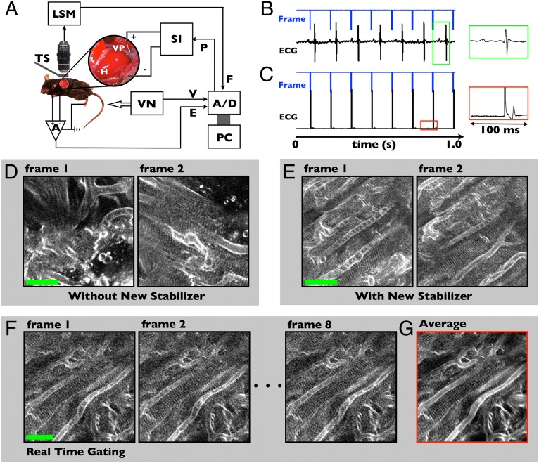
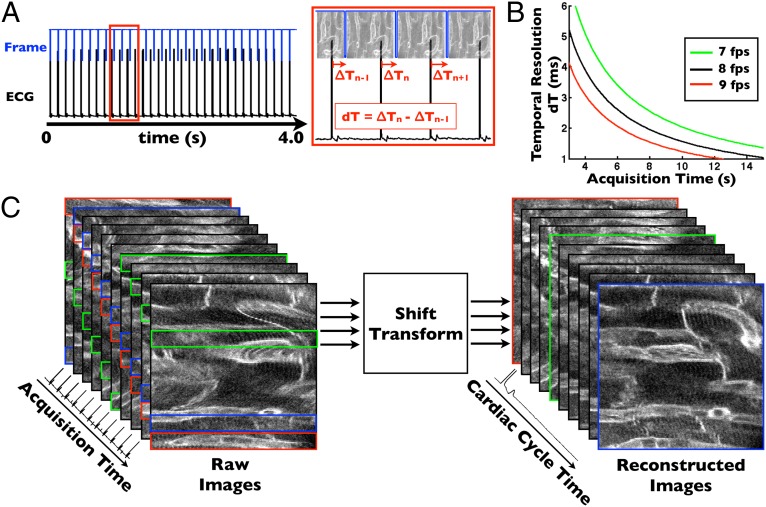
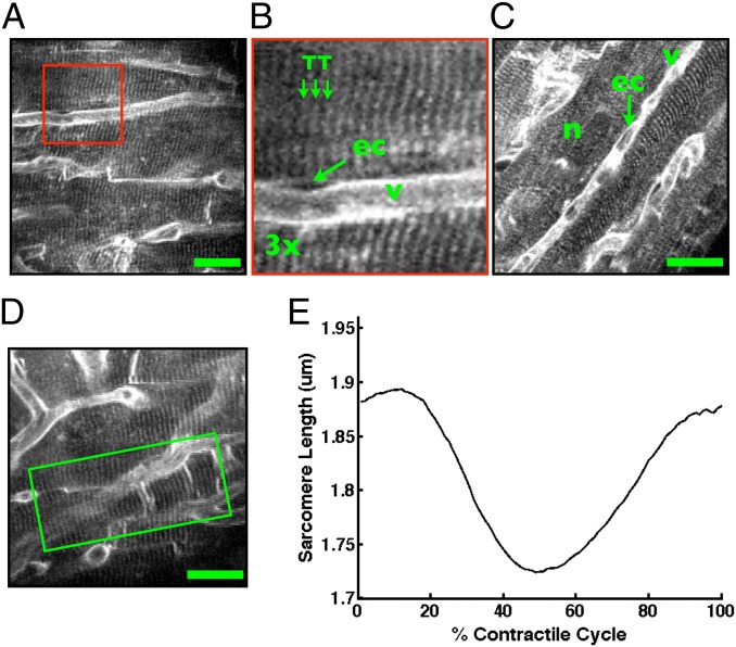
Knowledge of cardiomyocyte biology is limited by the lack of methods to interrogate single-cell physiology in vivo. Here we show that contracting myocytes can indeed be imaged with optical microscopy at high temporal and spatial resolution in the beating murine heart, allowing visualization of individual sarcomeres and measurement of the single cardiomyocyte contractile cycle. Collectively, this has been enabled by efficient tissue stabilization, a prospective real-time cardiac gating approach, an image processing algorithm for motion-artifact-free imaging throughout the cardiac cycle, and a fluorescent membrane staining protocol. Quantification of cardiomyocyte contractile function in vivo opens many possibilities for investigating myocardial disease and therapeutic intervention at the cellular level.
Keywords: cardiovascular imaging; fluorescence; intravital micoscopy; molecular imaging; pacing.
Conflict of interest statement
The authors declare no conflict of interest.
Figures
References
-
- Rubart M. Two-photon microscopy of cells and tissue. Circ Res. 2004;95(12):1154–1166. - PubMed
-
- Botcherby EJ, et al. Fast measurement of sarcomere length and cell orientation in Langendorff-perfused hearts using remote focusing microscopy. Circ Res. 2013;113(7):863–870. - PubMed
-
- Hama T, Takahashi A, Ichihara A, Takamatsu T. Real time in situ confocal imaging of calcium wave in the perfused whole heart of the rat. Cell Signal. 1998;10(5):331–337. - PubMed
Publication types
MeSH terms
Grants and funding
LinkOut - more resources
Full Text Sources
Other Literature Sources
Medical
