Review
. 2014 Jun;31(2):167-79.
doi: 10.1055/s-0034-1373791.

Bone and soft tissue ablation

Affiliations
Review

Bone and soft tissue ablation

Ryan C B Foster et al. Semin Intervent Radiol. 2014 Jun.

Abstract

Bone and soft tissue tumor ablation has reached widespread acceptance in the locoregional treatment of various benign and malignant musculoskeletal (MSK) lesions. Many principles of ablation learned elsewhere in the body are easily adapted to the MSK system, particularly the various technical aspects of probe/antenna design, tumoricidal effects, selection of image guidance, and methods to reduce complications. Despite the common use of thermal and chemical ablation procedures in bone and soft tissues, there are few large clinical series that show longitudinal benefit and cost-effectiveness compared with conventional methods, namely, surgery, external beam radiation, and chemotherapy. Percutaneous radiofrequency ablation of osteoid osteomas has been evaluated the most and is considered a first-line treatment choice for many lesions. Palliation of painful metastatic bone disease with thermal ablation is considered safe and has been shown to reduce pain and analgesic use while improving quality of life for cancer patients. Procedure-related complications are rare and are typically easily managed. Similar to all interventional procedures, bone and soft tissue lesions require an integrated approach to disease management to determine the optimum type of and timing for ablation techniques within the context of the patient care plan.

Keywords: bone and soft tissue; cryoablation; interventional radiology; microwave ablation; radiofrequency ablation; tumors.


Figures
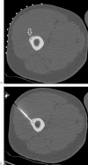
Figure 1
Figure 1
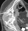
A 44-year-old man with recent onset radiating hip to mid-thigh pain. (A) Axial CT with cortical thickening and lucent lesion with central sclerotic nidus consistent with osteoid osteoma (arrow). Overlying skin grid used during lesion targeting for safe access window. (B) Initial biopsy needle for tissue sample was inserted, and the RFA probe tip was advanced into the central portion of the lesion. CT, computed tomography; RFA, radiofrequency ablation.
Figure 2
Figure 2
(A) A 60-year-old man with previous hemipelvectomy for large chondrosarcoma. Biopsy-proven recurrence near pseudoarticulation with large mucoid component. (B) Prone CT post-cryoablation depicts ice zone (arrows) after probe removal. Four probes were placed due to the large tumor size. (C) Two weeks later, the patient developed staphylococcal abscess at the treatment site requiring catheter drainage for an extended period. Contrast sinogram via pigtail catheter (arrow) shows residual cavity. CT, computed tomography.
Figure 3
Figure 3
A 55-year-old man with local colorectal bone metastasis, who failed external beam radiation therapy for pain control. He was receiving 15 mg/hour hydromorphone basal rate patient-controlled analgesia pump with breakthrough oral oxycodone. The patient was lethargic with severe pain (10/10). Two microwave antennae were placed adjacent to a destructive ilium soft tissue mass, and ablation was performed at 65 W for 10 minutes. After treatment, the patient's pain and medication requirements lessened with an improved quality of life during hospice.
Figure 4
Figure 4
Multimodality treatment in 65-year-old nonambulatory man with metastatic renal cell cancer and severe right hip pain. (A) Coronal reconstruction CT scan demonstrates an acetabular osteolytic lesion and intra-articular fracture (arrow). (B) Preembolization angiogram demonstrating a hypervascular lesion (arrow) treated with particle embolization. (C) The patient suffered recurrent pain, and subsequent bone RFA was performed along the superior aspect of the lesion. (D) Cementoplasty augmentation after RFA was performed in to stabilize the joint. (E) Six-month posttreatment plain film; the patient's pain resolved and he was able to walk. CT, computed tomography; RFA, radiofrequency ablation.
Figure 5
Figure 5
A 24-year-old woman professional dancer with recurring abdominal wall desmoid since the age of 12, now causing pain and inability to perform. She underwent multiple surgeries and chemotherapy. (A) Pretreatment MRI with large right rectus desmoid (arrow) with adjacent small bowel. (B) Cryoablation probes and ice ball formation (open arrow) with hydrodissection interface medially to protect bladder and bowel. Overlying gloves with warmed saline were placed for thermal skin insulation (solid arrow). (C) Six-month posttreatment scan demonstrating decreased tumor size and firmness, which allowed the patient to resume her dancing career. MRI, magnetic resonance imaging.
Figure 6
Figure 6
A 45-year-old with neurofibromatosis type 1 and new painful medial thigh neurofibroma (NF). (A) Coronal contrast-enhanced MRI shows NF enhancement. (B) PET/CT reveals FDG avid uptake in the lesion. (C) Biopsy results were benign, but the patient opted cryoablation for symptomatic control. CT scan performed during the procedure with ice ball formation at NF site. The patient's pain resolved posttreatment, and has now been symptom free for 3 years. CT, computed tomography; FDG, fluorodeoxyglucose; PET, positron emission tomography.
Figure 7
Figure 7
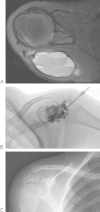
A 7-year-old patient with enlarging painful posterior right shoulder mass. (A) Axial MRI with multiple fluid-fluid levels and cortical thinning. On biopsy, this was proven to be an aneurysmal bone cyst (ABC). (B) The patient was placed prone under general anesthesia and bloody contents were aspirated from the ABC. Contrast injection showed absent vessel outflow and cyst communication. Foamed concentrated doxycycline was injected with double 18-gauge needles to allow the sclerosant to egress to prevent lesion overfilling. (C) Expected bony sclerosis posttreatment. MRI, magnetic resonance imaging.
Figure 8
Figure 8
Painful quadriceps desmoid tumor limiting function. (A) A Whitaker needle (arrow) was placed into the tumor for slow intralesional injection of 2 mL of 50% acetic acid. Real-time ultrasound was used to observe for peritumoral extravasation. (B) Two-month follow-up ultrasound demonstrating hollowed hypoechoic central necrosis. The patient's pain reduced from 9/10 to 3/10.
Figure 9
Figure 9
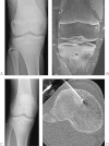
A 13-year-old boy with knee pain. (A) Radiography demonstrated a vague lucent area near growth plate. (B) Coronal MRI showing osteoid osteoma lesion and adjacent edema. (C) Due to risk of early growth plate closure with surgical and RFA treatment, he was treated conservatively. Follow-up plane film at the age of 16 years at the time of closure. (D) CT-guided RFA ablation at sclerotic OO zone. CT, computed tomography; MRI, magnetic resonance imaging; RFA, radiofrequency ablation.
Figure 10
Figure 10
Large soft tissue skin ulceration following thermal ablation of a painful ischial fibrous tumor. The skin ulceration was due to insufficient dermal temperature protection. A second small ulcer is noted more medially. The wound eventually healed follow eschar formation and surgical debridement.
Figure 11
Figure 11
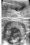
A 1-month-old baby with thigh mass. (A) MRI with diffuse vastus lateralis tissue thickening. A biopsy was performed, and the diagnosis of diffuse infantile fibromatosis was made. No therapy was undertaken, and the lesion became more discreet over time. (B) MRI at age 7 demonstrating a symptomatic enlargement of the mass. At this point, the family opted for intralesional injection treatment. (C) Ultrasound image during 50% acetic acid injection with immediate posttreatment fluid leakage around mass. Abdominal pain and nausea 3 days later required hospitalization for acute kidney injury from tumor lysis or acetic acid–induced crystal/cast nephropathy. Subsequent repeat biopsy showed a fibrosarcoma with tumor dedifferentiation. MRI, magnetic resonance imaging.

References

    1. Rosenthal D I, Alexander A, Rosenberg A E, Springfield D. Ablation of osteoid osteomas with a percutaneously placed electrode: a new procedure. Radiology. 1992;183(1):29–33. - PubMed
    1. Rosenthal D I, Hornicek F J, Wolfe M W, Jennings L C, Gebhardt M C, Mankin H J. Percutaneous radiofrequency coagulation of osteoid osteoma compared with operative treatment. J Bone Joint Surg Am. 1998;80(6):815–821. - PubMed
    1. Kurup A N, Callstrom M R. Image-guided percutaneous ablation of bone and soft tissue tumors. Semin Intervent Radiol. 2010;27(3):276–284. - PMC - PubMed
    1. Jaffe H L. Osteoid osteoma: a benign osteoblastic tumor composed of osteoid and atypical bone. Arch Surg. 1935;31:708–728.
    1. Greenspan A. Benign bone-forming lesions: osteoma, osteoid osteoma, and osteoblastoma. Clinical, imaging, pathologic, and differential considerations. Skeletal Radiol. 1993;22(7):485–500. - PubMed