Review
. 2014 Jan 29;2014(1):52-77.
doi: 10.5339/gcsp.2014.11. eCollection 2014.

The living aortic valve: From molecules to function

Affiliations
Review

The living aortic valve: From molecules to function

Adrian H Chester et al. Glob Cardiol Sci Pract. .

Abstract

The aortic valve lies in a unique hemodynamic environment, one characterized by a range of stresses (shear stress, bending forces, loading forces and strain) that vary in intensity and direction throughout the cardiac cycle. Yet, despite its changing environment, the aortic valve opens and closes over 100,000 times a day and, in the majority of human beings, will function normally over a lifespan of 70-90 years. Until relatively recently heart valves were considered passive structures that play no active role in the functioning of a valve, or in the maintenance of its integrity and durability. However, through clinical experience and basic research the aortic valve can now be characterized as a living, dynamic organ with the capacity to adapt to its complex mechanical and biomechanical environment through active and passive communication between its constituent parts. The clinical relevance of a living valve substitute in patients requiring aortic valve replacement has been confirmed. This highlights the importance of using tissue engineering to develop heart valve substitutes containing living cells which have the ability to assume the complex functioning of the native valve.

Keywords: Cells; calcification; developmental biology; endothelium; mechanobiology; nanostructure aortic stenosis; nerves.


Figures

Figure 1.
Figure 1.
Relationship between size and shape of the wings of a swift and the speed of lift during flight.
Figure 2.
Figure 2.
Photograph of an open aortic root showing its structural components (annulus, cusps, sinuses of Valsalva and sinotubular junction [dashed line]) and their relationship with the left ventricular outflow tract and the mitral valve. AML, anterior mitral leaflet; LC, left coronary cusp; LFT, left fibrous trigone; MS, membranous septum; Musc Sep, muscular septum; NC, non-coronary cusp; RC, right coronary cusp; RFT, right fibrous trigone. Adapted from Yacoub, El-Hamamsy et al.
Figure 3.
Figure 3.
Aortic annular deformation of left (L), right (R), and non-coronary cusp (NC) sectors of aortic annulus throughout different stages of the cardiac cycle (from Dagum et al. 1999).
Figure 4.
Figure 4.
Overview of cardiac development and formation of heart valves (from High & Epstein, 2008).
Figure 5.
Figure 5.
Porcine aortic valve endothelial cell (PAVEC) and porcine aortic endothelial cell (PAEC) morphology in static and fluid flow environments. Insets depict F-actin filament organization; arrow shows the direction of flow (from Butcher & Nerem, 2004).
Figure 6.
Figure 6.
Microstructure of aortic valve cusps showing the characteristic trilaminar architecture.
Figure 7.
Figure 7.
Presence of sympathetic nerves in the aortic root. Panel A shows the whole aortic root (stained for tyrosine hydroxylase), Panels B–E (stained for tyrosine hydroxylase) and F–I (stained for neurofilament protein) show nerve bundles at a higher magnification in the sinotubular junction (STJ) sinus area, hinge region of the cusp, and in the annular region (from Chester et al., 2008).
Figure 8.
Figure 8.
Porcine aortic cusp mounted on biaxial micromechanical testing device (top panel) and changes in the stiffness of the tissue induced by 5-HT, endothelial denudation, nitric oxide synthase inhibitor (L-NAME), endothelin-1, and cytochalasin B (CyB), an actin depolymerizing agent (bottom panel) (from El-Hamamsy et al., 2009).
Figure 9.
Figure 9.
Actuarial survival after Ross procedure (autograft) compared with homograft aortic root replacement. Survival over a 12 year period after a Ross procedure is indistinguishable from that of an aged matched population (from El-Hamamsy et al., 2010).
Figure 10.
Figure 10.
Effective orifice area. Velocity maps (color contours) and effective orifice areas (broken lines) visualized on cross sections above for autograft, xenograft, homograft native (normal) valve in the aortic position visualised at peak. Images are derived from 2-dimensional cine phase contrast magnetic resonance velocity mapping of valves approximately 10 years after implantation (from Torii et al., 2012).
Figure 11.
Figure 11.
Density-dependent color scanning electron micrographs of human aortic valve (AV) tissue. Orange color identifies denser material (calcium phosphate) and less dense structures are in green color (extracelular matrix). Micrographs acquired by secondary and backscatter electron detectors were combined in post-processing. A) Surface of AV with spherical calcified particles. B) Surface of AV with spherical calcified particles and calcified fibers. C) Surface of AV with spherical calcified particles and compact calcification. Scale bar = 2 μm.

References

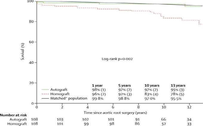
    1. El-Hamamsy I, Eryigit Z, Stevens LM, Sarang Z, George R, Clark L, Melina G, Takkenberg JJ, Yacoub MH. Long-term outcomes after autograft versus homograft aortic root replacement in adults with aortic valve disease: a randomised controlled trial. Lancet. 2010;376(9740):524–531. - PubMed
    1. Yacoub MH, Kilner PJ, Birks EJ, Misfeld M. The aortic outflow and root: a tale of dynamism and crosstalk. Ann Thorac Surg. 1999;68(3):S37–S43. - PubMed
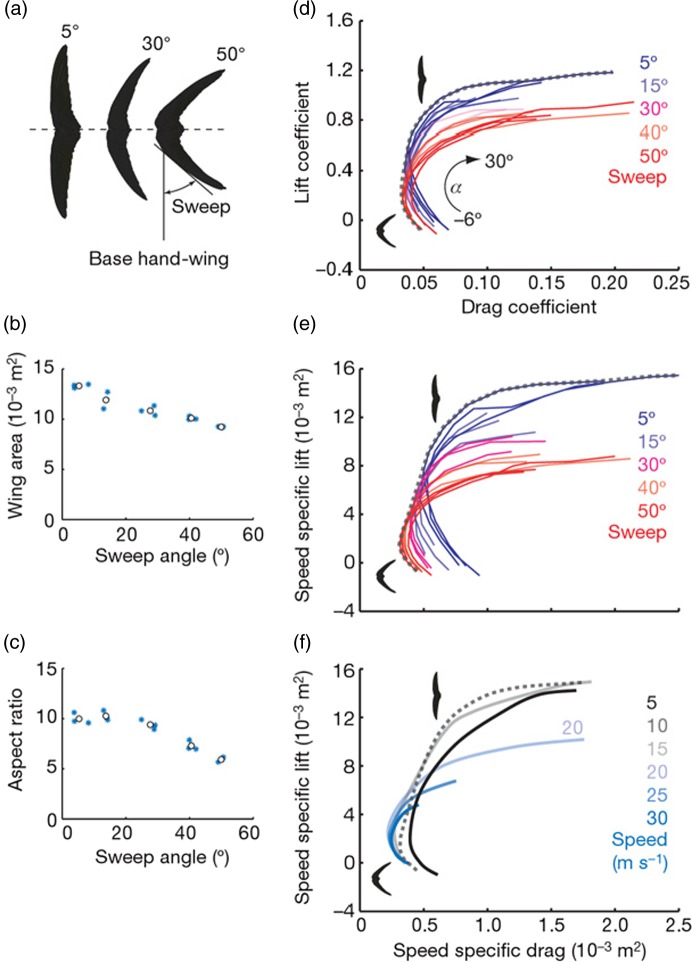
    1. Lentink D, Müller UK, Stamhuis EJ, de Kat R, van Gestel W, Veldhuis LLM, Henningsson P, Hedenström A, Videler JJ, van Leeuwen JL. How swifts control their glide performance with morphing wings. Nature. 2007;446(7139):1082–1085. - PubMed
    1. Rajamannan NM. Bicuspid aortic valve disease: the role of oxidative stress in Lrp5 bone formation. Cardiovasc Pathol. 2011;20(3):168–176. - PMC - PubMed
    1. Arjunon S, Rathan S, Jo H, Yoganathan AP. Aortic valve: mechanical environment and mechanobiology. Ann Biomed Eng. 2013;41(7):1331–1346. - PMC - PubMed