Review
. 2014:2014:837421.
doi: 10.1155/2014/837421. Epub 2014 Jun 23.

Hypoxia in diabetic kidneys

Affiliations
Review

Hypoxia in diabetic kidneys

Yumi Takiyama et al. Biomed Res Int. 2014.

Abstract

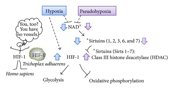
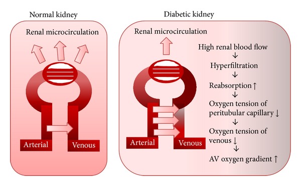
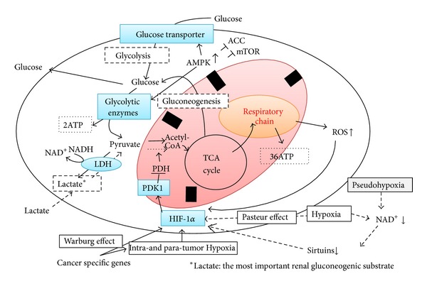
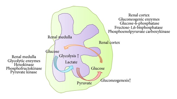
Diabetic nephropathy (DN) is now a leading cause of end-stage renal disease. In addition, DN accounts for the increased mortality in type 1 and type 2 diabetes, and then patients without DN achieve long-term survival compatible with general population. Hypoxia represents an early event in the development and progression of DN, and hypoxia-inducible factor- (HIF-) 1 mediates the metabolic responses to renal hypoxia. Diabetes induces the "fraternal twins" of hypoxia, that is, pseudohypoxia and hypoxia. The kidneys are susceptible to hyperoxia because they accept 20% of the cardiac output. Therefore, the kidneys have specific vasculature to avoid hyperoxia, that is, AV oxygen shunting. The NAD-dependent histone deacetylases (HDACs) sirtuins are seven mammalian proteins, SIRTs 1-7, which are known to modulate longevity and metabolism. Recent studies demonstrated that some isoforms of sirtuins inhibit the activation of HIF by deacetylation or noncatalyzing effects. The kidneys, which have a vascular system that protects them against hyperoxia, unfortunately experience extraordinary hypernutrition today. Then, an unexpected overload of glucose augments the oxygen consumption, which ironically results in hypoxia. This review highlights the primary role of HIF in diabetic kidneys for the metabolic adaptation to diabetes-induced hypoxia.


Figures

Figure 1
Figure 1
Metformin redistributes the intracellular oxygen. Metformin inhibits oxygen consumption and ATP production by inhibiting mitochondrial complex I. Subsequently, intracellular oxygen redistribution supplies oxygen for prolyl hydroxylase, which promotes the degradation of HIF-1α in the proteasome. ATP depletion caused by mitochondrial inhibition activates AMPK, which is a downstream signaling pathway of mitochondrial respiratory chain [21].
Figure 2
Figure 2
Oxygen regulates HIF-1α protein expression. HIF-1 is a heterodimeric transcription factor complex that is composed of an oxygen-regulated HIF-1α subunit and a constitutively expressed HIF-1β subunit; HIF-1 activity depends on the degradation of HIF-1α subunit; the half-life of HIF-1α protein on reoxygenation is less than one minutes [31].
Figure 3
Figure 3
NAD+-sirtuins modulate glucose metabolism via HIF-1 in all metazoan species from the simplest animal Trichoplax adhaerens to human. Sirtuins (SIRT) consist of seven mammalian proteins, SIRTs 1–7. Some isoforms of sirtuins inhibit HIF-1 activation by deacetylation or noncatalyzing effects [–38]. Under hypoxia or pseudohypoxia, decreased NAD+ levels downregulated SIRT, leading to upregulated HIF-1 activation which shift the glucose metabolism by promoting glycolysis and by inhibiting oxidative phosphorylation.
Figure 4
Figure 4
Diabetic kidney exhibits more AV oxygen shunting. Intrarenal oxygen tension is maintained at stable levels by hemodynamic and metabolic interactions of renal blood flow, GFR, oxygen consumption, and arteriovenous (AV) oxygen shunting. Originally, AV oxygen shunting develops as a unique system to rescue kidney from hyperoxia. However, in diabetic kidney, hyperglycemia induces hyperfiltration leading to more AV oxygen gradient which turns in increased AV oxygen shunting. The figure was modified from O'Connor et al. [42].
Figure 5
Figure 5
The essential role of HIF-1 is to switch the glucose metabolism from oxidative phosphorylation to glycolysis under hypoxic (Pasteur effect) or normoxic (pseudohypoxia or Warburg effect) conditions. Glycolysis: glucose + 2Pi + 2ADP + 2NAD+→ 2pyruvate + 2ATP + 2NADH + 2H2O, 2pyruvate + 2NADH → 2lactate + 2NAD+. Aerobic respiration: glucose + 6O2 + 36Pi + 36ADP → 6CO2 + 36ATP + 42H2O.
Figure 6
Figure 6
Glycolysis and gluconeogenesis in the kidney. A lactate shuttle may operate in the kidney between medulla and cortex as a main source of energy to sustain renal physiology. In diabetic kidneys, HIF may augment the medullary glycolysis rather than oxidative phosphorylation, leading to the production of lactate, which could be taken up by the cortical proximal tubules for gluconeogenesis. Then, renal glucose release in diabetic kidney is increased.

References

    1. Orchard TJ, Secrest AM, Miller RG, Costacou T. In the absence of renal disease, 20 year mortality risk in type 1 diabetes is comparable to that of the general population: a report from the Pittsburgh Epidemiology of Diabetes Complications Study. Diabetologia. 2010;53(11):2312–2319. - PMC - PubMed
    1. Afkarian M, Sachs MC, Kestenbaum B, et al. Kidney disease and increased mortality risk in type 2 diabetes. Journal of the American Society of Nephrology. 2013;24(2):302–308. - PMC - PubMed
    1. Nangaku M. Chronic hypoxia and tubulointerstitial injury: a final common pathway to end-stage renal failure. Journal of the American Society of Nephrology. 2006;17(1):17–25. - PubMed
    1. Haase VH. The VHL/HIF oxygen-sensing pathway and its relevance to kidney disease. Kidney International. 2006;69(8):1302–1307. - PubMed
    1. Eckardt K-U, Bernhardt W, Willam C, Wiesener M. Hypoxia-inducible transcription factors and their role in renal disease. Seminars in Nephrology. 2007;27(3):363–372. - PubMed