. 2014 Jul 23;9(7):e102993.
doi: 10.1371/journal.pone.0102993. eCollection 2014.

Platelet-rich plasma and skeletal muscle healing: a molecular analysis of the early phases of the regeneration process in an experimental animal model

Affiliations

Platelet-rich plasma and skeletal muscle healing: a molecular analysis of the early phases of the regeneration process in an experimental animal model

Ivan Dimauro et al. PLoS One. .

Abstract

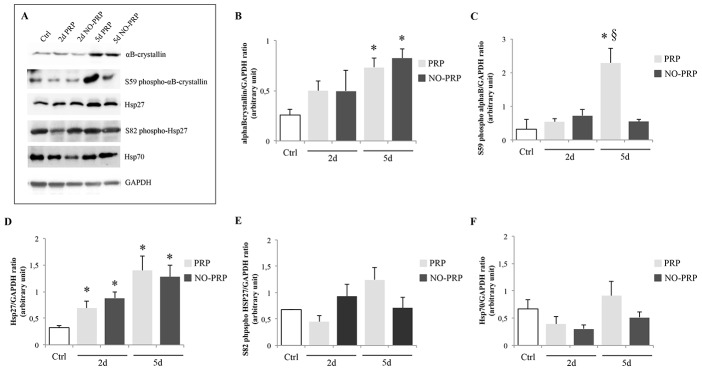
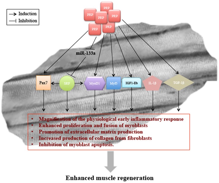
Platelet-rich plasma (PRP) has received increasing interest in applied medicine, being widely used in clinical practice with the aim of stimulating tissue healing. Despite the reported clinical success, there is still a lack of knowledge when considering the biological mechanisms at the base of the activity of PRP during the process of muscle healing. The aim of the present study was to verify whether the local delivery of PRP modulates specific molecular events involved in the early stages of the muscle regeneration process. The right flexor sublimis muscle of anesthetized Wistar rats was mechanically injured and either treated with PRP or received no treatment. At day 2 and 5 after surgery, the animals were sacrificed and the muscle samples evaluated at molecular levels. PRP treatment increased significantly the mRNA level of the pro-inflammatory cytokines IL-1β, and TGF-β1. This phenomenon induced an increased expression at mRNA and/or protein levels of several myogenic regulatory factors such as MyoD1, Myf5 and Pax7, as well as the muscular isoform of insulin-like growth factor1 (IGF-1Eb). No effect was detected with respect to VEGF-A expression. In addition, PRP application modulated the expression of miR-133a together with its known target serum response factor (SRF); increased the phosphorylation of αB-cristallin, with a significant improvement in several apoptotic parameters (NF-κB-p65 and caspase 3), indexes of augmented cell survival. The results of the present study indicates that the effect of PRP in skeletal muscle injury repair is due both to the modulation of the molecular mediators of the inflammatory and myogenic pathways, and to the control of secondary pathways such as those regulated by myomiRNAs and heat shock proteins, which contribute to proper and effective tissue regeneration.


Conflict of interest statement

Competing Interests: The authors have declared that no competing interests exist.

Figures

Figure 1
Figure 1. Changes in the fold increase expression levels of (A) cytokines (TNFα, IL-6, IL-10, IL-1β, and TGF-1β) (B) myogenic response factors (MyoD1, Myf5, Pax7, Myogenin and Mrf4) and (C) growth factors (VEGF-A and IGF-1Eb) mRNA in skeletal muscle treated or not with PRP during the experimental period (at day 2 and 5).
The y-axis for all graphs represents the fold-difference relative to the Ctrl group. * represents significant difference between injured compared with the Ctrl group (p<0.05). § represents a significant difference between the PRP-injury treated group and the NO PRP-injury treated group (p<0.05). Values are means ± SEM (n = 5 rats/group at each time point). Dashed line represents the base line control group.
Figure 2
Figure 2. Effects of PRP on total MyoD1 (A) and Myogenin (B) protein expression in uninjured skeletal muscle of rat (Ctrl), injured-PRP treated (PRP group) or not PRP treated (NO-PRP group) at different times post-injury (at day 2 and 5).
The relative protein expression was determined by the ratio of the sample value to an internal standard control (GAPDH). Values are means ± SEM (n = 5 rats/group at each time point). * represents a significant difference between injured groups and the Ctrl group (p<0.05). § represents a significant difference between PRP-injury treated groups and NO PRP-injury treated groups (p<0.05).
Figure 3
Figure 3. Immunohistochemical analysis of (A) MyoD1-positive or (C) Pax7-positive nuclei in skeletal muscle injury treated or not with PRP.
Representative double-immunoflorescence staining of skeletal muscle for (B) MyoD1 (green) and laminin (red) or (D) Pax7 (green) and laminin (red), at day 2- and 5, respectively. Myonuclei were counterstained by blue fluorescent dyes (Hoechst). The percentage of MyoD1-positive or Pax7-positive cells was calculated as the ratio of the number of nuclei in MyoD1- or Pax7 positive cells over that of Hoechst-positive nuclei. Results were presented as means ± SEM from n = 5 rats/group per time point and on five sections from each animal. Scale bars  = 50 µm. * p<0.05 vs. Ctrl group. § p<0.05 vs. NO-PRP group.
Figure 4
Figure 4. Real time-PCR analysis of miR-1, miR-133a and miR-206 expression using total RNA isolated from Ctrl-, PRP- and NO-PRP- group at 2 (A) and 5 (B) day post-injury.
C) Western blot analysis of SRF protein expression in skeletal muscle at 2- and 5-day post-injury. The histograms represent fold change expression calculated as means ± SEM (n = 5 rats/group at each time point) respect to the Ctrl group. *p<0.05 vs. Ctrl group. §p<0.05 vs. 5d NO-PRP group. Dashed line represents the base line Ctrl group.
Figure 5
Figure 5. Effects of PRP on (A) p38MAPK, (B) ERK activity and (C) AKT tot at 2- and 5-day post-injury in regenerating skeletal muscle.
Bar diagrams representing the densitometric intensities of p-p38MAPK, pERK1/2 and AKT tot normalized with those for p38MAPK, ERK and GAPDH content, respectively. Results were presented as means ± SEM from n = 5 rats/group per time point. *p<0.05 vs. Ctrl group. §p<0.05 vs. NO-PRP group.
Figure 6
Figure 6. Effect of PRP treatment on several HSPs during regeneration process (2- and 5-day post-injury).
(A) Representative immunoblot of each protein marker reported. (B) αB-crystallin; (C) S59 phospho-αB-crystallin; (D) Hsp27; (E) S82 phpspho-Hsp27; (F) Hsp70. Each bar represents mean value ± SEM (n = 5/group at each point). * p<0.05 vs. Ctrl group.§ p<0.05 vs. 5d NO-PRP group.
Figure 7
Figure 7. Effect of PRP treatment on several apoptotic markers during regeneration process (2- and 5-day postinjury).
(A) Representative immunoblot of each protein marker reported; (B) NF-κB-p65, and (C) Bax/Bcl-2 ratio. Values are means ± SEM (n  =  5/group at each point). * p< 0.05 vs. Ctrl group.§ p< 0.05 vs. 5d NO-PRP group.
Figure 8
Figure 8. Model of PRP-mediated regulation of skeletal muscle healing.
The presence of PRP modulated the expression of miR-133a and SRF protein as well as several myogenic response factors such as MyoD1, Pax7, and Myf5, the growth factor IGF-1Eb and both the cytokine IL-1β and TGF-1β. The modulation of these factors may affect important physiological processes such as the inflammatory response, myoblast proliferation and differentiation, production of extracellular matrix, and myoblast apoptosis.
Figure 9
Figure 9. Schematic representation of the role of αB-crystallin and Hsp27 in the myofiber stabilization and in cytoprotection following skeletal muscle injury.
The presence of PRP enhances phosphorylation of Ser-59 of αB-crystallin, which binds myofilaments and the inactive precursor of caspase 3, causing their stabilization and inhibition of apoptosis. Further, phospho Ser-59 αB-crystallin enhances NF-κB-p65 activation which may contribute to increased cell survival during regeneration process.

References

    1. Woolf AD, Pfleyer B (2003) Burden of major musculoskeletal conditions. Bull World Health Organ 81: 646–56. - PMC - PubMed
    1. Marx RE (2004) Platelet-rich plasma: evidence to support its use. J Oral Maxillofac Surg 62: 489–496. - PubMed
    1. Wright-Carpenter T, Opolon P, Appell HJ, Meijer H, et al. (2004) Treatment of muscle injuries by local administration of autologous conditioned serum: animal experiments using a muscle contusion model. Int J Sports Med 25: 582–587. - PubMed
    1. Cervelli V, Gentile P, Scioli MG, Grimaldi M, Casciani CU, et al. (2009) Application of platelet-rich plasma in plastic surgery: clinical and in vitro evaluation. Tissue Eng Part C Methods 15: 625–634. - PubMed
    1. Kazakos K, Lyras DN, Verettas D, Tilkeridis K, Tryfonidis M (2009) The use of autologous PRP gel as an aid in the management of acute trauma wounds. Injury 40: 801–805. - PubMed

Publication types

Substances