

A large response range reflectometric urea biosensor made from silica-gel nanoparticles
- PMID: 25054632
- PMCID: PMC4168459
- DOI: 10.3390/s140713186
A large response range reflectometric urea biosensor made from silica-gel nanoparticles
Abstract
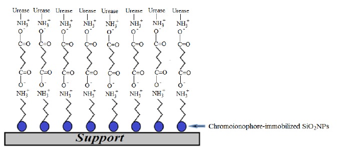
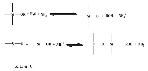
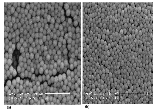
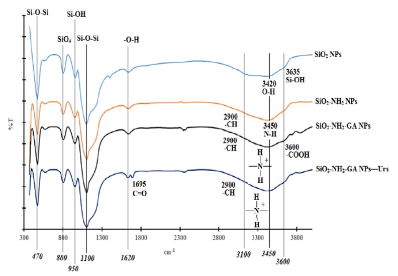
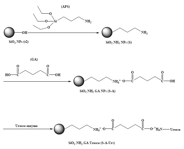
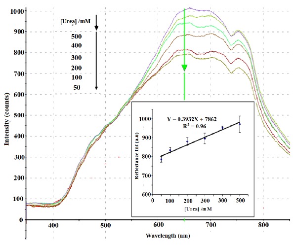
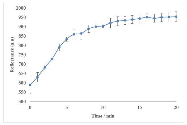
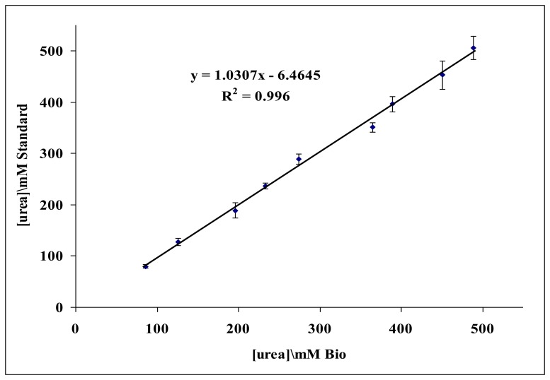
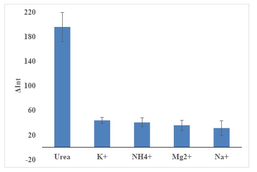
A new silica-gel nanospheres (SiO2NPs) composition was formulated, followed by biochemical surface functionalization to examine its potential in urea biosensor development. The SiO2NPs were basically synthesized based on sol-gel chemistry using a modified Stober method. The SiO2NPs surfaces were modified with amine (-NH2) functional groups for urease immobilization in the presence of glutaric acid (GA) cross-linker. The chromoionophore pH-sensitive dye ETH 5294 was physically adsorbed on the functionalized SiO2NPs as pH transducer. The immobilized urease determined urea concentration reflectometrically based on the colour change of the immobilized chromoionophore as a result of the enzymatic hydrolysis of urea. The pH changes on the biosensor due to the catalytic enzyme reaction of immobilized urease were found to correlate with the urea concentrations over a linear response range of 50-500 mM (R2 = 0.96) with a detection limit of 10 mM urea. The biosensor response time was 9 min with reproducibility of less than 10% relative standard deviation (RSD). This optical urea biosensor did not show interferences by Na+, K+, Mg2+ and NH4+ ions. The biosensor performance has been validated using urine samples in comparison with a non-enzymatic method based on the use of p-dimethylaminobenzaldehyde (DMAB) reagent and demonstrated a good correlation between the two different methods (R2 = 0.996 and regression slope of 1.0307). The SiO2NPs-based reflectometric urea biosensor showed improved dynamic linear response range when compared to other nanoparticle-based optical urea biosensors.
Figures
References
-
- Lee W.Y., Kim S.R., Kim T.H., Lee K.S., Shin M.C., Park J.K. Sol-gel-derived thick-film conductometric biosensor for urea determination in serum. Anal. Chim. Acta. 2000;404:195–203.
-
- Sawicka K., Gouma P., Sanford S. Electrospun biocomposite nanofibers for urea biosensing. Sens. Actuators B. Chem. 2005;108:585–588.
-
- Rajesh B.V., Wataru T., Keiichi K. An amperometric urea biosensor based on covalent immobilization of urease onto an electrochemically prepared copolymer poly(N-3-aminopropyl pyrrole-co-pyrrole) film. Biomaterials. 2005;26:3683–3690. - PubMed
-
- Rajesh B.V., Takashima W., Kaneto K. A novel thin film urea biosensor based on copolymer poly(N-3-aminopropylpyrrole-co-pyrrole) film. Surface Coat. Technol. 2005;198:231–236.
-
- Rajesh B.V., Takashima W., Kaneto K. Development of a potentiometric urea biosensor based on copolymer poly(N-3-aminopropyl pyrrole-co-pyrrole) film. React. Funct. Polym. 2005;62:51–59. - PubMed
Publication types
MeSH terms
Substances
LinkOut - more resources
Full Text Sources
Other Literature Sources
