Review
. 2014 Jul 22;6(7):2826-57.
doi: 10.3390/v6072826.

Membranous replication factories induced by plus-strand RNA viruses

Affiliations
Review

Membranous replication factories induced by plus-strand RNA viruses

Inés Romero-Brey et al. Viruses. .

Abstract

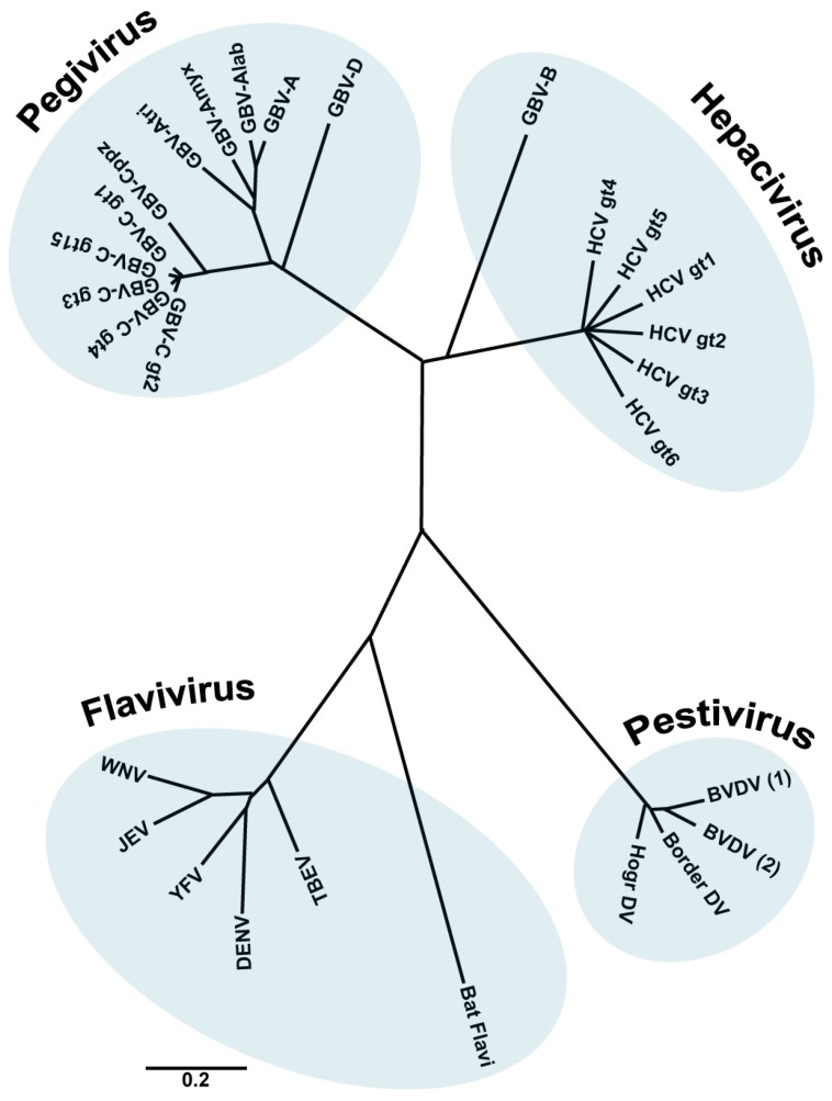
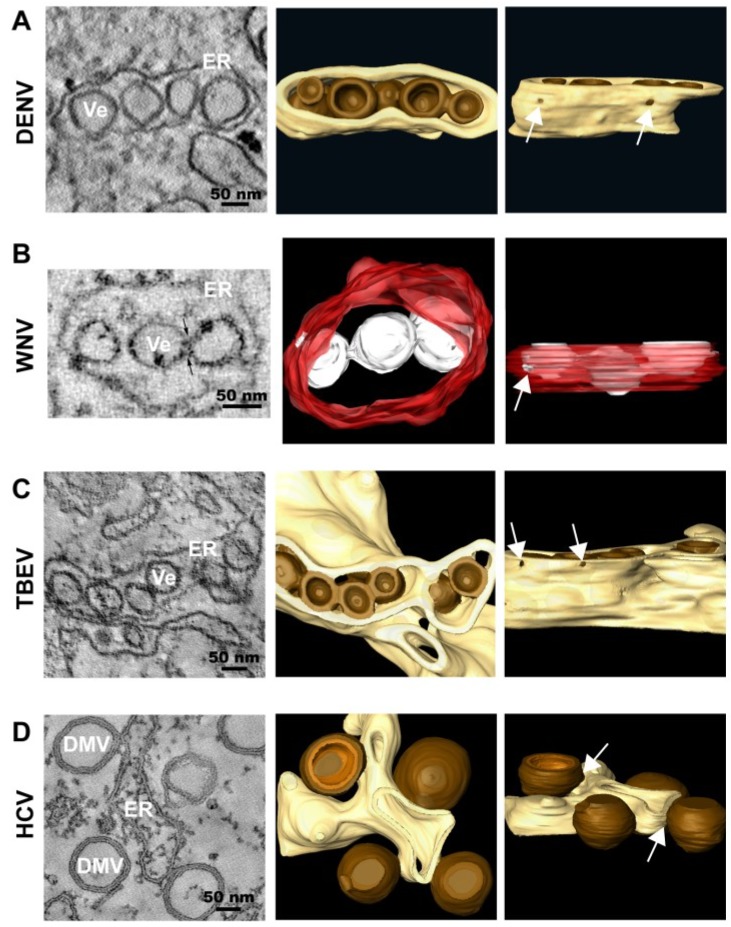
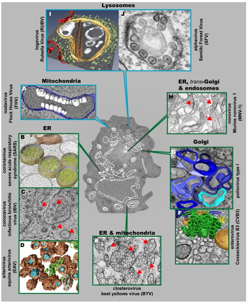
In this review, we summarize the current knowledge about the membranous replication factories of members of plus-strand (+) RNA viruses. We discuss primarily the architecture of these complex membrane rearrangements, because this topic emerged in the last few years as electron tomography has become more widely available. A general denominator is that two "morphotypes" of membrane alterations can be found that are exemplified by flaviviruses and hepaciviruses: membrane invaginations towards the lumen of the endoplasmatic reticulum (ER) and double membrane vesicles, representing extrusions also originating from the ER, respectively. We hypothesize that either morphotype might reflect common pathways and principles that are used by these viruses to form their membranous replication compartments.


Figures

Figure 1
Figure 1
The family Flaviviridae. Phylogenetic tree based on the analysis (neighbour joining) of aligned conserved motifs of the RNA dependent RNA polymerase (RdRp). Shown are selected members of the family. A distance scale corresponding to amino acid substitutions per position is shown. (Figure adapted from [1], with permission).
Figure 2
Figure 2
Representative images of membrane rearrangements induced by different members of the family Flaviviridae. (A) Dengue Virus (DENV); (B) West Nile Virus (WNV); (C) Tick-borne Encephalitis Virus (TBEV); (D) Hepatitis C Virus (HCV). Slices through tomograms of infected cells (on the left) and 3D top and lateral (90° rotation) views of the same tomograms (on the right) are depicted, showing the characteristic virus-induced structures. The replication vesicles (Ve) of DENV, WNV and TBEV (genus Flavivirus) correspond to invaginations of ER membranes that remain connected to the cytosol via 10 nm-pores (highlighted with white arrows in the 3D lateral views), forming vesicle packets (VPs). The replication factory of HCV (genus Hepacivirus) is primarily composed of double membrane vesicles (DMVs) that seem to be formed asER protusions connected to ER membranes via neck-like structures (highlighted with white arrows in the 3D lateral view). The ER is shown in yellow (DENV, TBEV and HCV) or in red (WNV) and the replication organelles in brown (DENV, TBEV and HCV) or in white (WNV). The outer and inner membranes of DMVs are depicted in different shades of brown (outer membrane in dark brown and inner membrane in light brown). Figure 2B is reproduced with permission from [20].
Figure 3
Figure 3
Positive strand RNA viruses usurp and modify cell membranes of different origins to replicate their genomes. (A) Mitochondrial membranes are targeted by Flock House Virus (FHV), which induces the formation of vesicles (white) at the outer mitochondrial membrane (blue); (B) 3D surface-rendered model of SARS-CoV–infected Vero E6 cells containing large double membrane vesicles (DMVs) (outer membrane, gold; inner membrane, silver) that remain connected to their source, the ER (in bronze); (C) Zippered ER, found in cells infected with the gammacoronavirus Infectious Bronchitis Virus (IBV), is connected to spherules (red arrows). DMVs (red arrowheads) are also found, but to a lesser extent; (D) Equine Arteriviruses (EAV) infection of HeLa cells results in the formation of DMVs (brown), depicting a core (blue) that is associated with the ER (beige) and close to ER tubules (green); (E) Closterovirus rearranges ER and mitochondrial membranes to form DMVs (red arrowheads) and vesicles packets (VPs) (red arrows); (F) Coxsackie B3 Virus (CVB3) usurps donor membranes most likely derived from the Golgi, appearing early in infection as single-membrane tubules (green), open (orange) and closed (yellow) DMVs and ER (blue); (G) 3D architecture of poliovirus membranous replication factories at intermediate stages of development, originating from cis-Golgi membranes. Single-membrane structures are depicted in different shades of blue. Note that these single membrane vesicles undergo secondary invaginations giving rise to DMVs at the late stages of infection; (H) Murine Norovirus 1 (MNV-1) infection results in the formation of vesiculated areas (VAs, red arrowheads) within aggregates of MNV-1 particles (red arrows). These VAs seem to originate from ER, trans-Golgi and endosomes; (I) 3D model of a cytopathic vacuole (CPV) found in Rubella Virus-infected cells. This CPV (yellow) is surrounded by the rER (light green) and contains a number of vacuoles, vesicles (white) and a rigid straight sheet (brown) that is connected with the periphery of the CPV. Mitochondria are depicted in red; (J) Typical cytoplasmic vacuoles induced by Semliki Forest Virus (SFV) in baby hamster kidney cells. Color code is as follows: blue-framed images depict single-membrane vesicle inducers; green-framed images correspond to double-membrane vesicle inducers. (The different parts were reproduced with permission: see acknowledgement section)

References

    1. Stapleton J.T., Foung S., Muerhoff A.S., Bukh J., Simmonds P. The GB viruses: A review and proposed classification of GBV-A, GBV-C (HGV), and GBV-D in genus Pegivirus within the family Flaviviridae. J. Gen. Virol. 2011;92:233–246. doi: 10.1099/vir.0.027490-0. - DOI - PMC - PubMed
    1. Simmonds P. The Origin of Hepatitis C Virus. In: Bartenschlager R., editor. Hepatitis C Virus: From Molecular Virology to Antiviral Therapy. 82nd ed. Volume 369 Springer Berlin Heidelberg; Berlin, Heidelberg, Germany: 2013.
    1. Choo Q.L., Kuo G., Weiner A.J., Overby L.R., Bradley D.W., Houghton M. Isolation of a cDNA clone derived from a blood-borne non-A, non-B viral hepatitis genome. Science. 1989;244:359–362. doi: 10.1126/science.2523562. - DOI - PubMed
    1. Feinstone S.M., Kapikian A.Z., Purcell R.H., Alter H.J., Holland P.V. Transfusion-associated hepatitis not due to viral hepatitis type A or B. N. Engl. J. Med. 1975;292:767–770. doi: 10.1056/NEJM197504102921502. - DOI - PubMed
    1. Lindenbach B.D., Thiel H.-J., Rice C.M. Flaviviridae: The Viruses and Their Replication. In: Fields B.N., Knipe D.M., Howley P., editors. Fields Virology. 5th ed. Raven Press; New York, NY, USA: 2001. pp. 1001–1152.

Publication types

MeSH terms

LinkOut - more resources