Review
. 2014 Jul;36(4):461-78.
doi: 10.1007/s00281-014-0436-6. Epub 2014 Jul 24.

The immunopathology of ANCA-associated vasculitis

Affiliations
Review

The immunopathology of ANCA-associated vasculitis

Eoin F McKinney et al. Semin Immunopathol. 2014 Jul.

Abstract

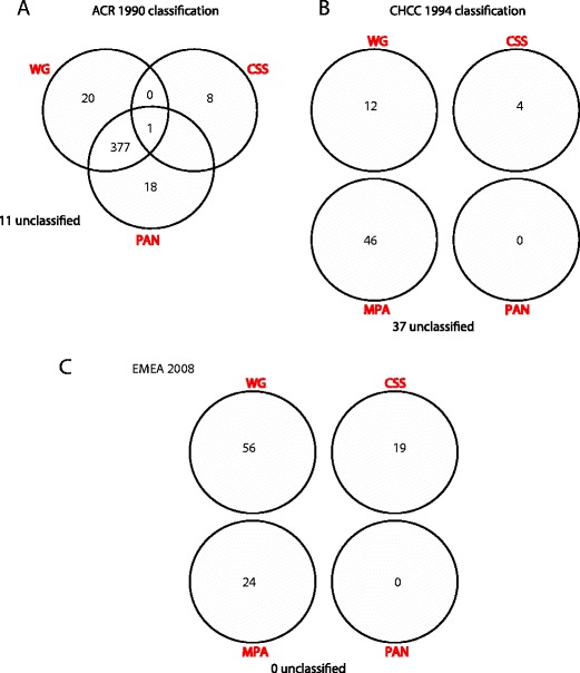
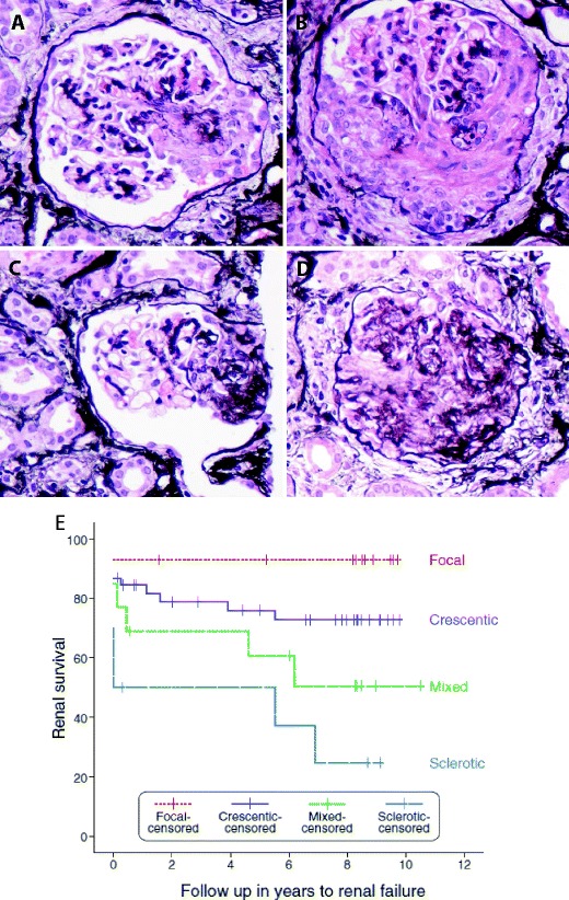
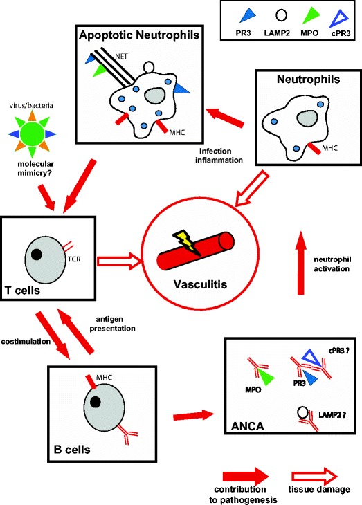
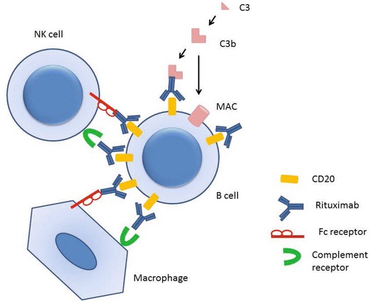
The small-vessel vasculitides are a group of disorders characterised by variable patterns of small blood vessel inflammation producing a markedly heterogeneous clinical phenotype. While any vessel in any organ may be involved, distinct but often overlapping sets of clinical features have allowed the description of three subtypes associated with the presence of circulating anti-neutrophil cytoplasmic antibodies (ANCA), namely granulomatosis with polyangiitis (GPA, formerly known as Wegener's Granulomatosis), microscopic polyangiitis (MPA) and eosinophilic granulomatosis with polyangiitis (eGPA, formerly known as Churg-Strauss syndrome). Together, these conditions are called the ANCA-associated vasculitidies (AAV). Both formal nomenclature and classification criteria for the syndromes have changed repeatedly since their description over 100 years ago and may conceivably do so again following recent reports showing distinct genetic associations of patients with detectable ANCA of distinct specificities. ANCA are not only useful in classifying the syndromes but substantial evidence implicates them in driving disease pathogenesis although the mechanism by which they develop and tolerance is broken remains controversial. Advances in our understanding of the pathogenesis of the syndromes have been accompanied by some progress in treatment, although much remains to be done to improve the chronic morbidity associated with the immunosuppression required for disease control.


Figures

Fig. 1
Fig. 1
The changing classification of AAV (adapted from Watts et al [20].). A single cohort of 99 patients with AAV (from Lane et al [4].) were assigned diagnostic labels using a the American College of Rheumatology 1990 criteria [15], b the Chapel Hill Consensus Conference definitions [16] and c the EMEA algorithm [20]. WG Wegener’s granulomatosis, CSS Churg-Strauss syndrome, MPA microscopic polyangiitis, PAN polyarteritis nodosa
Fig. 2
Fig. 2
Histopathology of AAV. All pictures show glomerular lesions from one patient with an ANCA-associated vasculitis. (Jones-Methamine-Silver stain, original magnification ×400) a Cellular crescent involving <50 % of the glomerular tuft defined (according to the histopathologic criteria for AAV [73]) as a lesion filling the Bowman’s space containing a cellular component of >10 %, regardless of the proportion of the glomerular tuft that is involved (<50 % in this picture). The amount of fibrin or fibrosis does not affect the classification, as long as the required cellular component is seen. b Cellular crescent involving >50 % of the glomerular tuft. A case is classified as “crescentic” if ≥50 % of all glomeruli in the biopsy show crescents (regardless of the proportion involved in the individual glomerulus). If <50 % of all glomeruli show crescents, the classification depends on the proportion of normal and sclerotic glomeruli present in the biopsy: any biopsy with ≥50 % normal glomeruli is designated as “focal”. In contrast, any biopsy showing ≥50 % of sclerotic glomeruli is classified as “sclerotic” and a biopsy not fulfilling any of the aforementioned criteria is called “mixed”. c Glomerulus with a fibrous crescent defined as a lesion filling the Bowman’s space containing a cellular component of <10 %, affecting up to 80 % of the tuft. The lesion is largely composed of fibrous tissue. d Globally sclerosed glomerulus, >80 % of the tuft is sclerotic. This is not specific for AAV. e Renal survival (the absence of end-stage renal failure) in a cohort of 82 patients with AAV by histological classification (from Berden et al.) [73]. Inclusion in the classification schema requires a pauci-immune staining pattern on immunofluorescence microscopy and at least one glomerulus with necrotising or crescentic glomerulonephritis on light microscopy
Fig. 3
Fig. 3
Pathogenesis of AAV. Schematic illustration of our current understanding of the pathogenesis of AAV. TCR T cell receptor, PR3 proteinase-3, MPO myeloperoxidase, LAMP-2 lysosome-associated membrane protein-2, rPR3 reverse PR3
Fig. 4
Fig. 4
Mechanisms of B cell depletion with rituximab therapy

References

    1. Jennette JC, Falk RJ. Small-vessel vasculitis. N Engl J Med. 1997;337:1512–1523. - PubMed
    1. Hagen EC, et al. Diagnostic value of standardized assays for anti-neutrophil cytoplasmic antibodies in idiopathic systemic vasculitis. EC/BCR project for ANCA assay standardization. Kidney Int. 1998;53:743–753. - PubMed
    1. Hoffman GS. Classification of the systemic vasculitides: antineutrophil cytoplasmic antibodies, consensus and controversy. Clin Exp Rheumatol. 1998;16:111–115. - PubMed
    1. Lane SE, Watts RA, Shepstone L, Scott DG. Primary systemic vasculitis: clinical features and mortality. QJM. 2005;98:97–111. - PubMed
    1. McBride P. Photographs of a case of rapid destruction of the nose and face. 1897. J Laryngol Otol. 1991;105:1120. - PubMed

MeSH terms

Substances