. 2014 Sep;42(15):10061-72.
doi: 10.1093/nar/gku663. Epub 2014 Jul 23.

New insights into the incorporation of natural suppressor tRNAs at stop codons in Saccharomyces cerevisiae

Affiliations

New insights into the incorporation of natural suppressor tRNAs at stop codons in Saccharomyces cerevisiae

Sandra Blanchet et al. Nucleic Acids Res. 2014 Sep.

Abstract

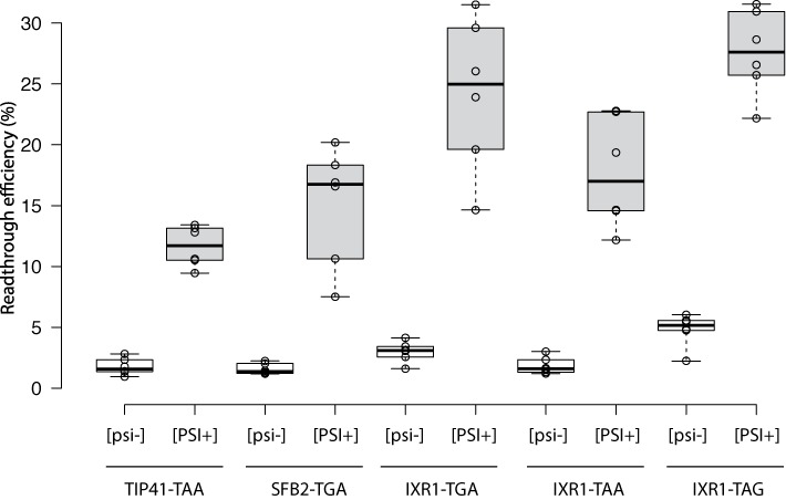
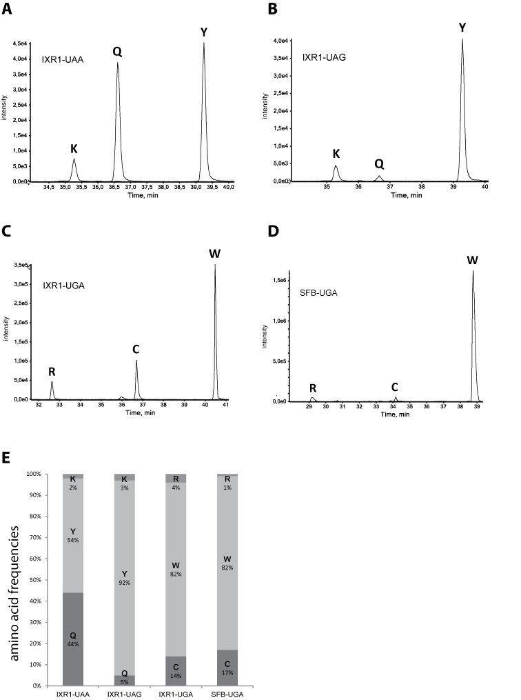
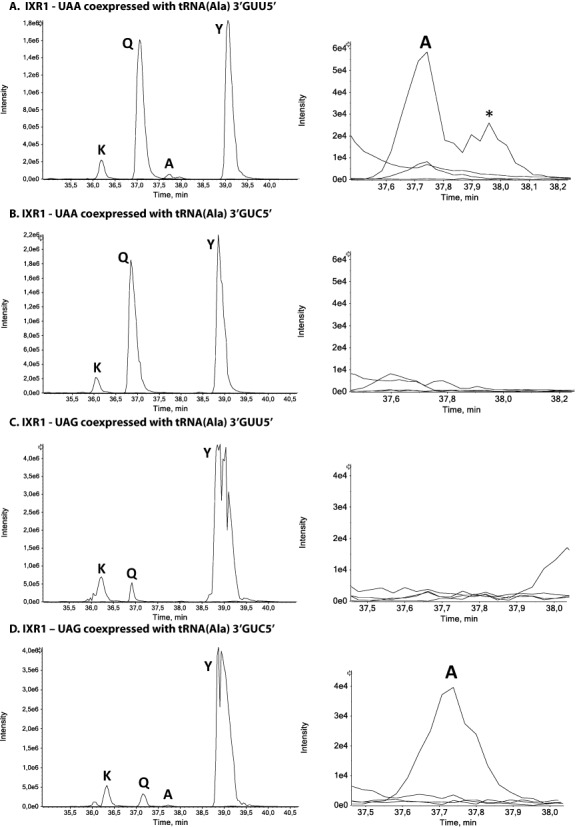
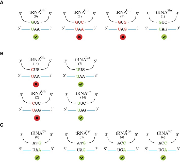
Stop codon readthrough may be promoted by the nucleotide environment or drugs. In such cases, ribosomes incorporate a natural suppressor tRNA at the stop codon, leading to the continuation of translation in the same reading frame until the next stop codon and resulting in the expression of a protein with a new potential function. However, the identity of the natural suppressor tRNAs involved in stop codon readthrough remains unclear, precluding identification of the amino acids incorporated at the stop position. We established an in vivo reporter system for identifying the amino acids incorporated at the stop codon, by mass spectrometry in the yeast Saccharomyces cerevisiae. We found that glutamine, tyrosine and lysine were inserted at UAA and UAG codons, whereas tryptophan, cysteine and arginine were inserted at UGA codon. The 5' nucleotide context of the stop codon had no impact on the identity or proportion of amino acids incorporated by readthrough. We also found that two different glutamine tRNA(Gln) were used to insert glutamine at UAA and UAG codons. This work constitutes the first systematic analysis of the amino acids incorporated at stop codons, providing important new insights into the decoding rules used by the ribosome to read the genetic code.


Figures

Figure 1.
Figure 1.
Readthrough efficiency for the sequences used for the GST reporter system. The lines in the center of the boxes indicate the median values; the box limits indicate the 25th and 75th percentiles calculated with R software; the whiskers extend to the minimum and maximum values; data points are plotted as open circles. n = 6 sample points. [psi] values are indicated in white boxes, [PSI+] values are indicated in gray boxes.
Figure 2.
Figure 2.
Purification and verification of readthrough GST proteins. (A) The chromatograms show absorbance at 280 nm. The red curve corresponds to in-frame GST purification; the blue curve corresponds to GST-IXRI-TAG purification. The elution peaks appears in fraction 3. (B) SDS-PAGE gel stained by Coomassie Blue. Lane 1: whole-cell extract; lane 2: flowthrough after binding of the extract; lane 3: in-frame GST, and lane 4: purified readthrough protein, GST-IXRI-TAG. For lanes 3 and 4, 1.8 μg of protein was loaded on the gel. (C) The presence of the GST protein was checked by western blotting with an antibody against GST. Lane 1 corresponds to the whole-cell extract, lane 2 corresponds to the flowthrough after binding of the extract, lane 3 to in-frame GST, and lane 4 to purified readthrough protein, GST-IXRI-TAA. For lanes 3 and 4, 0.5 μg of protein was loaded on the gel.
Figure 3.
Figure 3.
Relative quantification of readthrough peptides. (A)–(D) MS extracted-ion chromatograms (XIC) of readthrough peptides in the IXR1 and SFB contexts. The X- and Y-axis correspond to LC elution time and absolute MS signal intensity, respectively. (E) Relative frequencies of the readthrough amino acids incorporated at UAA, UAG and UGA stop codons in the IXR1 and SFB2 contexts. Quantification data were processed and adjusted as described in the Materials and Methods.
Figure 4.
Figure 4.
Alanine insertion at stop codons after the coexpression of IXR1-UAA and IXR1-UAG readthrough proteins with modified 3′GUU5′ and 3′GUC5′ tRNAsAla. MS extracted-ion chromatograms (XIC) of readthrough peptides with lysine, glutamine, tyrosine and alanine incorporated at the UAA and UAG stop codons are shown in left panels. To better visualize alanine incorporation at UAA and UAG stop codons, a zoom of the elution region is shown on the right side of each XIC. The alanine incorporated readthrough peptide (KNEAQINNLSPILGYW) is detected only when IXR1-UAA and IXR1-UAG are coexpressed with tRNAAla3′GUU5′ (panel A) or tRNAAla3′GUC5′ (panel D), respectively. The asterisk (*) indicates a peak corresponding to the alanine isobaric peptide with alanine incorporated at the glutamine site downstream from the stop codon (KNEQAINNLSPILGYW).
Figure 5.
Figure 5.
Proposed rules for stop codon decoding. This figure shows matching between potential suppressor tRNAs and stop codons. For each tRNA, the number of gene copies is indicated in brackets. The non-canonical base pairings appear in color green when they are accepted, red when they are not. Panel A corresponds to glutamine tRNAs, panel B to glutamate and lysine tRNAs, panel C to tyrosine, cysteine and tryptophan tRNAs. S represents a uridine modified with mcm5S2 and Ψ represents a uridine converted into a pseudouridine.

References

    1. Bertram G., Bell H.A., Ritchie D.W., Fullerton G., Stansfield I. Terminating eukaryote translation: domain 1 of release factor eRF1 functions in stop codon recognition. RNA. 2000;6:1236–1247. - PMC - PubMed
    1. Chavatte L., Seit-Nebi A., Dubovaya V., Favre A. The invariant uridine of stop codons contacts the conserved NIKSR loop of human eRF1 in the ribosome. EMBO J. 2002;21:5302–5311. - PMC - PubMed
    1. Zhouravleva G., Frolova L., Le Goff X., Le Guellec R., Inge-Vechtomov S., Kisselev L., Philippe M. Termination of translation in eukaryotes is governed by two interacting polypeptide chain release factors, eRF1 and eRF3. EMBO J. 1995;14:4065–4072. - PMC - PubMed
    1. Frolova L., Le Goff X., Zhouravleva G., Davydova E., Philippe M., Kisselev L. Eukaryotic polypeptide chain release factor eRF3 is an eRF1- and ribosome-dependent guanosine triphosphatase. RNA. 1996;2:334–341. - PMC - PubMed
    1. Loh P.G., Song H. Structural and mechanistic insights into translation termination. Curr. Opin. Struct. Biol. 2010;20:98–103. - PubMed

Publication types

MeSH terms