. 2015 Dec;25(12):4653-66.
doi: 10.1093/cercor/bhu147. Epub 2014 Jul 23.

Abnormal Neural Activation to Faces in the Parents of Children with Autism

Affiliations

Abnormal Neural Activation to Faces in the Parents of Children with Autism

G H Yucel et al. Cereb Cortex. 2015 Dec.

Abstract

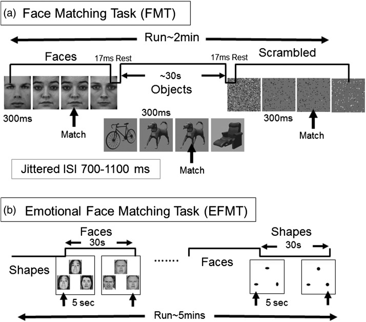
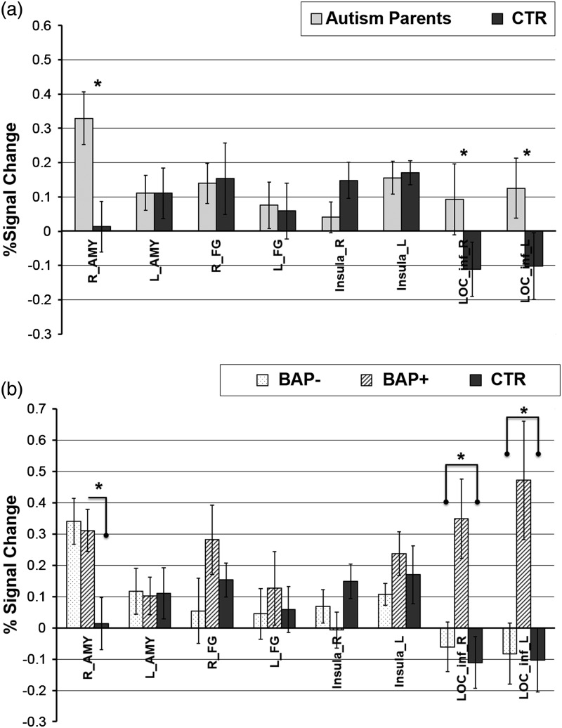
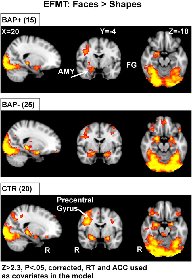
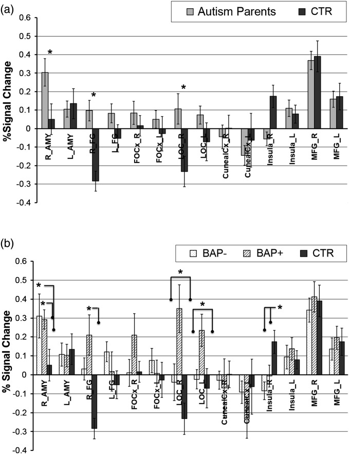
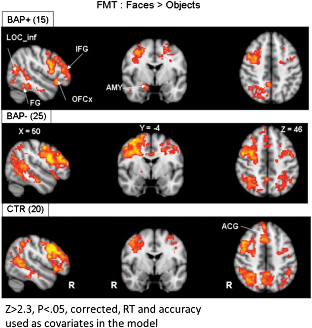
Parents of children with an autism spectrum disorder (ASD) show subtle deficits in aspects of social behavior and face processing, which resemble those seen in ASD, referred to as the "Broad Autism Phenotype " (BAP). While abnormal activation in ASD has been reported in several brain structures linked to social cognition, little is known regarding patterns in the BAP. We compared autism parents with control parents with no family history of ASD using 2 well-validated face-processing tasks. Results indicated increased activation in the autism parents to faces in the amygdala (AMY) and the fusiform gyrus (FG), 2 core face-processing regions. Exploratory analyses revealed hyper-activation of lateral occipital cortex (LOC) bilaterally in autism parents with aloof personality ("BAP+"). Findings suggest that abnormalities of the AMY and FG are related to underlying genetic liability for ASD, whereas abnormalities in the LOC and right FG are more specific to behavioral features of the BAP. Results extend our knowledge of neural circuitry underlying abnormal face processing beyond those previously reported in ASD to individuals with shared genetic liability for autism and a subset of genetically related individuals with the BAP.

Keywords: amygdala; autism; broad autism phenotype; face perception; fusiform gyrus.


Figures

Figure 1.
Figure 1.
Task designs. (a) Face Match Task: a blocked design was used with 3 categories of stimuli: faces, objects, and scrambled objects. Each block was selarated by a rest period of 17 ms; stimulus duration was 300 ms; ISI was jittered between 700 and 1100 ms; block duration was 30 s. Participants performed a 1-back matching task to maintain attention. (b) EFMT as a localizer task: For the amygdala localizer task, a well-established blocked-design task was used with 2 categories of stimuli: faces and shapes. Block duration was 30 s; stimulus duration was 5 s; run duration about 5 min. Participants performed a choice-response button press matching task to indicate which of the bottom 2 items (right or left) matched the top item for each trial.
Figure 2.
Figure 2.
ROI-based results from the EFMT. (a) 2-group results (Autism Parents, CTR. (b) 3-group results (BAP−, BAP+, CTR). AMY, amygdala; FG, fusiform gyrus; LOC_inf, Inferior lateral occipital cortex; L, left; R, right. * indicates P < 0.05.
Figure 3.
Figure 3.
Voxel-based whole-brain analysis during the EFMT. The EFMT localizer reliably identified a set of brain regions involved in visual processing and emotion. Highlighted here are areas of increased activation (faces > shapes) along broad regions of temporal and occipital cortex, as well as the amygdala in all subject groups. AMY, amygdala; FG, fusiform gyrus; RT, reaction time; ACC, accuracy. Areas of activation passed a cluster-significance threshold of z > 2.3, with whole-brain cluster-correction at P ≤ 0.05. R indicates right.
Figure 4.
Figure 4.
ROI-based results from the FMT. (a) depicts 2-group analyses (Autism Parents, CTR), (b) depicts 3-group analysis (BAP−, BAP+, CTR). Our primary analysis focused on group differences in activation within hypothesized brain regions known to be important for processing faces. AMY, amygdala; FG, fusiform gyrus; OFC, orbital frontal cortex; LOC_inf, inferior lateral occipital cortex; MFG, middle frontal gyrus. L indicates left; R, right. * indicates P < 0.05.
Figure 5.
Figure 5.
Voxel-based whole-brain analysis during the FMT. Highlighted here are areas of increased activation (faces > objects) along broad regions of temporal and occipital cortex, as well as the amygdala in all subject groups. AMY, amygdala; FG, fusiform gyrus; OFC, orbital frontal cortex; IFG, inferior frontal gyrus; LOC_inf, inferior lateral occipital cortex; ACG, anterior cingulate gyrus; RT, reaction time; ACC, accuracy. Areas of activation passed a cluster-significance threshold of z > 2.3, with whole-brain cluster-correction at P ≤ 0.05. R indicates right.

References

    1. Adolphs R. 2001. The neurobiology of social cognition. Curr Opin Neurobiol. 11:231–239. - PubMed
    1. Adolphs R. 2008. Fear, faces, and the human amygdala. Curr Opin Neurobiol. 18:166–172. - PMC - PubMed
    1. Adolphs R. 2010. What does the amygdala contribute to social cognition? Ann N Y Acad Sci. 1191:42–61. - PMC - PubMed
    1. Adolphs R, Gosselin F, Buchanan TW, Tranel D, Schyns P, Damasio AR. 2005. A mechanism for impaired fear recognition after amygdala damage. Nature. 433:68–72. - PubMed
    1. Adolphs R, Spezio ML, Parlier M, Piven J. 2008. Distinct face-processing strategies in parents of autistic children. Curr Bio. 18:1090–1093. - PMC - PubMed

Publication types