. 2014 Oct;88(19):11034-44.
doi: 10.1128/JVI.01505-14. Epub 2014 Jul 23.

Virus-specific memory CD8 T cells provide substantial protection from lethal severe acute respiratory syndrome coronavirus infection

Affiliations

Virus-specific memory CD8 T cells provide substantial protection from lethal severe acute respiratory syndrome coronavirus infection

Rudragouda Channappanavar et al. J Virol. 2014 Oct.

Abstract

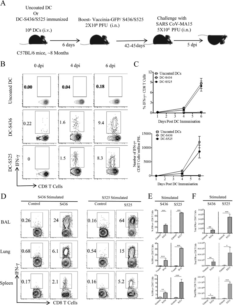
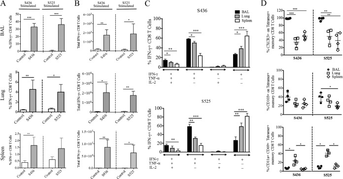
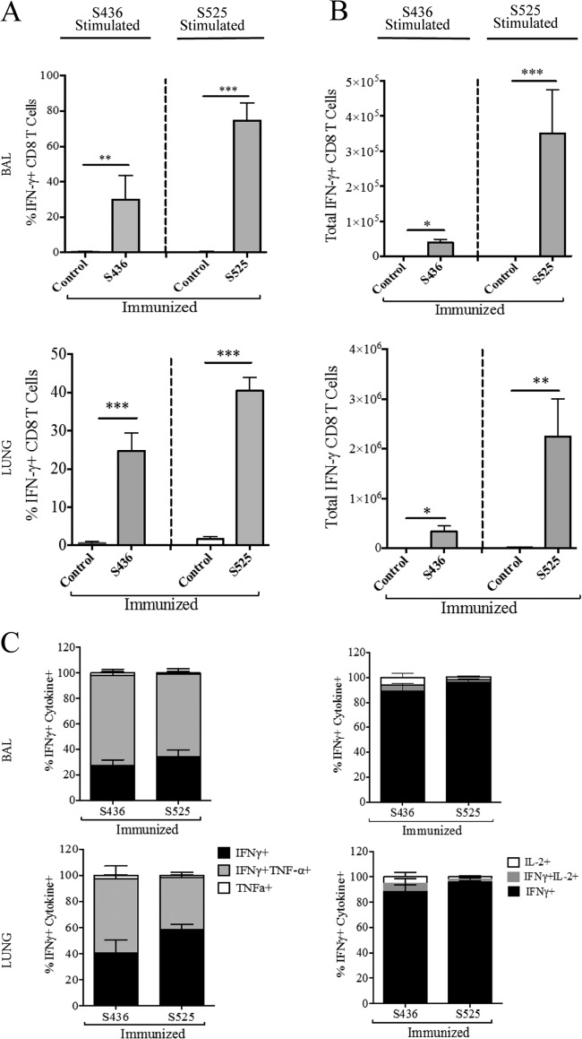
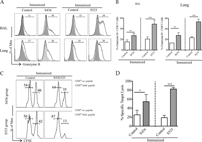
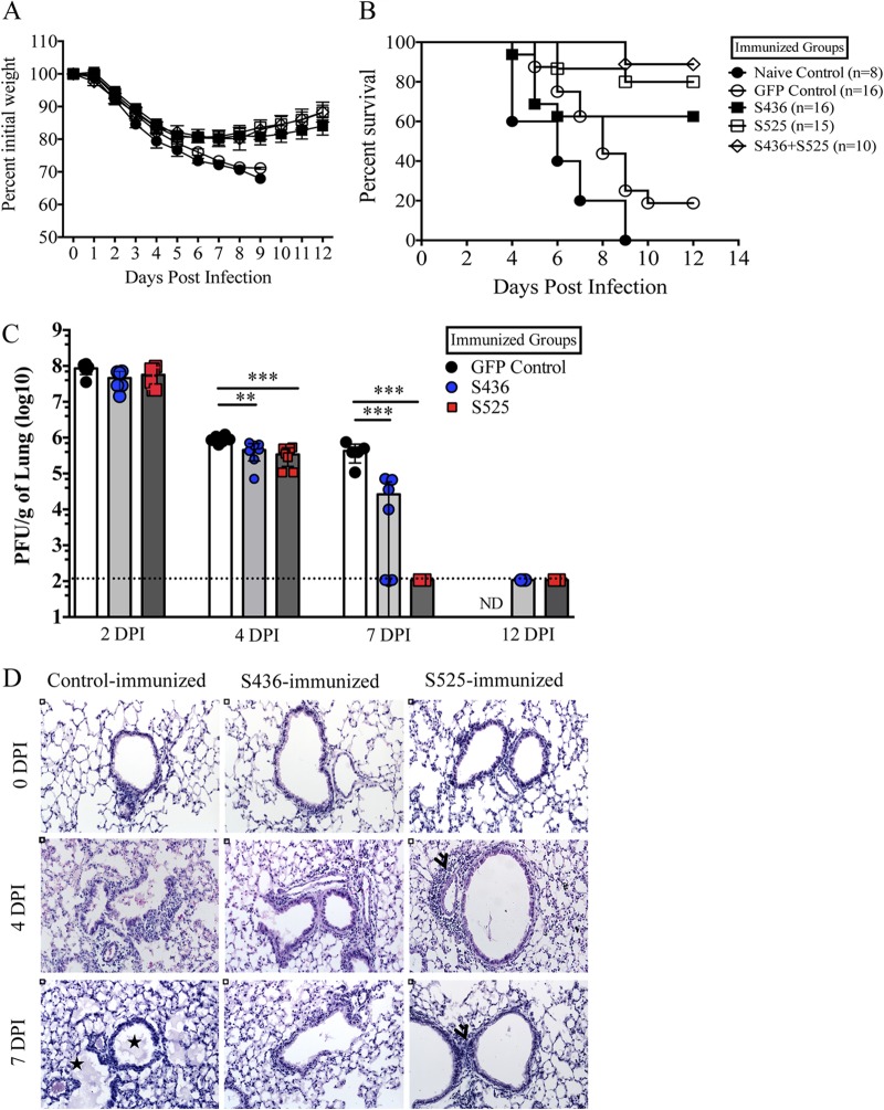
Severe acute respiratory syndrome coronavirus (SARS-CoV) caused an acute human respiratory illness with high morbidity and mortality in 2002-2003. Several studies have demonstrated the role of neutralizing antibodies induced by the spike (S) glycoprotein in protecting susceptible hosts from lethal infection. However, the anti-SARS-CoV antibody response is short-lived in patients who have recovered from SARS, making it critical to develop additional vaccine strategies. SARS-CoV-specific memory CD8 T cells persisted for up to 6 years after SARS-CoV infection, a time at which memory B cells and antivirus antibodies were undetectable in individuals who had recovered from SARS. In this study, we assessed the ability of virus-specific memory CD8 T cells to mediate protection against infection in the absence of SARS-CoV-specific memory CD4 T or B cells. We demonstrate that memory CD8 T cells specific for a single immunodominant epitope (S436 or S525) substantially protected 8- to 10-month-old mice from lethal SARS-CoV infection. Intravenous immunization with peptide-loaded dendritic cells (DCs) followed by intranasal boosting with recombinant vaccinia virus (rVV) encoding S436 or S525 resulted in accumulation of virus-specific memory CD8 T cells in bronchoalveolar lavage fluid (BAL), lungs, and spleen. Upon challenge with a lethal dose of SARS-CoV, virus-specific memory CD8 T cells efficiently produced multiple effector cytokines (gamma interferon [IFN-γ], tumor necrosis factor alpha [TNF-α], and interleukin 2 [IL-2]) and cytolytic molecules (granzyme B) and reduced lung viral loads. Overall, our results show that SARS-CoV-specific memory CD8 T cells protect susceptible hosts from lethal SARS-CoV infection, but they also suggest that SARS-CoV-specific CD4 T cell and antibody responses are necessary for complete protection.

Importance: Virus-specific CD8 T cells are required for pathogen clearance following primary SARS-CoV infection. However, the role of SARS-CoV-specific memory CD8 T cells in mediating protection after SARS-CoV challenge has not been previously investigated. In this study, using a prime-boost immunization approach, we showed that virus-specific CD8 T cells protect susceptible 8- to 10-month-old mice from lethal SARS-CoV challenge. Thus, future vaccines against emerging coronaviruses should emphasize the generation of a memory CD8 T cell response for optimal protection.


Figures

FIG 1
FIG 1
Prime-boost immunization induces a strong CD8 T cell response in 8-month-old mice. (A) Eight-month-old B6 mice were immunized intravenously (i.v.) with DC-loaded peptides and boosted 6 days later with rVV-S436 or rVV-S525 intranasally. Mice were rested for 42 to 45 days and then challenged with a lethal dose (5 × 10⁴ PFU) of MA15 (i.n.). Lungs and BAL were harvested 5 days postchallenge for further analysis. dpi, days postinfection. (B) FACS plots show percentages of S436- and S525-specific IFN-γ+ CD8 T cells in the blood (after direct ex vivo stimulation with respective peptides) on 0, 4, and 6 days after DC-peptide immunization. (C) Mean percentages (top) and numbers (bottom) of S436- and S525-specific CD8 T cells in the blood are shown. (D) FACS plots represent percentages of S436- and S525-specific IFN-γ+ CD8 T cells (after direct ex vivo stimulation with respective peptides) in BAL, lungs, and spleen 8 days after rVV-minigene boosting. (E and F) Bar graphs show mean percentages (E) and numbers (F) of S436- and S525-specific IFN-γ+ CD8 T cells (after in vitro stimulation with respective peptides) in BAL, lungs, and spleen 8 days after rVV-minigene boosting. Data are representative of 2 independent experiments with 3 or 4 mice/group. *, P < 0.05; **, P < 0.01; ***, P < 0.001 (by unpaired two-tailed Student's t test).
FIG 2
FIG 2
Identification of SARS-CoV-specific memory CD8 T cells. Prime-boost-immunized mice were rested for 42 to 45 days, and the percentages and numbers of S436 and S525-specific memory CD8 T cells were determined in BAL, lungs, and spleen. (A and B) The bar graphs show mean percentages (A) and numbers (B) of S436- and S525-specific IFN-γ+ CD8 T cells in the BAL, lungs, and spleen 42 to 45 days after rVV-minigene boosting. (C) The bar graphs show mean percentage of polyfunctional S436- and S525-specific IFN-γ+ CD8 T cells in the BAL, lungs, and spleen. Data are means of cytokine-positive CD8 T cells obtained by Boolean gating. (D) Scatter plots represent the percentages of S436 and S525 CD8 T cells that express the indicated marker. Data are representative of 2 or 3 independent experiments with 3 or 4 mice/group. *, P < 0.05; **, P < 0.01; ***, P < 0.001 (by unpaired two-tailed Student's t test).
FIG 3
FIG 3
Immunization induces polyfunctional secondary effector CD8 T cells. Prime-boost-immunized mice were rested for 42 to 45 days and then challenged with a lethal dose (5 × 10⁴ PFU) of MA15 (i.n.). Lungs and BAL were harvested 5 days postchallenge, and the percentage and number of epitope-specific CD8 T cells were determined. (A and B) The bar graphs show mean percentages (A) and numbers (B) of S436- and S525-specific IFN-γ+ CD8 T cells (after direct ex vivo stimulation with respective peptides) in the BAL and lungs 5 days after MA15 challenge. (C) IFN-γ+ CD8 T cells were further gated for TNF-α and IL-2 expression to determine polyfunctionality in the BAL and lungs. Data are representative of 3 independent experiments with 3 or 4 mice/group. *, P < 0.05; **, P < 0.01; ***, P < 0.001 (by unpaired two-tailed Student's t test).
FIG 4
FIG 4
Increased granzyme B production and cytotoxicity after challenge. (A and B) Histograms represent percent granzyme B+ CD8 T cells in the BAL and lungs at day 5 after MA15 challenge (A). (B) Bar graphs represent mean percentage of granzyme B+ CD8 T cells (after direct ex vivo stimulation with respective peptide). (C and D) In vivo cytotoxicity assays were performed 5 days after MA15 challenge, and the percent killing was calculated as described in Materials and Methods (numbers represent the percentage of cells labeled with different concentrations of CFSE). n = 4 or 5 mice/group/experiment. Data are representative of 2 independent experiments. *, P < 0.05; **, P < 0.01; ***, P < 0.001 (by unpaired two-tailed Student's t test).
FIG 5
FIG 5
Virus-specific memory CD8 T cells protect mid-aged mice from lethal MA15 infection. Prime-boost-immunized mice were rested for 42 to 45 days and then challenged with a lethal dose (5 × 10⁴ PFU) of MA15 intranasally. Percent initial body weight (A) and survival curves (B) are shown. The experiment shows data combined from three independent experiments, with 8 mice in the naive group and 15 or 16 mice in all other groups. Data are expressed as the percent initial weight ± the SEM. (C) Virus titers in lungs. Data correspond to means for 4 mice per group ± SEM and are representative of 2 or 3 independent experiments. (D) Lung sections from control and immunized mice are shown at days 0, 4, and 7 postchallenge. Stars represent alveolar and bronchiolar edema and arrowheads show peribronchial lymphocyte infiltration. DPI, days postinfection. *, P < 0.05; ** P < 0.01; ***, P < 0.001 (by unpaired Student's t test).

References

    1. Fouchier RA, Kuiken T, Schutten M, van Amerongen G, van Doornum GJ, van den Hoogen BG, Peiris M, Lim W, Stohr K, Osterhaus AD. 2003. Aetiology: Koch's postulates fulfilled for SARS virus. Nature 423:240. 10.1038/423240a - DOI - PMC - PubMed
    1. Kuiken T, Fouchier RA, Schutten M, Rimmelzwaan GF, van Amerongen G, van Riel D, Laman JD, de Jong T, van Doornum G, Lim W, Ling AE, Chan PK, Tam JS, Zambon MC, Gopal R, Drosten C, van der Werf S, Escriou N, Manuguerra JC, Stohr K, Peiris JS, Osterhaus AD. 2003. Newly discovered coronavirus as the primary cause of severe acute respiratory syndrome. Lancet 362:263–270. 10.1016/S0140-6736(03)13967-0 - DOI - PMC - PubMed
    1. Peiris JS, Guan Y, Yuen KY. 2004. Severe acute respiratory syndrome. Nat. Med. 10:S88–S97. 10.1038/nm1143 - DOI - PMC - PubMed
    1. Zaki AM, van Boheemen S, Bestebroer TM, Osterhaus AD, Fouchier RA. 2012. Isolation of a novel coronavirus from a man with pneumonia in Saudi Arabia. N. Engl. J. Med. 367:1814–1820. 10.1056/NEJMoa1211721 - DOI - PubMed
    1. World Health Organization. 2014. Middle East respiratory syndrome coronavirus (MERS-CoV)—update. World Health Organization, Geneva, Switzerland: http://www.who.int/csr/don/2014_03_25/en/

Publication types

MeSH terms