. 2014 Oct;88(19):11540-55.
doi: 10.1128/JVI.01745-14. Epub 2014 Jul 23.

Characterization of the mode of action of a potent dengue virus capsid inhibitor

Affiliations

Characterization of the mode of action of a potent dengue virus capsid inhibitor

Pietro Scaturro et al. J Virol. 2014 Oct.

Abstract

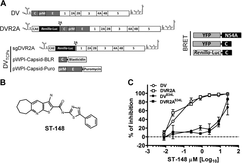
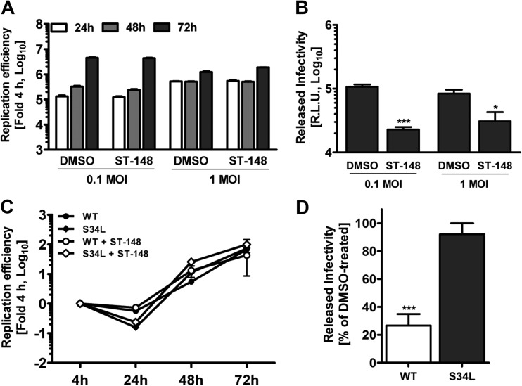
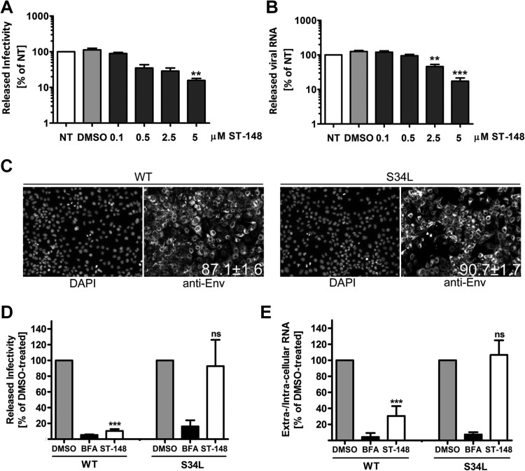
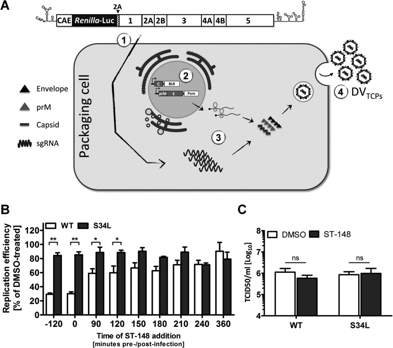
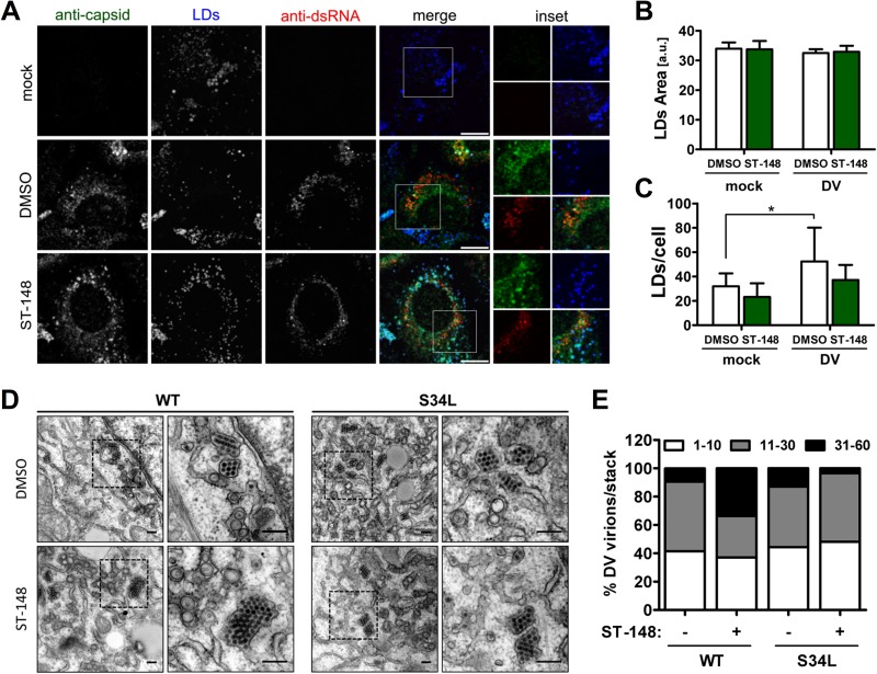
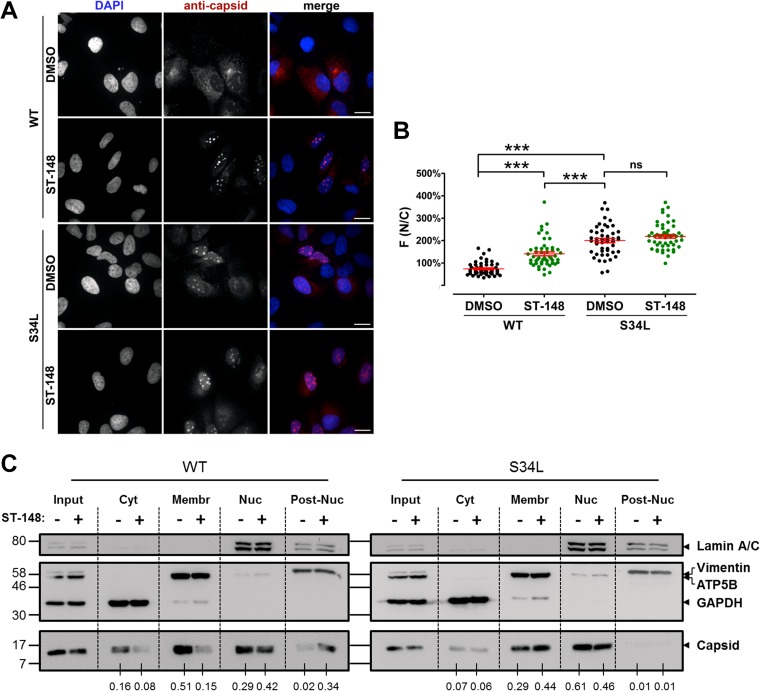
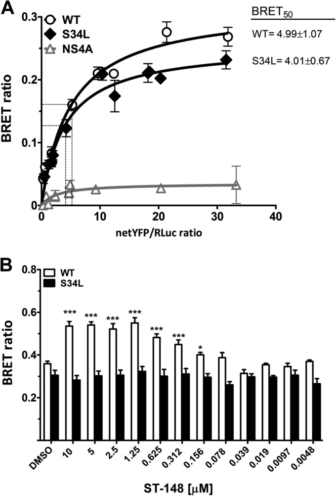
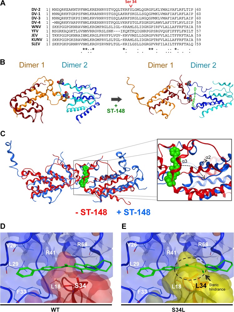
Dengue viruses (DV) represent a significant global health burden, with up to 400 million infections every year and around 500,000 infected individuals developing life-threatening disease. In spite of attempts to develop vaccine candidates and antiviral drugs, there is a lack of approved therapeutics for the treatment of DV infection. We have previously reported the identification of ST-148, a small-molecule inhibitor exhibiting broad and potent antiviral activity against DV in vitro and in vivo (C. M. Byrd et al., Antimicrob. Agents Chemother. 57:15-25, 2013, doi:10 .1128/AAC.01429-12). In the present study, we investigated the mode of action of this promising compound by using a combination of biochemical, virological, and imaging-based techniques. We confirmed that ST-148 targets the capsid protein and obtained evidence of bimodal antiviral activity affecting both assembly/release and entry of infectious DV particles. Importantly, by using a robust bioluminescence resonance energy transfer-based assay, we observed an ST-148-dependent increase of capsid self-interaction. These results were corroborated by molecular modeling studies that also revealed a plausible model for compound binding to capsid protein and inhibition by a distinct resistance mutation. These results suggest that ST-148-enhanced capsid protein self-interaction perturbs assembly and disassembly of DV nucleocapsids, probably by inducing structural rigidity. Thus, as previously reported for other enveloped viruses, stabilization of capsid protein structure is an attractive therapeutic concept that also is applicable to flaviviruses.

Importance: Dengue viruses are arthropod-borne viruses representing a significant global health burden. They infect up to 400 million people and are endemic to subtropical and tropical areas of the world. Currently, there are neither vaccines nor approved therapeutics for the prophylaxis or treatment of DV infections, respectively. This study reports the characterization of the mode of action of ST-148, a small-molecule capsid inhibitor with potent antiviral activity against all DV serotypes. Our results demonstrate that ST-148 stabilizes capsid protein self-interaction, thereby likely perturbing assembly and disassembly of viral nucleocapsids by inducing structural rigidity. This, in turn, might interfere with the release of viral RNA from incoming nucleocapsids (uncoating) as well as assembly of progeny virus particles. As previously reported for other enveloped viruses, we propose the capsid as a novel tractable target for flavivirus inhibitors.


Figures

FIG 1
FIG 1
Schematic representation of constructs used in this study and antiviral potency of ST-148. (A) Schematic representation of genomic and subgenomic constructs. The DV2 full-length genome is shown at the top (DV). The 5′ and 3′ N terminal regions are depicted with their putative secondary structures. Polyprotein cleavage products are separated by vertical lines and labeled as specified in the introduction. DVR2A is derived from the DV2 full-length genome by insertion of a Renilla luciferase coding sequence preceded by the capsid cyclization sequence (CAE) and followed by a Tosea asigna virus 2A cleavage site. The middle panel depicts the constructs used for production of trans-complemented DV particles (DVTCP). sgDVR2A is a subgenomic reporter replicon that is packaged into virus-like particles by the capsid and prM/envelope proteins transiently expressed in trans via two different lentiviruses (pWPI capsid-BLR and pWPI prME-Puro, respectively). All constructs are derived from the DVs2 16681 isolate (17). The right panel depicts the constructs used for BRET experiments. YFP and Renilla luciferase proteins were N-terminally fused to either capsid or NS4A of DV2 (strain 16681). (B) Structure of ST-148. (C) Antiviral potency of ST-148. Huh7 cells were infected at a DV or DVR2A reporter virus MOI of 0.1, with the virus containing or not containing the resistance mutation S34L, for 60 min at 37°C in the presence of various concentrations of the compound. After removal of the inoculum and several washings with PBS, cells were incubated with fresh medium containing various concentrations of ST-148 or DMSO. After 48 h, virus amounts released into the supernatant were determined by PFU assay with VeroE6 cells. Data represent averages and error ranges from two independent experiments.
FIG 2
FIG 2
ST-148 does not affect viral RNA replication but reduces production of infectious DV particles. (A) No effect of ST-148 on replication kinetics of the DVR2A reporter virus. Huh7 cells were infected with DVR2A at an MOI of 0.1 or 1 for 4 h at 37°C, washed with PBS, and incubated with fresh medium containing 5 μM ST-148 or DMSO. At the indicated time points after infection, luciferase activity was measured in the lysates as described in Materials and Methods. Note that error bars are poorly visible due to the size of the graph. (B) Reduction of virus production by ST-148. Supernatants of infected cells treated with 5 μM ST-148 were harvested 72 h postinfection and used to infect naive Huh7 cells. After 48 h, cells were harvested and luciferase activity was determined. (C) Comparison of replication kinetics of DVR2A and DVR2AS34L in the presence or absence of ST-148. Huh7 cells were electroporated with 10 μg of capped in vitro transcripts of WT DVR2A or DVR2AS34L (S34L), and 4 h later, 5 μM ST-148 or DMSO was added to the medium. At time points specified at the bottom, luciferase activity was determined. (D) Inhibition of virus production by DVR2A- and DVR2AS34L-transfected cells upon ST-148 treatment. Supernatants of transfected and ST-148-treated cells were harvested 72 h postinfection and used to infect naive Huh7 cells. After 48 h, cells were harvested and luciferase activity was determined. Data shown in each panel represent averages and standard deviations from three independent experiments (***, P < 0.001; *, P < 0.05).
FIG 3
FIG 3
Inhibition of the release of infectious DV particles by ST-148 as revealed by single-cycle experiments. (A) Production of infectious extracellular DV upon ST-148 treatment. (B) Quantification of released viral RNA. Huh7 cells were infected with DVR2A at an MOI of 1 for 4 h at 37°C, washed with PBS, and incubated with fresh medium (nontreated [NT]) or medium containing DMSO or the given concentrations of ST-148. After 48 h, clarified supernatants were used for TCID50 assay to quantify infectivity titers or two-step qRT-PCR for quantification of viral RNA amounts released into the supernatant. Data were analyzed using one-way ANOVA and Dunnett's post hoc test (**, P < 0.01; ***, P < 0.001). (C) Determination of infection efficiency. Cells were infected with DV or DVS34L and 48 h later were fixed and analyzed by immunofluorescence using a DV E-specific antibody. Nuclear DNA was stained with DAPI. Numbers in the lower right of the panels refer to the percentage of infected cells (means from at least 1,500 cells ± standard deviations). (D and E) Effect of ST-148 on single-round DV particle release. Huh7 cells were infected with DV or DVS34L for 48 h, washed extensively with PBS, and incubated for 12 h with medium containing 0.1% DMSO, 1 μg/ml brefeldin A (BFA), or 10 μM ST-148. Released infectivity and viral RNA were quantified by TCID50 assay and qRT-PCR, respectively. Results represent means and standard deviations from at least 3 independent experiments. (ns, nonsignificant; *, P < 0.05; ***, P < 0.001).
FIG 4
FIG 4
ST-148 additionally inhibits DV particle entry. (A) Schematic representation of the system used to produce trans-complemented DV particles (DVTCP). 293T cells were electroporated with 10 μg of capped in vitro transcripts of the subgenomic replicon sgDVR2A (segment 1). The next day, cells were transiently transduced with lentiviruses expressing the structural proteins capsid (wild type or containing the ST-148 resistance mutation S34L) and prM-E (segment 2). Replicating viral RNA is packaged in trans (segment 3), giving rise to DVTCP stocks that are released into the culture supernatant and harvested 4 to 6 days after transduction (segment 4). (B) Effect of ST-148 on entry of DVTCP. Huh7 cells were infected with DVTCP (WT or the S34L mutant) for 90 min at 37°C. The inoculum was removed and fresh medium was added. At time points specified at the bottom of the graph, medium was replaced by medium containing 10 μM ST-148 or 0.1% DMSO. In one case (−120 min), cells were pretreated with ST-148 for 2 h prior to infection. Cells were harvested 48 h postinfection, and luciferase activities in the lysates were measured. (C) No virucidal effect of ST-148. Stocks of DV WT or DVS34L were incubated at 37°C for 1 h with 10 μM ST-148 or 0.1% DMSO and used to determine infectivity titers by limiting-dilution assay (TCID50). Data represent averages and standard deviations from three independent experiments (ns, nonsignificant; *, P < 0.05; **, P < 0.001).
FIG 5
FIG 5
Effect of ST-148 on subcellular localization of viral RNA and protein and accumulation of DV particles in ER stacks. (A) Colocalization of capsid with dsRNA and LDs in ST-148 treated cells. Huh7 cells were infected with DV WT at an MOI of 1, and 4 h later, DMSO or 10 μM ST-148 was added to the medium. After 48 h, cells were fixed with 4% PFA in 4% sucrose. To visualize cytosolic LD-associated capsid protein, cells were permeabilized with 0.1% Triton X-100 and incubated with a rabbit polyclonal anti-capsid antibody, mouse anti-dsRNA antibody, and Bodipy-488 (to detect LDs). Scale bars represent 10 μm. (B and C) Impact of ST-148 on LD size and number, respectively. The mean area and number of LDs per cell has been calculated by using the Fiji plug-in of ImageJ. Each data set represents averages and standard deviations from at least 500 cells per condition (a.u., arbitrary units; *, P < 0.05). (D) Accumulation of DV particles in ER stacks upon ST-148 treatment. Huh7 cells were infected with DV WT or the DVS34L mutant (S34L) at an MOI of 3. Four hours after infection, the inoculum was removed and cells were incubated for 44 h in the presence of 10 μM ST-148 or DMSO. Cells were fixed, processed, and analyzed by transmission electron microscopy as described in Materials and Methods. The areas boxed in the left panels are shown at higher magnification on the right. Scale bars in each panel represent 200 nm. (E) Quantification of intracellular DV particle distribution. The number of virions present in ER stacks was quantified and assigned to 3 categories according to the number of virions per stack. Each column represents the mean of at least 1,000 virions from at least 15 infected cells per condition.
FIG 6
FIG 6
ST-148 induces accumulation of capsid protein in nuclear insoluble aggregates. (A) Intracellular distribution of capsid protein upon ST-148 treatment. Huh7 cells were infected with DV or DVS34L at an MOI of 1, and 4 h later, DMSO or 10 μM ST-148 was added to the medium. After 48 h, cells were fixed with 4% paraformaldehyde, permeabilized with 0.5% Triton X-100, and immune stained for capsid with a mouse monoclonal antibody. Scale bars represent 20 μm. (B) Image-based quantification of intracellular distribution of capsid protein. The extent of nuclear localization of capsid was determined by quantifying capsid-specific signals detected in the nucleus (N) and the cytoplasm (C), respectively, and calculating the ratio of mean fluorescence (F) in these two compartments [F(N/C)]. Results represent the means ± standard errors of the means (n ≥ 50). Data were analyzed using one-way ANOVA with Tukey's post hoc test (***, P < 0.001; ns, nonsignificant). (C) Subcellular fractionation of DV proteins. Huh7 cells were infected with DV or DVS34L at an MOI of 1, and 4 h later, cells were treated with DMSO or 10 μM ST-148. Intracellular distribution of capsid in soluble cytosolic (Cyt), membrane-associated (Membr), nuclear (Nuc), and postnuclear insoluble (Post-Nuc) fractions was evaluated by Western blotting. GAPDH, ATP5B, lamin A/C, and vimentin served as specificity controls for the various fractions. Numbers below each lane refer to the relative amounts of C proteins as determined by densitometry. After subtraction of the background, capsid-specific signals in each fraction were normalized to the corresponding input and expressed as a fold of total signal in the four fractions. Numbers on the left refer to the positions of molecular weight standards (in thousands). One result from two independent experiments is shown.
FIG 7
FIG 7
ST-148 increases DV capsid protein self-interaction. (A) Self-interaction kinetics of WT and S43L capsid as determined by BRET. DSA were performed in live 293T cells cotransfected with increasing amounts of YFP-tagged and a constant amount of Rluc-tagged plasmids encoding WT or mutant (S34L) capsid or NS4A, which served as negative controls. Forty-eight hours later, energy transfer was induced by the addition of the Renilla luciferase substrate coelenterazine H. The x axis shows the ratio between the normalized fluorescence of the acceptor (netYFP) measured before coelenterazine addition and the luminescence of the donor. BRET50 values (netYFP/Renilla ratio at which 50% of maximal BRET is occurring), which reflect the relative affinity of the acceptor protein for the donor protein, are given in the top right of the panel. Curves represent the means ± standard deviations of results from one representative experiment carried out in triplicate. The curves were fitted using a nonlinear regression equation in which a single binding site was assumed. (B) Effect of ST-148 on capsid self interaction. 293T cells were transfected with 1 μg of YFP capsid and 20 ng of Rluc capsid (WT and S34L). Prior to transfection, different concentrations of ST-148 or DMSO were added to the medium, and 48 h later BRET was measured (***, P < 0.001; *, P < 0.01).
FIG 8
FIG 8
Docking model of ST-148 and DV capsid and possible stabilization of interdimer capsid protein interaction. (A) Sequence comparison of capsid amino acid sequences of DV serotypes 1 to 4 and selected flaviviruses. The position of serine 34 is highlighted in red. Sequence alignment was carried out using the ClustalW algorithm, available at the Uniprot webserver, on the following isolates: DV-2 (Thailand/16681-PDK53), DV-1 (Brazil/97-11/1997), DV-3 (Martinique/1243/1999), DV-4 (Thailand/0348/1991), West-Nile virus (WNV; H442), yellow fever virus (YFV; Ivory Coast/1999), Japanese encephalitis virus (JEV; SA-14), Kunjin virus (KUNV; MRM61C), and St. Louis encephalitis virus (SLEV; MS1-7). UniprotKB accession numbers are given in Materials and Methods. (B) Results from molecular dynamics (MD) simulations. Stable conformations of the WT capsid tetramer in the absence (left) or presence (right) of ST-148. Capsid dimers are represented in blue and orange, with each monomer given in different shades of the same color. ST-148 is shown with green sticks, and the serine residue at position 34 is represented with the Corey-Pauling-Koltun (CPK) model. Movies of the MD simulations are given in the supplemental material. (C) Results of superimposition of the MD simulation of the WT C tetramer in the absence (red ribbon) or presence (blue ribbon) of ST-148 are presented at the left. ST-148 is shown in green with the CPK model. A closer view of the same superimposition is presented in the right panel with the same color code. The dashed arrows indicate the structural rearrangements, which occur at α2 and α3 helices upon ST-148 binding and allow accommodating the ligand. (D and E) Close-up view of the docking pose of ST-148. (D) WT capsid dimer-dimer interface complexed with ST-148. (E) Same interface as that shown in panel D, with the serine residue superimposed on Leu34. The dashed circle highlights the steric hindrance induced by the leucine residue at position 34, clarifying how the mutation impedes this binding pose. In both panels one dimer is depicted in light blue, while the other capsid dimer is in red in the case of the WT or in yellow in the case of the S34L resistance mutant. Protein residues are displayed as lines, while Ser34 and Leu34 are drawn with stick representations.

References

    1. Bhatt S, Gething PW, Brady OJ, Messina JP, Farlow AW, Moyes CL, Drake JM, Brownstein JS, Hoen AG, Sankoh O, Myers MF, George DB, Jaenisch T, Wint GR, Simmons CP, Scott TW, Farrar JJ, Hay SI. 2013. The global distribution and burden of dengue. Nature 496:504–507. 10.1038/nature12060 - DOI - PMC - PubMed
    1. Sabchareon A, Wallace D, Sirivichayakul C, Limkittikul K, Chanthavanich P, Suvannadabba S, Jiwariyavej V, Dulyachai W, Pengsaa K, Wartel TA, Moureau A, Saville M, Bouckenooghe A, Viviani S, Tornieporth NG, Lang J. 2012. Protective efficacy of the recombinant, live-attenuated, CYD tetravalent dengue vaccine in Thai schoolchildren: a randomised, controlled phase 2b trial. Lancet 380:1559–1567. 10.1016/S0140-6736(12)61428-7 - DOI - PubMed
    1. Nowak T, Farber PM, Wengler G, Wengler G. 1989. Analyses of the terminal sequences of West Nile virus structural proteins and of the in vitro translation of these proteins allow the proposal of a complete scheme of the proteolytic cleavages involved in their synthesis. Virology 169:365–376. 10.1016/0042-6822(89)90162-1 - DOI - PubMed
    1. Gollins SW, Porterfield JS. 1986. The uncoating and infectivity of the flavivirus West Nile on interaction with cells: effects of pH and ammonium chloride. J. Gen. Virol. 67(Part 9):1941–1950. 10.1099/0022-1317-67-9-1941 - DOI - PubMed
    1. Heinz FX, Stiasny K, Puschner-Auer G, Holzmann H, Allison SL, Mandl CW, Kunz C. 1994. Structural changes and functional control of the tick-borne encephalitis virus glycoprotein E by the heterodimeric association with protein prM. Virology 198:109–117. 10.1006/viro.1994.1013 - DOI - PubMed

Publication types

MeSH terms

Associated data