. 2014 Oct;88(19):11304-14.
doi: 10.1128/JVI.00306-14. Epub 2014 Jul 23.

Molecular characterization of a lizard adenovirus reveals the first atadenovirus with two fiber genes and the first adenovirus with either one short or three long fibers per penton

Affiliations

Molecular characterization of a lizard adenovirus reveals the first atadenovirus with two fiber genes and the first adenovirus with either one short or three long fibers per penton

Judit J Pénzes et al. J Virol. 2014 Oct.

Abstract

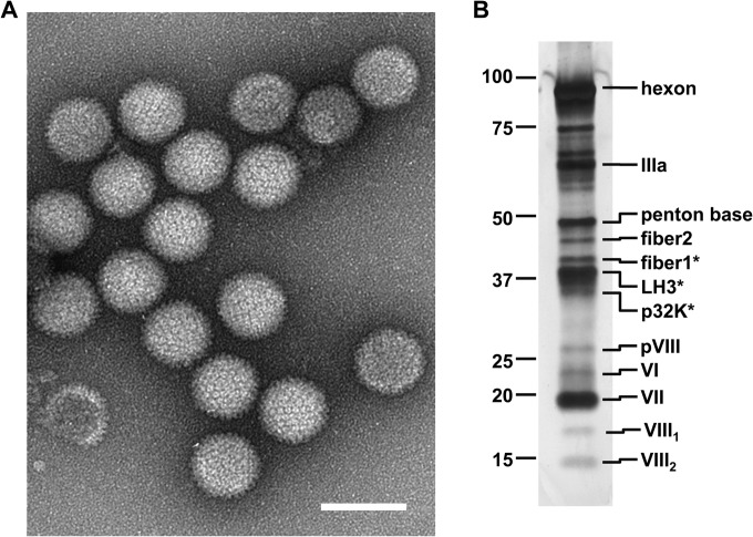
Although adenoviruses (AdVs) have been found in a wide variety of reptiles, including numerous squamate species, turtles, and crocodiles, the number of reptilian adenovirus isolates is still scarce. The only fully sequenced reptilian adenovirus, snake adenovirus 1 (SnAdV-1), belongs to the Atadenovirus genus. Recently, two new atadenoviruses were isolated from a captive Gila monster (Heloderma suspectum) and Mexican beaded lizards (Heloderma horridum). Here we report the full genomic and proteomic characterization of the latter, designated lizard adenovirus 2 (LAdV-2). The double-stranded DNA (dsDNA) genome of LAdV-2 is 32,965 bp long, with an average G+C content of 44.16%. The overall arrangement and gene content of the LAdV-2 genome were largely concordant with those in other atadenoviruses, except for four novel open reading frames (ORFs) at the right end of the genome. Phylogeny reconstructions and plesiomorphic traits shared with SnAdV-1 further supported the assignment of LAdV-2 to the Atadenovirus genus. Surprisingly, two fiber genes were found for the first time in an atadenovirus. After optimizing the production of LAdV-2 in cell culture, we determined the protein compositions of the virions. The two fiber genes produce two fiber proteins of different sizes that are incorporated into the viral particles. Interestingly, the two different fiber proteins assemble as either one short or three long fiber projections per vertex. Stoichiometry estimations indicate that the long fiber triplet is present at only one or two vertices per virion. Neither triple fibers nor a mixed number of fibers per vertex had previously been reported for adenoviruses or any other virus.

Importance: Here we show that a lizard adenovirus, LAdV-2, has a penton architecture never observed before. LAdV-2 expresses two fiber proteins-one short and one long. In the virion, most vertices have one short fiber, but a few of them have three long fibers attached to the same penton base. This observation raises new intriguing questions on virus structure. How can the triple fiber attach to a pentameric vertex? What determines the number and location of each vertex type in the icosahedral particle? Since fibers are responsible for primary attachment to the host, this novel architecture also suggests a novel mode of cell entry for LAdV-2. Adenoviruses have a recognized potential in nanobiomedicine, but only a few of the more than 200 types found so far in nature have been characterized in detail. Exploring the taxonomic wealth of adenoviruses should improve our chances to successfully use them as therapeutic tools.


Figures

FIG 1
FIG 1
Genomic characterization of LAdV-2. (A) Schematics of the LAdV-2 genetic map. Shading of the arrows marks the specificity of the genes. The G+C content of the genomic DNA is shown under the genome map. (B) Phylogeny reconstructions based on the hexon and protease amino acid sequences. Unrooted calculations were done by the ML method.
FIG 2
FIG 2
Molecular composition of purified LAdV-2. (A) Negative-staining EM image showing the general morphology of the LAdV-2 capsid. The bar represents 100 nm. (B) SDS-PAGE analysis of purified virions in a 4 to 20% gradient gel. Labels on the left-hand side indicate the positions of standard molecular mass markers (kDa). Labels on the right indicate the positions of virion proteins. Asterisks denote bands where protein identification was carried out by MS/MS.
FIG 3
FIG 3
LAdV-2 penton architecture. (A) SDS-PAGE analysis of the vertex-enriched preparation obtained after mild disruption using hypotonic dialysis and centrifugation. Asterisks indicate protein identification by MS/MS of excised gel bands. (B) Gallery of negative-staining EM images showing examples of a single fiber bound to one penton base, as represented by the drawing in panel C. (D) Gallery of pentons with three fibers attached to a single penton base, as illustrated by the drawing in panel E. In panels B and D, the scale bar represents 20 nm, and white arrowheads point to kinks in the fiber shafts. (F) Examples of negatively stained viral particles showing a fiber triplet. In the top row, arrows indicate the fiber knobs, while the trajectory of the shafts is highlighted with white curves in the bottom row. The bar represents 50 nm. (G and H) Prediction of structural domains in fiber1 (G) and fiber2 (H). The shaft pseudorepeats are aligned, and those with the largest departures from the repeating pattern that could originate kinks are highlighted in gray. The putative penton base binding peptide is underlined. On the right-hand side of each panel, a drawing shows the predicted number of structural repeats in the fiber shafts, and arrows indicate the locations of the predicted kinks.
FIG 4
FIG 4
Symmetry mismatches in the LAdV-2 penton organization. (A) Gallery of negative-staining EM images showing examples of triple fibers forming a complex in the absence of the penton base, observed in purified LAdV-2 preparations after heating at 50°C. The size bar represents 20 nm. (B) Drawings depicting different ways to fulfill the fiber-penton base symmetry mismatches. For a single fiber, each of the three N-terminal tails binds to one of the five equivalent interfaces between penton base monomers. For the LAdV-2 triple fibers, two possibilities are envisaged. First, each fiber uses two N-terminal tails to bind to its two partners and the third to bind to the penton base in a similar way as for the single fiber. Alternatively, all N-terminal tails associate as triplets, and each triplet binds to the penton base. Zigzag lines (continuous, dashed, or dotted) represent the three N-terminal tails of each trimeric fiber. Short transversal lines between zigzags indicate interactions between N-terminal tails of different fibers. The penton base pentamer is represented as a pentagon. (C) Example of purified LAdV-2 protein bands in a 10% acrylamide gel used for estimation of the fiber1/fiber2 ratio by densitometry. (D) A model for the distribution of the two different types of vertices in the LAdV-2 virion with a fiber1/fiber2 ratio of 1.6.

References

    1. Harrach B, Benkő M, Both G, Brown M, Davison A, Echavarría M, Hess M, Jones M, Kajon A, Lehmkuhl H, Mautner V, Mittal S, Wadell G. 2011. Family Adenoviridae, p 95–111 In King A, Adams M, Carstens E, Lefkowitz E. (ed), Virus taxonomy: classification and nomenclature of viruses. Ninth report of the International Committee on Taxonomy of Viruses. Elsevier, San Diego, CA
    1. Benkő M, Harrach B. 2003. Molecular evolution of adenoviruses. Curr. Top. Microbiol. Immunol. 272:3–36. 10.1007/978-3-662-05597-7_1 - DOI - PubMed
    1. Benkő M, Doszpoly A. 2011. Ichtadenovirus. Adenoviridae, p 29–32 In Tidona CA, Darai G. (ed), The Springer index of viruses. Springer, New York, NY
    1. Kovács ER, Benkő M. 2011. Complete sequence of raptor adenovirus 1 confirms the characteristic genome organization of siadenoviruses. Infect. Genet. Evol. 11:1058–1065. 10.1016/j.meegid.2011.03.021 - DOI - PubMed
    1. Farkas SL, Harrach B, Benkő M. 2008. Completion of the genome analysis of snake adenovirus type 1, a representative of the reptilian lineage within the novel genus Atadenovirus. Virus Res. 132:132–139. 10.1016/j.virusres.2007.11.009 - DOI - PubMed

Publication types

LinkOut - more resources