. 2014 Oct;88(19):11529-39.
doi: 10.1128/JVI.01712-14. Epub 2014 Jul 23.

Exosomes from human immunodeficiency virus type 1 (HIV-1)-infected cells license quiescent CD4+ T lymphocytes to replicate HIV-1 through a Nef- and ADAM17-dependent mechanism

Affiliations

Exosomes from human immunodeficiency virus type 1 (HIV-1)-infected cells license quiescent CD4+ T lymphocytes to replicate HIV-1 through a Nef- and ADAM17-dependent mechanism

Claudia Arenaccio et al. J Virol. 2014 Oct.

Abstract

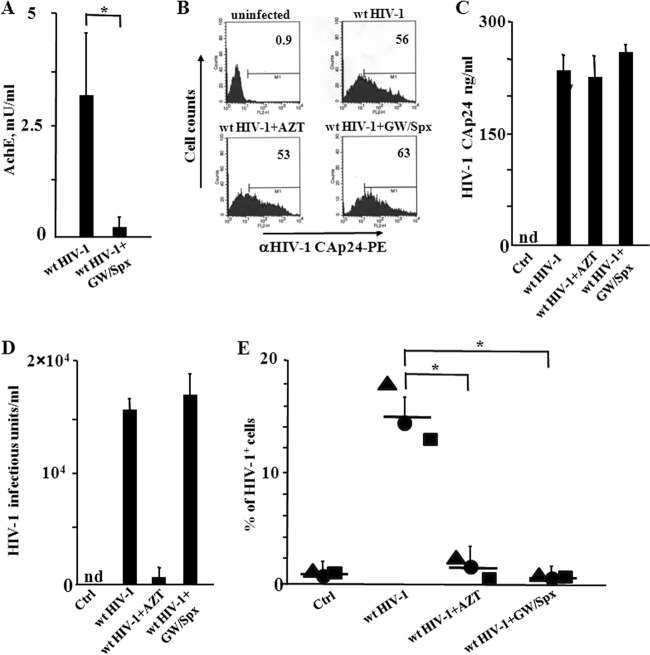
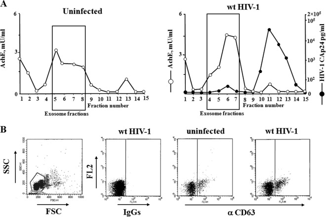
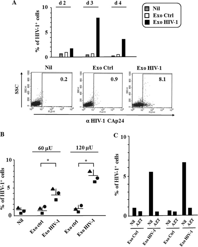
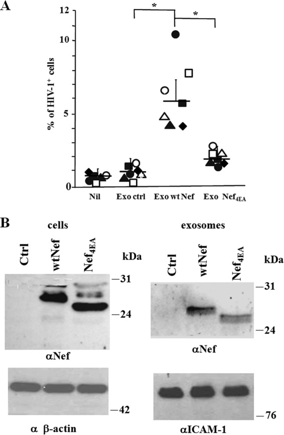
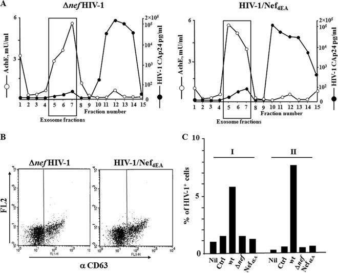
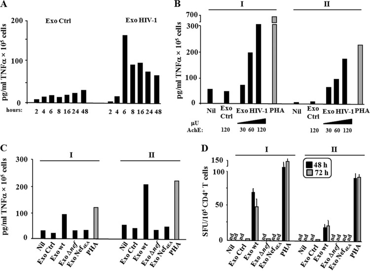
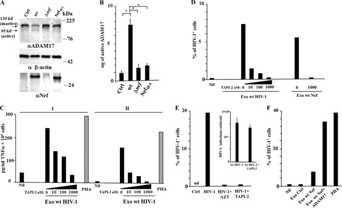
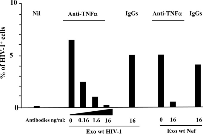
Resting CD4+ T lymphocytes resist human immunodeficiency virus (HIV) infection. Here, we provide evidence that exosomes from HIV-1-infected cells render resting human primary CD4+ T lymphocytes permissive to HIV-1 replication. These results were obtained with transwell cocultures of HIV-1-infected cells with quiescent CD4+ T lymphocytes in the presence of inhibitors of exosome release and were confirmed using exosomes purified from supernatants of HIV-1-infected primary CD4+ T lymphocytes. We found that the expression of HIV-1 Nef in exosome-producing cells is both necessary and sufficient for cell activation as well as HIV-1 replication in target CD4+ T lymphocytes. We also identified a Nef domain important for the effects we observed, i.e., the 62EEEE65 acidic cluster domain. In addition, we observed that ADAM17, i.e., a disintegrin and metalloprotease converting pro-tumor necrosis factor alpha (TNF-α) in its mature form, associates with exosomes from HIV-1-infected cells, and plays a key role in the HIV-1 replication in quiescent CD4+ T lymphocytes. Treatment with an inhibitor of ADAM17 abolished both activation and HIV-1 replication in resting CD4+ T lymphocytes. TNF-α is the downstream effector of ADAM17 since the treatment of resting lymphocytes with anti-TNF-α antibodies blocked the HIV-1 replication. The data presented here are consistent with a model where Nef induces intercellular communication through exosomes to activate bystander quiescent CD4+ T lymphocytes, thus stimulating viral spread.

Importance: Overall, our findings support the idea that HIV evolved to usurp the exosome-based intercellular communication network to favor its spread in infected hosts.


Figures

FIG 1
FIG 1
HIV-1 replicates in quiescent CD4+ T lymphocytes cocultured in transwell plates with activated CD4+ T lymphocytes infected by HIV-1. (A) Inhibition of exosome release from HIV-1-infected cells treated with GW4869 and spiroepoxide. A total of 3 ×10⁶ CD4+ T lymphocytes were activated with 2 μg/ml of PHA and, 2 days later, infected with VSV-G wt HIV-1. After an additional 2 days, the cells were treated with GW4869 and spiroepoxide for 16 h. Finally, exosomes in the supernatants were isolated and quantified in terms of mU of AchE. Values are the means + SD of results for duplicates from three independent experiments. *, P < 0.05. (B) FACS analysis for the HIV-1 CAp24 expression in activated CD4+ T lymphocytes from cocultures with resting lymphocytes. CD4+ T lymphocytes were activated and infected as described for panel A. Two days after infection, the cells were put in the upper chambers of transwell plates, and quiescent CD4+ T lymphocytes were seeded in the bottom chambers. The cocultures were run in the presence or not of either AZT or inhibitors of exosome release. Three days later, supernatants and cells from both the upper and bottom chambers were analyzed. The FACS analysis has been carried out in activated CD4+ T lymphocytes uninfected or infected with VSV-G wt HIV-1 in the presence or not of either AZT or the inhibitors of exosome release, GW4869 and spiroepoxide. The results are representative of data from three independent experiments. (C) HIV-1 CAp24 levels in supernatants of cocultures. nd, not detectable. Values are the means + SD of results for triplicates from one experiment representative of three independent experiments. (D) Infectivity of HIV-1 released by activated CD4+ T lymphocytes in the presence of GW4869 and spiroepoxide as measured on Rev-CEM indicator cells. Values are the means + SD of results for triplicates from one experiment representative of two independent experiments. (E) HIV-1 replication in resting CD4+ T lymphocytes. The cells were washed, treated with trypsin, and then scored for the expression of HIV-1 by means of intracellular FACS analysis. Shown are the mean percentages of HIV-1-positive cells as calculated from duplicate conditions using resting CD4+ T lymphocytes from three healthy donors. The interdonor mean percentages + SD are also shown. *, P < 0.05.
FIG 2
FIG 2
Characterization of exosomes from supernatants of activated CD4+ T lymphocytes either uninfected or HIV-1 infected. (A) AchE activity and, for HIV-1-infected cells only, HIV-1 Gag CAp24 contents were measured in fractions from 6 to 18% iodixanol gradients loaded with vesicles obtained by differential centrifugations of supernatants from activated CD4+ T lymphocytes either uninfected or infected with wt HIV-1. The results are the mean values from duplicate conditions and are representative of results from two independent experiments. (B) Detection by FACS of CD63 on pools of AchE-positive fractions from iodixanol gradients loaded with vesicles from uninfected or HIV-1-infected cells. Vesicles were bound to aldehyde/sulfate latex beads and then labeled with FITC-conjugated anti-CD63 monoclonal antibody or, as a control, FITC-conjugated isotype IgGs. On the left, a representative forward and side scatter dot plot of the exosome-bead complexes is shown. The results are representative of results from three independent experiments carried out on vesicles from two iodixanol gradient preparations.
FIG 3
FIG 3
HIV-1 replication in quiescent CD4+ T lymphocytes treated with exosomes from HIV-1-infected CD4+ T lymphocytes. (A) Kinetic of HIV-1 infection in resting CD4+ T lymphocytes. A total of 105 quiescent CD4+ T lymphocytes were challenged with 120 μU of exosomes from either uninfected (Ctrl) or HIV-1-infected CD4+ T lymphocytes and then infected with 50 ng of a T-tropic HIV-1 strain. As a control, quiescent cells were challenged with HIV-1 alone (Nil). From 2 to 4 days later, the cells were analyzed for HIV-1 expression by FACS analysis. Shown are the mean percentages of infected cells from duplicate conditions representative of results from two independent experiments carried out with cells from two healthy donors. On the bottom, shown are the dot plots of the FACS analysis from a representative experiment at day 3 postchallenge. (B) Dose-response effect of exosomes from HIV-1-infected cells on HIV-1 susceptibility of resting CD4+ T lymphocytes. A total of 105 quiescent CD4+ T lymphocytes were challenged with 60 or 120 μU of exosomes from either uninfected (Ctrl) or HIV-1-infected cells and then infected with 50 ng of a T-tropic HIV-1 strain. As a control, quiescent cells were challenged with HIV-1 alone (Nil). Three days later, the cells were analyzed for HIV-1 expression. Shown are the mean percentages of HIV-1-positive cells as calculated from duplicate conditions using resting CD4+ T lymphocytes from three healthy donors. The interdonor mean percentages + SD are also shown. *, P < 0.05. (C) Effects of AZT on HIV-1 replication in resting CD4+ T cells. A total of 105 quiescent CD4+ T lymphocytes were challenged with 120 μU of exosomes from either uninfected (Ctrl) or HIV-1-infected cells and then infected with 50 ng of a T-tropic HIV-1 strain. Cell cultures were run in the absence (Nil) or in the presence of 10 μM AZT. Three days later, the cells were analyzed for HIV-1 expression. Shown are the mean percentages of HIV-1-positive cells as calculated from duplicate conditions using resting CD4+ T lymphocytes from two (I, II) healthy donors.
FIG 4
FIG 4
HIV-1 replicates in quiescent CD4+ T lymphocytes treated with exosomes from Nef-expressing cells. (A) HIV-1 replication in resting CD4+ T lymphocytes treated with exosomes from cells expressing either wt Nef or Nef4EA. A total of 105 quiescent CD4+ T lymphocytes were challenged with 120 μU of exosomes from 293T cells transfected with wt Nef- or Nef4EA-expressing vector, or the empty vector (Ctrl), and then infected with 50 ng of a T-tropic HIV-1 strain. As a control, quiescent cells were challenged with HIV-1 alone (Nil). Three days later, the cells were analyzed for HIV-1 expression. Shown are the mean percentages of HIV-1 positive cells as calculated from duplicate conditions using resting CD4+ T lymphocytes from seven healthy donors. The interdonor mean percentages + SD are also shown. *, P < 0.05. (B) Detection of Nef in cells transfected with either wt Nef or Nef4EA and in exosomes purified from the respective supernatants. Shown is the anti-Nef Western blot analysis of both cells and exosomes from 293T cells transiently transfected with vectors expressing wt Nef, Nef4EA, or the empty vector (Ctrl). Signals from cellular Nef were normalized with β-actin detection, while anti-ICAM-1 analysis served to normalize exosome signals. The molecular markers are given in kDa. Results are representative of results from two independent experiments.
FIG 5
FIG 5
HIV-1 does not replicate in resting CD4+ T lymphocytes challenged with exosomes from cells infected by either the Δnef HIV-1 strain or the Nef4EA HIV-1 strain. (A) Characterization of exosomes from supernatants of activated CD4+ T lymphocytes infected by either the Δnef HIV-1 strain or the Nef4EA HIV-1 strain. AchE activity and HIV-1 Gag CAp24 contents were measured in fractions from 6 to 18% iodixanol gradients loaded with vesicles obtained by differential centrifugations of supernatants from activated CD4+ T lymphocytes infected by the Δnef HIV-1 strain or the Nef4EA HIV-1 strain. The results are the mean values for duplicate conditions and are representative of results from two independent experiments. (B) Detection by FACS of CD63 on pools of AchE-positive fractions from the iodixanol gradients described for panel A. (C) HIV-1 replication in resting CD4+ T lymphocytes treated with exosomes from cells infected by the wt, Δnef, or Nef4EA HIV-1 strain. A total of 105 quiescent CD4+ T lymphocytes were challenged with 120 μU of exosomes from the indicated infected cell populations, as well as from uninfected cells (Ctrl). As a control, cells were challenged with HIV-1 alone (Nil). Three days later, the cells were analyzed for HIV-1 expression. Shown are the mean percentages of HIV-1-positive cells as calculated from duplicate conditions using resting CD4+ T lymphocytes from two (I, II) healthy donors.
FIG 6
FIG 6
Resting CD4+ T lymphocytes release TNF-α and IL-2 when treated with exosomes from HIV-1-infected cells. (A) Kinetic of TNF-α release. A total of 105 quiescent CD4+ T lymphocytes were challenged with 120 μU of exosomes from HIV-1-infected or uninfected (Ctrl) cells. After extensive washes, the cells were seeded in complete medium, and supernatants were harvested at the indicated time points. Shown are the mean concentrations of TNF-α as calculated from duplicate conditions using resting CD4+ T lymphocytes from a representative of two healthy donors. (B) Detection of TNF-α on supernatants of 105 quiescent CD4+ T lymphocytes challenged with either increasing amounts (i.e., from 30 to 120 μU) of exosomes from HIV-1-infected cells or 120 μU of exosomes from uninfected cells (Ctrl). As a control, cells were treated with 2 μg/ml of PHA or left untreated (Nil). After extensive washes, the cells were seeded in complete medium, and supernatants were harvested 6 h later. Shown are the mean concentrations of TNF-α as calculated from duplicate conditions using resting CD4+ T lymphocytes from two (I, II) healthy donors. (C) TNF-α detection in supernatants from resting CD4+ T lymphocytes challenged with exosomes from cells infected by the Δnef HIV-1 strain or the Nef4EA HIV-1 strain. A total of 105 quiescent CD4+ T lymphocytes were challenged with 120 μU of exosomes from cells infected with the wt, Δnef HIV-1, or Nef4EA HIV-1 strain. As a control, cells were challenged with equal amounts of exosomes from uninfected cells (Ctrl), treated with 2 μg/ml of PHA, or left untreated (Nil). Supernatants were harvested 6 h later, and TNF-α contents were determined by ELISA. Shown are the mean concentrations of TNF-α as calculated from duplicate conditions using resting CD4+ T lymphocytes from two (I, II) healthy donors. (D) Detection of IL-2-producing cells. A total of 105 resting CD4+ T lymphocytes were challenged with 120 μU of exosomes from cells infected with the wt, Δnef, or Nef4EA HIV-1 strain. As a control, cells were challenged with equal amounts of exosomes from uninfected cells (Ctrl), treated with 2 μg/ml of PHA, or left untreated (Nil). Afterwards, the cells were washed and seeded in ELISPOT microwells previously coated with an anti-IL-2 monoclonal antibody. The spots were counted after both 48 and 72 h. Shown are the mean number of spots + SD as calculated from duplicate conditions evaluated in triplicate wells using resting CD4+ T lymphocytes from two (I, II) healthy donors. nd, no spots found.
FIG 7
FIG 7
Active ADAM17 associates with exosomes from wt HIV-1-infected cells and is important for both TNF-α release and induction of HIV-1 susceptibility in resting CD4+ T lymphocytes. (A) Western blot analysis for the expression of ADAM17 in cells infected with the wt, Δnef, or Nef4EA HIV-1 strain or mock infected (Ctrl). On the left of blots for ADAM17, arrows identify both inactive and active ADAM17 forms. Signals from cellular ADAM17 were normalized with both β-actin and Nef signals. On the right of each panel, molecular mass markers are given in kDa. The results are representative of results from two independent experiments. (B) ADAM17 activity detected in 1 mU of exosomes purified from the supernatants of lymphocytes either uninfected (Ctrl) or infected with the wt, Δnef, or Nef4EA HIV-1 strain. Mean values + SD of ng of active ADAM17 detected in duplicate samples from three exosome preparations are shown. *, P < 0.05. (C) Effect of TAPI-2 on TNF-α release. A total of 105 quiescent CD4+ T lymphocytes were challenged with 120 μU of exosomes from HIV-1-infected cells in the presence of increasing concentrations of TAPI-2. As a control, quiescent cells were challenged with HIV-1 alone (Nil) or in the presence of 2 μg/ml of PHA. The cells were left in culture for 6 h, and then TNF-α contents in supernatants were determined. Shown are the mean concentrations of TNF-α as calculated from duplicate conditions using resting CD4+ T lymphocytes from two (I, II) healthy donors. (D) Effects of TAPI-2 on HIV-1 replication. A total of 105 quiescent CD4+ T lymphocytes were challenged with 120 μU of exosomes from HIV-1-infected cells in the presence of increasing concentrations of TAPI-2. Alternatively, the cells were challenged with the same amounts of exosomes from wt Nef-expressing 293T cells in the presence of the highest TAPI-2 concentration. As a control, quiescent cells were challenged with HIV-1 alone (Nil). Cells were then infected with HIV-1 and 3 days later were analyzed for HIV-1 expression. Shown are the mean percentages of HIV-1-positive cells as calculated from duplicate conditions using resting CD4+ T lymphocytes from a representative of two healthy donors analyzed. (E) Transwell cocultures of resting CD4+ T lymphocytes with HIV-1-infected cells in the presence of AZT or TAPI-2. CD4+ T lymphocytes were activated with 2 μg/ml of PHA and 2 days later infected with VSV-G wt HIV-1. After an additional 2 days, the cells were put in the upper chamber of transwell plates, and quiescent CD4+ T lymphocytes were seeded in the bottom chamber. The cocultures were run in the presence or not of either AZT or 1 μM TAPI-2. Three days later, resting cells were harvested and analyzed for the percentage of HIV-1-infected cells by FACS. Shown are the mean percentages of HIV-1-positive cells as calculated from duplicate conditions using cells from a representative of two healthy donors analyzed. In the inset, shown is the infectivity of HIV-1 released by activated CD4+ T lymphocytes in the presence or not of TAPI-2 as measured in Rev-CEM cells. Shown are the mean values + SD of the infectious units as calculated from triplicate conditions using the supernatants from a representative of two independent experiments. (F) Effects of exosomes from ADAM17-transfected cells. A total of 120 μU of exosomes from 293T cells cotransfected with vectors expressing wt Nef and ADAM17 was used to challenge quiescent CD4+ T lymphocytes, which were then infected by HIV-1. As a control, cells were left untreated (Nil), activated with 2 μg/ml of PHA, or treated with exosomes from cells transfected with either an empty vector (Ctrl) or a wt Nef-expressing vector. Three days later, resting cells were analyzed for the percentage of HIV-1-infected cells by FACS. Shown are the mean percentages of HIV-1-positive cells as calculated from duplicate conditions using cells from a representative of two healthy donors analyzed.
FIG 8
FIG 8
TNF-α neutralization blocks the HIV-1 replication in quiescent CD4+ T lymphocytes treated with exosomes from HIV-1-infected cells. A total of 105 quiescent CD4+ T lymphocytes were challenged with 120 μU of exosomes from HIV-1-infected cells. Then, the cells were incubated for 6 h in the presence of the indicated amounts of anti-TNF-α neutralizing antibodies or unrelated, isotype-specific IgGs. Alternatively, the cells were treated with equal amounts of exosomes from 293T cells and then incubated with the highest dose of antibodies. Afterwards, the cells were infected with HIV-1 and then washed and reseeded in the presence of the antibodies. As a control, resting CD4+ T cells were treated with HIV-1 alone (Nil). Three days after challenges, the cells were analyzed for HIV-1 expression by FACS. Shown are the mean percentages of HIV-1 positive cells as calculated from duplicate conditions using cells from a representative of two healthy donors analyzed.

References

    1. Gyorgy B, Szabo TG, Pasztoi M, Pal Z, Misjak P, Aradi B, Laszlo V, Pallinger E, Pap E, Kittel A, Nagy G, Falus A, Buzas EI. 2011. Membrane vesicles, current state-of-the-art: emerging role of extracellular vesicles. Cell. Mol. Life Sci. 68:2667–2688. 10.1007/s00018-011-0689-3 - DOI - PMC - PubMed
    1. Booth AM, Fang Y, Fallon JK, Yang JM, Hildreth JEK, Gould SJ. 2006. Exosomes and HIV Gag bud from endosome-like domains of the T cell plasma membrane. J. Cell Biol. 172:923–935. 10.1083/jcb.200508014 - DOI - PMC - PubMed
    1. Mathivanan S, Ji H, Simpson RJ. 2010. Exosomes: extracellular organelles important in intercellular communication. J. Proteomics 73:1907–1920. 10.1016/j.jprot.2010.06.006 - DOI - PubMed
    1. Skog J, Wurdinger T, van Rijn S, Meijer DH, Gainche L, Sena-Esteves M, Curry WT, Carter BS, Krichevsky AM, Breakefield XO. 2008. Glioblastoma microvesicles transport RNA and proteins that promote tumour growth and provide diagnostic biomarkers. Nat. Cell Biol. 10:1470–1476. 10.1038/ncb1800 - DOI - PMC - PubMed
    1. Fang Y, Wu N, Gan X, Yan WH, Morrell JC, Gould SJ. 2007. Higher-order oligomerization targets plasma membrane proteins and HIV Gag to exosomes. PLoS Biol. 5:1267–1283. 10.1371/journal.pbio.0050158 - DOI - PMC - PubMed

Publication types

MeSH terms