Clinical Trial
. 2014 Sep 1;193(5):2373-83.
doi: 10.4049/jimmunol.1400486. Epub 2014 Jul 23.

Modulation of TNF-induced macrophage polarization by synovial fibroblasts

Affiliations
Clinical Trial

Modulation of TNF-induced macrophage polarization by synovial fibroblasts

Laura T Donlin et al. J Immunol. .

Abstract

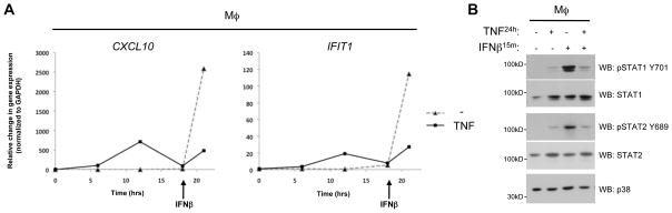
Mesenchymal stromal cells have emerged as powerful modulators of the immune system. In this study, we explored how the human macrophage response to TNF is regulated by human synovial fibroblasts, the representative stromal cell type in the synovial lining of joints that become activated during inflammatory arthritis. We found that synovial fibroblasts strongly suppressed TNF-mediated induction of an IFN-β autocrine loop and downstream expression of IFN-stimulated genes (ISGs), including chemokines CXCL9 and CXCL10 that are characteristic of classical macrophage activation. TNF induced the production of soluble synovial fibroblast factors that suppressed the macrophage production of IFN-β, and cooperated with TNF to limit the responsiveness of macrophages to IFN-β by suppressing activation of Jak-STAT signaling. Genome-wide transcriptome analysis showed that cocultured synovial fibroblasts modulate the expression of approximately one third of TNF-regulated genes in macrophages, including genes in pathways important for macrophage survival and polarization toward an alternatively activated phenotype. Pathway analysis revealed that gene expression programs regulated by synovial fibroblasts in our coculture system were also regulated in rheumatoid arthritis synovial macrophages, suggesting that these fibroblast-mediated changes may contribute to rheumatoid arthritis pathogenesis. This work furthers our understanding of the interplay between innate immune and stromal cells during an inflammatory response, one that is particularly relevant to inflammatory arthritis. Our findings also identify modulation of macrophage phenotype as a new function for synovial fibroblasts that may prove to be a contributing factor in arthritis pathogenesis.


Figures

FIGURE 1
FIGURE 1
Co-cultured synovial fibroblasts suppress the macrophage IFN signature induced by TNF. (A–B) Human macrophages (Mϕ) and RA synovial fibroblasts (Fib) cultured alone or together (Mϕ + Fib) and stimulated with TNF for 16 h were analyzed for mRNA levels by qPCR. Genes analyzed are listed above each graph. Gene expression values represent the mean of 4 experiments; error bars represent standard error (SE). In each experiment, macrophages were derived from a unique blood donor and the synovial fibroblasts were generated from a unique patient, such that n=4 for both cell types. The amount of transcript was normalized to GAPDH levels in the sample and all samples for a given gene are shown relative to the TNF-induced macrophage monoculture sample, which was set to 100. The mean level of mRNA encoded by the indicated genes relative to the internal standard GAPDH mRNA (% of GAPDH) in the TNF-induced samples was: CXCL9, 10%; CXCL10, 600%; IFIT1, 60%; MX1, 50%; NKG7, 1%; CXCL5, 750%; IL1B, 40%. **<p=0.001; NS, no significance.
FIGURE 2
FIGURE 2
Synovial fibroblasts stimulated by TNF produce soluble mediators responsible for restricting the macrophage TNF-induced IFN response. (A) qPCR mRNA analysis of human macrophages stimulated by TNF for 16h in the presence of synovial fibroblasts suspended in porous transwell chambers. Mϕ (Fib), transwell cultures of macrophages with synovial fibroblasts, in which only the macrophage mRNA was analyzed. (B) qPCR mRNA analysis of macrophages stimulated by TNF for 16h in the presence of supernatant media. Supernatants were collected from independent cultures of synovial fibroblast treated with TNF (FibTNF Supe) and without TNF (Fib Supe) for 16h. (C) qPCR mRNA analysis of macrophages cultured in a similar manner to part B except that the supernatants from TNF-treated fibroblasts were fractionated by molecular weight (left panel) or boiled (right panel) and individually incubated with the macrophages. kDA, kilodalton. (D) qPCR mRNA analysis of macrophages treated with neutralizing antibodies were incubated alone or with synovial fibroblasts in a transwell chamber (Fib). mRNA levels were normalized to GAPDH and made relative to the TNF-induced macrophage monoculture sample, which was set to 100. The mean level of mRNA encoded by the indicated genes relative to GAPDH mRNA (% GAPDH) for the TNF-induced samples in the Transwell cultures in part (A) was: CXCL10, 300%; IFIT1, 20%; and in the supernatant experiments in part (B–D) was: CXCL10, 1400%; IFIT1, 40%. Mean of n=3 in part A, n=4 in part B and n=2 in parts C–D; error bars represent SE; ***<p=0.0001; **<p=0.001; *<p=0.02; NS, no significance.
FIGURE 3
FIGURE 3
Synovial fibroblast products suppress IFN-β expression and responsiveness in TNF-stimulated macrophages. (A) Macrophages stimulated by TNF for 16 h with synovial fibroblasts in standard co-cultures, transwell cultures or with synovial fibroblast supernatants were analyzed for mRNA levels by qPCR. The supernatant media (FibTNF Supe) was collected from independent fibroblast cultures stimulated with TNF for 16 h. (B) qPCR mRNA analysis of macrophages cultured with control media (media) or supernatant media (FibTNF Supe) from fibroblasts independently cultured with TNF for 16h. Supernatants obtained from TNF-induced fibroblast cultures were first treated with Infliximab (Ifx) to eliminate TNF activity or isotype control immunoglobulin (IgG). Following the 16h supernatant incubation, the macrophages were stimulated with IFN-β for 3h. mRNA levels were normalized to GAPDH and made relative to the TNF-induced macrophage alone sample (part A) or IFN-induced macrophage with control media and IgG (part B), which were set to 100. The mean level of IFNB mRNA relative to GAPDH mRNA (% GAPDH) in part (A) was: coculture, 0.1%; transwell, 0.1%; supernatant, 0.1%; and the IFN-β-induced responses in part (B): CXCL10, 800%; IFIT1, 140%. Mean of n=4 donor experiments; error bars represent standard error (SE); ***<p=0.0001; **<p=0.001; *<p=0.05.
FIGURE 4
FIGURE 4
Synovial fibroblast products suppress the production of type I IFN response protein effectors in the presence of TNF. (A) Western blot analysis of human macrophages stimulated by TNF for 16h in the presence of synovial fibroblasts suspended in porous transwell chambers. Mϕ (Fib), transwell cultures of macrophages with synovial fibroblasts, in which only the macrophage lysate was analyzed. Blots represent one of three experiments, with the average densitometry value and SE for phospho-STAT1 from all three experiments plotted to the right. (B) ELISA protein measurements for the CXCL10/IP10 chemokine found in supernatants from human macrophages and synovial fibroblasts cultured alone or in transwell cultures (as in part A) and treated with TNF at Day 0. Data points represent the average of 3 independent experiments, with error bars representing the standard error.
FIGURE 5
FIGURE 5
In macrophages, TNF induces an IFN signature but also limits IFN responsiveness. (A) qPCR mRNA analysis of macrophages cultured with and without TNF for 18h and subsequently stimulated with IFN-β for 3h. Graphs depict a representative experiment from 3 independent donors. Gene expression changes were normalized to GAPDH and calculated relative to uninduced. (B) Western blot analysis of macrophages treated with or without TNF for 24h followed by IFN-β for 15min. Blots represent one of three experiments.
FIGURE 6
FIGURE 6
Transcriptome-wide analysis of the synovial fibroblast influence over macrophage TNF responses. (A) Venn diagrams representing the overlap of macrophage genes that TNF induces (left diagram, red circle) or represses (right diagram, green circle), which in the presence of synovial fibroblast are opposingly regulated (left diagram, green circle represents macrophage genes repressed by fibroblasts upon TNF treatment)(right diagram, red circle represents macrophage genes induced by fibroblasts in TNF conditions). Human blood-derived macrophages were treated for 2 days with TNF with or without synovial fibroblasts suspended above the macrophages in transwell chambers and the macrophage RNA was sequenced (RNAseq). TNF-regulated genes included those which became greater than 2-fold induced or repressed in comparison to untreated macrophages. Fibroblast-regulated genes included those with greater than 2-fold differences when comparing macrophages treated with TNF alone versus treated with TNF in the presence of fibroblasts. (B) Heat map depiction of all macrophage genes repressed (left) or upregulated (right) by co-cultured synovial fibroblasts upon TNF treatment (by at least 2-fold) in comparison to macrophages treated with TNF and cultured alone. Colored bars represent the gene expression levels (log2 of FPKM), with red representing higher and green representing lower levels. (C) Proteins and small molecules predicted to mediate the gene expression changes brought on by synovial fibroblasts in TNF-induced macrophages. The dataset of macrophage genes regulated by fibroblasts by at least 2-fold were analyzed by the Ingenuity IPA program. Based on published findings, the Upstream Regulator analytic predicted upstream molecules whose change in expression or function could explain the observed gene expression changes (the p-value relates to the extent of overlap between known targets genes for an upstream regulator and genes altered in the present dataset). A greater absolute z-score value indicates the upstream regulator target genes are mostly altered in a direction consistent with either reduced or increased activity of the upstream regulator, with negative (left panel) or positive (right panel) values indicating predicted inhibition or activation of the upstream regulator, respectively. (D) Target gene networks for the upstream regulators MYC (left) and EGF (right) include genes regulated by fibroblasts more than 4-fold in the presence of TNF and whose direction of expression is consistent with reduced MYC and increased EGF activity. Red versus green shapes indicate increased versus decreased expression of the target genes by fibroblasts, while arrow heads versus inhibitory lines indicate whether the upstream regulator (MYC or EGF) is known to induce or repress the expression of each gene, respectively. The target gene shapes represent the molecular classification or function of the protein encoded by the gene: square, growth factor/cytokine; triangle, transcription regulator; diamond, enzyme; oval, transmembrane receptor; rectangle, nuclear receptor; trapezoid, transporter; circle, other.

References

    1. Frenette PS, Pinho S, Lucas D, Scheiermann C. Mesenchymal Stem Cell: Keystone of the Hematopoietic Stem Cell Niche and a Stepping-Stone for Regenerative Medicine. Annu Rev Immunol. 2013;31:285–316. - PubMed
    1. Le Blanc K, Mougiakakos D. Multipotent mesenchymal stromal cells and the innate immune system. Nat Rev Immunol. 2012;12:383–396. - PubMed
    1. Kalluri R, Zeisberg M. Fibroblasts in cancer. Nat Rev Cancer. 2006;6:392–401. - PubMed
    1. Kraman M, Bambrough PJ, Arnold JN, Roberts EW, Magiera L, Jones JO, Gopinathan A, Tuveson DA, Fearon DT. Suppression of Antitumor Immunity by Stromal Cells Expressing Fibroblast Activation Protein. Science. 2010;330:827–830. - PubMed
    1. Straussman R, Morikawa T, Shee K, Barzily-Rokni M, Qian ZR, Du J, Davis A, Mongare MM, Gould J, Frederick DT, Cooper ZA, Chapman PB, Solit DB, Ribas A, Lo RS, Flaherty KT, Ogino S, Wargo JA, Golub TR. Tumour micro-environment elicits innate resistance to RAF inhibitors through HGF secretion. Nature. 2013;487:500–504. - PMC - PubMed

Publication types

MeSH terms

Associated data