. 2014 Sep 15;25(18):2853-65.
doi: 10.1091/mbc.E14-05-0971. Epub 2014 Jul 23.

The Caenorhabditis elegans SUN protein UNC-84 interacts with lamin to transfer forces from the cytoplasm to the nucleoskeleton during nuclear migration

Affiliations

The Caenorhabditis elegans SUN protein UNC-84 interacts with lamin to transfer forces from the cytoplasm to the nucleoskeleton during nuclear migration

Courtney R Bone et al. Mol Biol Cell. .

Abstract

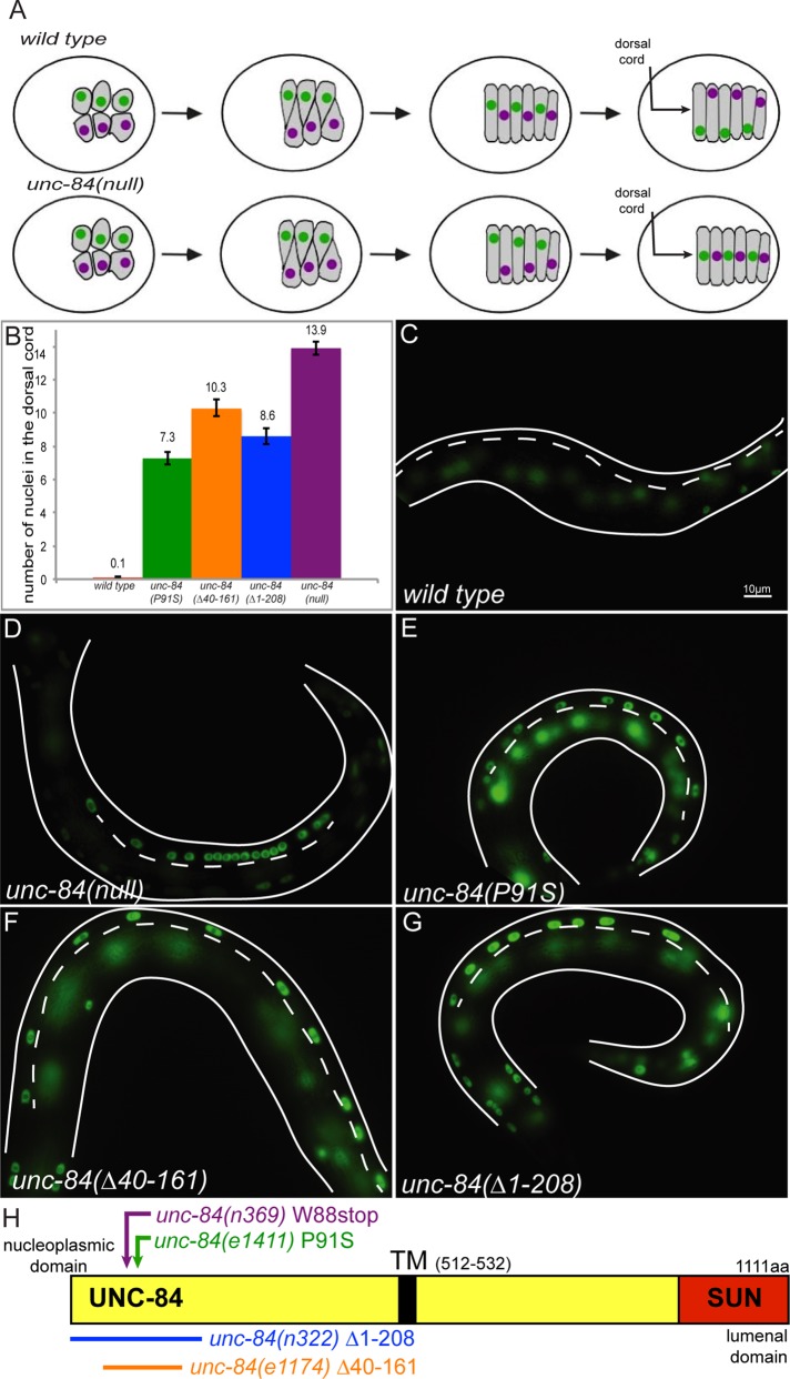
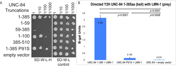
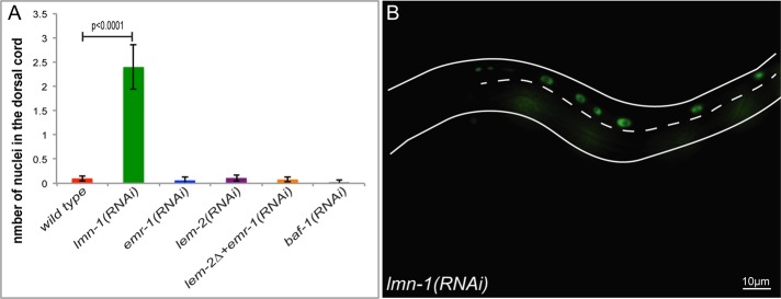
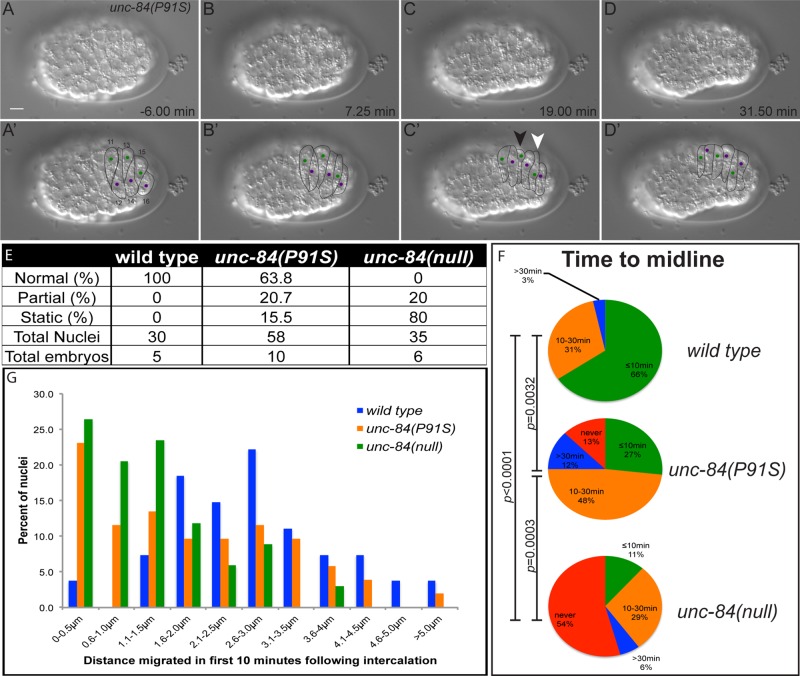
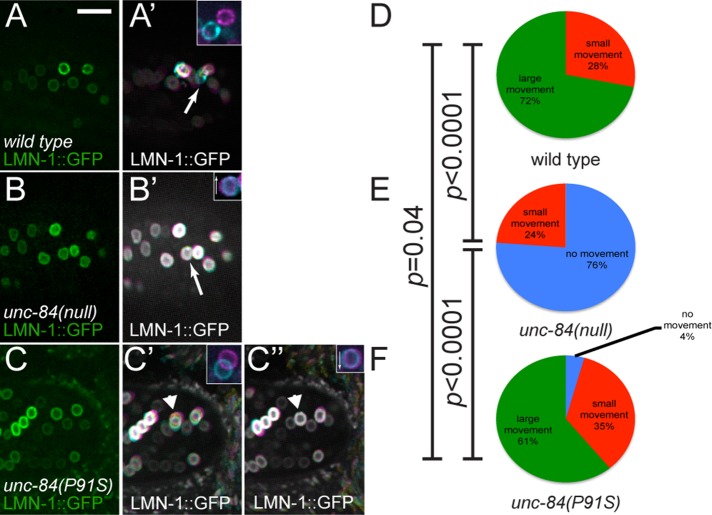
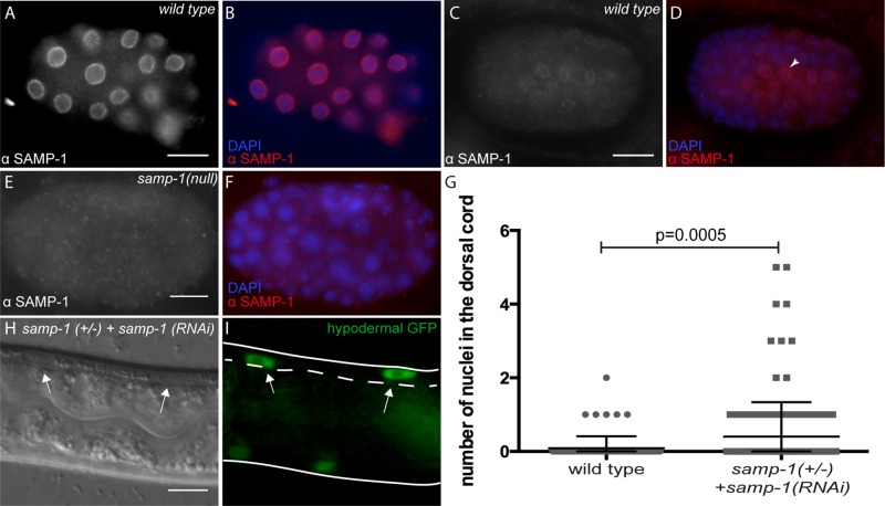
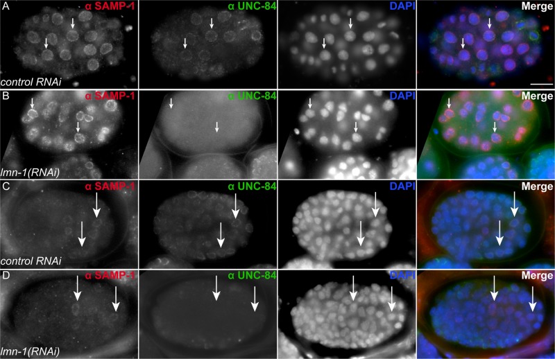
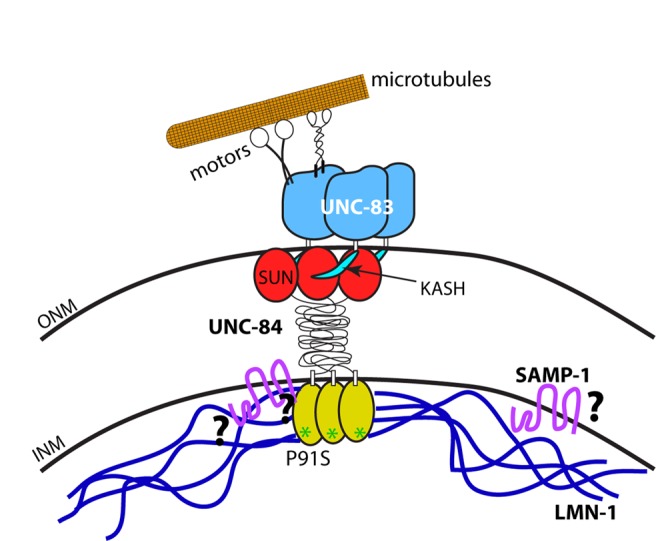
Nuclear migration is a critical component of many cellular and developmental processes. The nuclear envelope forms a barrier between the cytoplasm, where mechanical forces are generated, and the nucleoskeleton. The LINC complex consists of KASH proteins in the outer nuclear membrane and SUN proteins in the inner nuclear membrane that bridge the nuclear envelope. How forces are transferred from the LINC complex to the nucleoskeleton is poorly understood. The Caenorhabditis elegans lamin, LMN-1, is required for nuclear migration and interacts with the nucleoplasmic domain of the SUN protein UNC-84. This interaction is weakened by the unc-84(P91S) missense mutation. These mutant nuclei have an intermediate nuclear migration defect-live imaging of nuclei or LMN-1::GFP shows that many nuclei migrate normally, others initiate migration before subsequently failing, and others fail to begin migration. At least one other component of the nucleoskeleton, the NET5/Samp1/Ima1 homologue SAMP-1, plays a role in nuclear migration. We propose a nut-and-bolt model to explain how forces are dissipated across the nuclear envelope during nuclear migration. In this model, SUN/KASH bridges serve as bolts through the nuclear envelope, and nucleoskeleton components LMN-1 and SAMP-1 act as both nuts and washers on the inside of the nucleus.


Figures

FIGURE 1:
FIGURE 1:
Mutations in the nucleoplasmic domain of UNC-84 lead to an intermediate nuclear migration defect. (A) Cartoon describing hyp7 precursor nuclear migration on the dorsal surface of the pre–comma-stage embryo. In wild-type embryos (top), two rows of hyp7 precursors (gray) intercalate to form a row of column-shaped cells. Nuclei then migrate from right to left (green) or left to right (purple). In unc-84(null) mutant embryos, intercalation occurs normally, but the nuclei fail to migrate. Instead, underlying body wall muscle migrations push unc-84 nuclei to the dorsal cord (arrow). The dorsal surface is shown; anterior is left. (B) Average number of nuclei present in the dorsal cord of L1 larvae, which approximates the number of failed nuclear migrations. Error bars, 95% CI. (C–G) The number of nuclei in the larval dorsal cord was counted following hypodermal nuclei that express a nucleoplasmic GFP from ycIs10[pcol-10nls::gfp::lacZ]. Lateral views of L1 larvae. Dorsal is up, and anterior is left; the dorsal cord (arrow in A) is demarcated by the white dotted line. Scale bar, 10 μm. Representative images of (C) wild type, (D) unc-84(null), (E) unc-84(P91S), (F) unc-84(Δ40-161), and (G) unc-84(Δ1-208). (H) Schematic of the domain structure of UNC-84. The conserved SUN domain is red, and the transmembrane span is black. The mutants discussed in the text are indicated.
FIGURE 2:
FIGURE 2:
UNC-84 and LMN-1 interact in a yeast two-hybrid assay. (A) Yeast growing in a directed yeast two-hybrid assay. All yeast express the LMN-1::Gal4AD prey construct and the UNC-84::Gal4BD bait construct indicated on the left. Yeast were grown to the same concentration, serially diluted (as indicated at the top), and plated on SD−Trp−Leu−His medium, which requires an interaction to grow (left), or SD−Trp−Leu medium as control (right). (B) Activity of the lacZ gene as activated by a liquid o-nitrophenyl-β-galactoside assay that represents a two-hybrid interaction. Average β-galactosidase units (ΔOD420/min/ml of cells) from three different experiments, each done in triplicate, and the associated 95% CI error bars. Significant statistical differences as determined by Student's t test are noted at the top.
FIGURE 3:
FIGURE 3:
lmn-1(RNAi) animals have a nuclear migration defect. (A) Mean number of nuclei present in the dorsal cord of wild type, lmn-1(RNAi), emr-1(RNAi), lem-2(RNAi), lem-2(tm1582); emr-1(RNAi), and baf-1(RNAi). Error bars, 95% CI. (B) A representative lmn-1(RNAi) L1 larva depicting nuclei in the dorsal cord that have failed to migrate. The dashed line marks the dorsal cord, anterior is to the left, and dorsal is up. Scale bar, 10 μm.
FIGURE 4:
FIGURE 4:
Time-lapse imaging of unc-84 mutant nuclear migration events. (A–D) A time-lapse series showing nuclear migration in an unc-84(P91S) mutant embryo. Dorsal views with anterior on the left. Top row, raw DIC images; bottom row, marked cell–cell boundaries and nuclei. The green nuclei in cells 11, 13, and 15 migrate from right to left, and the purple nuclei in cells 12, 14, and 16 migrate left to right. The nucleus of cell 13 (white arrowhead) fails to migrate, whereas the nucleus in cell 15 (black arrowhead) migrated halfway across and then stopped. Scale bar, 5 µm. This is the same embryo as in Supplemental Movie S3. (E) Quantification of percentage of nuclei that migrated normally, initiated nuclear migration but failed to complete it (partial), or failed to move at all (static). (F) Quantification of the time it took nuclei to reach the dorsal midline of the embryo. Nuclei were categorized into those that reached the midline within 10 min of the completion of intercalation (green), at 10–30 min (orange), at >30 min (blue), or never (red). Significant statistical differences as determined by χ2 contingency tests are noted on the left. (G) The distance a nucleus traveled in the first 10 min after completion of intercalation plotted in a histogram. Each individual nucleus was binned into 0.5-μm increments.
FIGURE 5:
FIGURE 5:
LMN-1::GFP shows dynamic nuclear morphology during nuclear migration. (A–C) Images of embryos expressing LMN-1::GFP specifically in hypodermal cells at the start of time-lapse imaging. Dorsal views; anterior is left. Insets show the identified nucleus at the beginning (magenta) and end (cyan) of the 8 min, 20 s film. Arrows in insets show the direction the nucleus is supposed to be moving. (A) Wild-type, (B) unc-84(null), and (C) unc-84(P91S) embryos. (A′–C′) Time projections of 500 frames taken at 1-s intervals. In these projections, frames 1–166 are colored magenta, 167–333 are yellow, and 334–500 are cyan to show the direction of movement (A′–C′). A second time-lapse projection of the same embryo for unc-84(P91S) (C′′). The arrowheads in C′ and C′′ mark a unc-84(P91S) nucleus that was migrating normally in time-lapse 1 (C′) but then failed to continue migration in time lapse 2 (C′′). Scale bar, 10 μm. (D–F) Nuclei were classified into three categories: no movement, small movement, and large movement. The percentage in each category is depicted. Significant statistical differences as determined by χ2 contingency tests are noted on the left. The arrow in A′ is an example of a large movement, and the arrow in B′ demonstrates no movement.
FIGURE 6:
FIGURE 6:
samp-1(RNAi) animals have a weak nuclear migration defect. (A–F) Embryos were stained for SAMP-1 localization. Lateral views, with anterior left and dorsal up. Scale bars, 10 μm. For each pair of images, SAMP-1 immunostaining is shown in white on the left and in red on the right when it is merged with DAPI staining of nuclei in blue. (A, B) An early wild-type embryo. (C, D) A later, pre–comma-stage embryo. Arrowhead points to a hyp7 precursor nucleus. (E, F) A samp-1(tm2710)-null embryo is shown to demonstrate specificity of the antibody. (G) Numbers of nuclei in the dorsal cords of wild-type or samp-1(tm2710)/(+); samp-1(RNAi) L1 larvae. Each gray dot represents an individual animal. The mean and 95% CI error bars are shown. (H, I) DIC and GFP images showing two hyp7 nuclei abnormally in the dorsal cord (arrows) of a samp-1(tm2710)/(+); samp-1(RNAi) L1 larva. The dorsal cord is up and is demarcated by the dotted line. Scale bar, 10 μm.
FIGURE 7:
FIGURE 7:
SAMP-1 localizes independently of LMN-1. (A–D) Embryos were stained for SAMP-1 and UNC-84 localization. Lateral views, with anterior left and dorsal up. For each row, SAMP-1 immunostaining is shown in white in the left column and in red on the right when all channels are merged. UNC-84 is shown in white in the second column from the left and in green when merged. DAPI staining of nuclei is shown in white in the third column and in blue when merged. (A) An early embryo fed bacteria containing the empty L4440 vector as control. (B) An early embryo fed lmn-1(RNAi). (C) A later, pre–comma-stage embryo fed bacteria containing the empty L4440 vector as control. (D) A later, pre–comma-stage embryo fed lmn-1(RNAi). Arrows highlight specific nuclei to provide reference points in all four columns. Scale bar, 10 μm.
FIGURE 8:
FIGURE 8:
Nut-and-bolt model for nuclear migration. Cartoon of the KASH/SUN nuclear envelope bridge during nuclear migration. UNC-83 is shown in blue, with the KASH peptide in teal. UNC-84 is shown with the SUN domain in red, the domain spanning the perinuclear space in black, and the nucleoplasmic domain in yellow. The green asterisks indicate the P91S mutation in UNC-84. LMN-1 is shown in dark blue and SAMP-1 in fuchsia. Microtubule motors are shown in black and white interacting with a single microtubule in orange. Question marks symbolize open questions regarding protein interactions.

References

    1. Altun ZF, Hall DH. Epithelial system, hypodermis. 2009 In: WormAtlas, DOI:10.3908/wormatlas.1.13.
    1. Borrego-Pinto J, Jegou T, Osorio DS, Aurade F, Gorjanacz M, Koch B, Mattaj IW, Gomes ER. Samp1 is a component of TAN lines and is required for nuclear movement. J Cell Sci. 2012;125:1099–1105. - PubMed
    1. Brenner S. The genetics of Caenorhabditis elegans. Genetics. 1974;77:71–94. - PMC - PubMed
    1. Bruusgaard JC, Liestol K, Gundersen K. Distribution of myonuclei and microtubules in live muscle fibers of young, middle-aged, and old mice. J Appl Physiol. 2006;100:2024–2030. - PubMed
    1. Buch C, Lindberg R, Figueroa R, Gudise S, Onischenko E, Hallberg E. An integral protein of the inner nuclear membrane localizes to the mitotic spindle in mammalian cells. J Cell Sci. 2009;122:2100–2107. - PubMed

Publication types

MeSH terms