. 2015 Mar 15;211(6):889-97.
doi: 10.1093/infdis/jiu407. Epub 2014 Jul 23.

Inhibition of proprotein convertases abrogates processing of the middle eastern respiratory syndrome coronavirus spike protein in infected cells but does not reduce viral infectivity

Affiliations

Inhibition of proprotein convertases abrogates processing of the middle eastern respiratory syndrome coronavirus spike protein in infected cells but does not reduce viral infectivity

Stefanie Gierer et al. J Infect Dis. .

Abstract

Middle East respiratory syndrome coronavirus (MERS-CoV) infection is associated with a high case-fatality rate, and the potential pandemic spread of the virus is a public health concern. The spike protein of MERS-CoV (MERS-S) facilitates viral entry into host cells, which depends on activation of MERS-S by cellular proteases. Proteolytic activation of MERS-S during viral uptake into target cells has been demonstrated. However, it is unclear whether MERS-S is also cleaved during S protein synthesis in infected cells and whether cleavage is required for MERS-CoV infectivity. Here, we show that MERS-S is processed by proprotein convertases in MERS-S-transfected and MERS-CoV-infected cells and that several RXXR motifs located at the border between the surface and transmembrane subunit of MERS-S are required for efficient proteolysis. However, blockade of proprotein convertases did not impact MERS-S-dependent transduction of target cells expressing high amounts of the viral receptor, DPP4, and did not modulate MERS-CoV infectivity. These results show that MERS-S is a substrate for proprotein convertases and demonstrate that processing by these enzymes is dispensable for S protein activation. Efforts to inhibit MERS-CoV infection by targeting host cell proteases should therefore focus on enzymes that process MERS-S during viral uptake into target cells.

Keywords: MERS-coronavirus; TMPRSS2; activation; proprotein convertase; protease; spike; trypsin.


Figures

Figure 1.
Figure 1.
The Middle East respiratory syndrome coronavirus (MERS-CoV) spike protein (MERS-S) is cleaved in transfected and infected cells. 293T cells were transfected with a plasmid encoding the MERS-S protein or with empty plasmid (pcDNA). Vero B4 cells were either infected with MERS-CoV at a multiplicity of infection of 5 or mock infected. Subsequently, the cells were lysed and analyzed by Western blot, using a polyclonal antibody directed against the S2 subunit of MERS-S. A β-actin antibody served as a loading control. Similar results were obtained in 2 separate experiments.
Figure 2.
Figure 2.
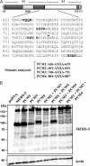
RXXR motifs located at the border between S1 and S2 are required for efficient processing of the Middle East respiratory syndrome coronavirus spike protein (MERS-S). A, The domain organization of the MERS-S protein is schematically depicted. The MERS-S sequence at the border between the S1 and S2 subunits is shown. RXXR motifs, which constitute potential cleavage sites, are highlighted, and the predicted start of the S2 subunit is underlined. The mutations introduced into the potential cleavage sites in MERS-S are shown. B, 293T cells were transfected with expression plasmids coding for MERS-S wild type and the indicated MERS-S mutants equipped with a C-terminal V5 tag. Transfection of empty plasmid (pcDNA) served as negative control. Expression of S proteins in cell lysates was determined by Western blot, using a V5 tag–specific monoclonal antibody. Expression of β-actin in cell lysates was assessed as a loading control. The results shown are representative for at least 3 independent experiments. Abbreviations: CT, cytoplasmic tail; PCM, potential cleavage site mutant; RBD, receptor binding domain; SP, signal peptide; TM, transmembrane domain.
Figure 3.
Figure 3.
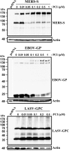
The Middle East respiratory syndrome coronavirus spike protein (MERS-S) is cleaved by proprotein convertases. 293T cells were transfected with expression plasmids encoding MERS-S, Zaire ebolavirus glycoprotein (EBOV-GP), or Lassa virus glycoprotein (LASV-GPC), all equipped with a C-terminal V5 tag. Cells transfected with empty plasmid (pcDNA) served as negative control. Subsequently, cells were incubated with the indicated concentrations of the proprotein convertase inhibitor (PCI). At 48 hours after transfection, glycoprotein expression was analyzed by Western blot, using a V5 tag–specific monoclonal antibody. Detection of β-actin served as loading control. The results shown are representative of three independent experiments.
Figure 4.
Figure 4.
Proprotein convertase activity is dispensable for Middle East respiratory syndrome coronavirus spike protein (MERS-S)–driven cell-cell and virus-cell fusion. A, Fusion of 293T effector cells transfected to express MERS-S with target cells transfected to express DPP4 and/or TMPRSS2 or control transfected with empty plasmid (pcDNA) was assessed. Both effector and target cells were incubated with 1 µM of proprotein convertase inhibitor (PCI) as indicated and, 1 day later, were mixed for cocultivation. The effector/target cell mixtures were incubated with phosphate-buffered saline (PBS) or PCI, and cell-cell fusion was quantified by determination of luciferase activities in cell lysates. The results of a representative experiment performed with triplicate samples are shown. Error bars indicate standard deviation (SD). Two separate experiments yielded similar results. B, Lentiviral pseudotypes carrying MERS-S, Lassa virus glycoprotein (LASV-GPC), Zaire ebolavirus glycoprotein (EBOV-GP), or the glycoprotein of vesicular stomatitis virus (VSV-G), as well as pseudotypes bearing no viral glycoprotein (pcDNA), were generated in the presence or absence of PCI (1 µM). Subsequently, target 293T cells transfected with empty plasmid or DPP4 expression plasmid were preincubated with dimethyl sulfoxide (DMSO), PBS, or PCI at a final concentration of 0.5 µM for 30–60 minutes, followed by transduction with the pseudotypes specified above. At 72 hours after transduction, luciferase activities in cell lysates were measured. The average of 5–7 independent experiments performed with triplicate samples is shown. Transduction with pseudotypes bearing VSV-G in the absence of inhibitor was set as 100%. Error bars indicate standard error of the mean (SEM). A 2-tailed Student t test was used to assess statistical significance.
Figure 5.
Figure 5.
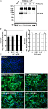
Processing of Middle East respiratory syndrome coronavirus (MERS-CoV) spike protein (MERS-S) by proprotein convertases is dispensable for MERS-CoV infectivity. Caco-2 cells were incubated with the indicated concentrations of proprotein convertase inhibitor (PCI) for 1 hour and then inoculated with MERS-CoV at a multiplicity of infection (MOI) of 0.01 and 0.001 for 30 minutes at 4°C. Thereafter, virus was removed, inhibitor was replenished, and cells were lysed 24 hours after infection for S protein detection by Western blot analysis (A, shown only for the MOI of 0.01). In parallel, viral genomic RNA copies (B, left panel) or infectious MERS-CoV particles (B, right panel) in supernatants of MERS-CoV–infected Caco-2 cells (with and without PCI treatment) were determined by quantitative reverse-transcription polymerase chain reaction (upE assay) or plaque assay. The experiment was performed in quadruplicates and repeated twice with 2 different MOIs, giving comparable results. Shown is 1 representative experiment (MOI, 0.001). C, Cytopathogenic effects of MERS-CoV (MOI, 0.1) infected Vero B4 cells 42 hours after infection in the presence and absence of PCI. Viral antigen was detected with a specimen from a patient with MERS (diluted 1:100), followed by a cyanine 2–labeled goat-anti human immunoglobulin G. Nuclei were stained with DAPI-containing mounting medium. Samples were analyzed by immunofluorescence microscopy (Zeiss), and pictures were taken at the same microscopic settings at a magnification of 200×. Similar results were obtained in 2 independent experiments performed with duplicate samples.

References

    1. Graham RL, Donaldson EF, Baric RS. A decade after SARS: strategies for controlling emerging coronaviruses. Nat Rev Microbiol. 2013;11:836–48. - PMC - PubMed
    1. Hilgenfeld R, Peiris M. From SARS to MERS: 10 years of research on highly pathogenic human coronaviruses. Antiviral Res. 2013;100:286–95. - PMC - PubMed
    1. Drexler JF, Corman VM, Drosten C. Ecology, evolution and classification of bat coronaviruses in the aftermath of SARS. Antiviral Res. 2014;101:45–56. - PMC - PubMed
    1. Ge XY, Li JL, Yang XL, et al. Isolation and characterization of a bat SARS-like coronavirus that uses the ACE2 receptor. Nature. 2013;503:535–8. - PMC - PubMed
    1. Zaki AM, van Boheemen S, Bestebroer TM, Osterhaus AD, Fouchier RA. Isolation of a novel coronavirus from a man with pneumonia in Saudi Arabia. N Engl J Med. 2012;367:1814–20. - PubMed

Publication types

MeSH terms