Case Reports
. 2015 Apr 15;211(8):1241-50.
doi: 10.1093/infdis/jiu412. Epub 2014 Jul 23.

Inherited CARD9 deficiency in 2 unrelated patients with invasive Exophiala infection

Affiliations
Case Reports

Inherited CARD9 deficiency in 2 unrelated patients with invasive Exophiala infection

Fanny Lanternier et al. J Infect Dis. .

Abstract

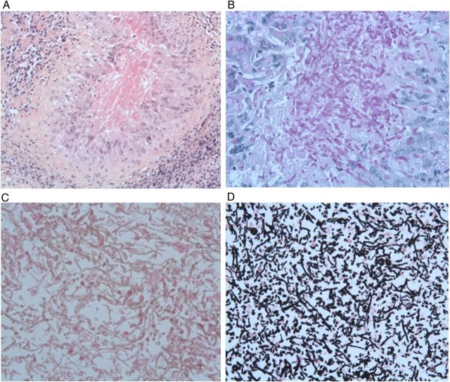
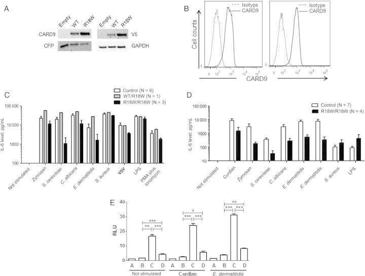
Background: Exophiala species are mostly responsible for skin infections. Invasive Exophiala dermatitidis disease is a rare and frequently fatal infection, with 42 cases reported. About half of these cases had no known risk factors. Similarly, invasive Exophiala spinifera disease is extremely rare, with only 3 cases reported, all in patients with no known immunodeficiency. Autosomal recessive CARD9 deficiency has recently been reported in otherwise healthy patients with severe fungal diseases caused by Candida species, dermatophytes, or Phialophora verrucosa.

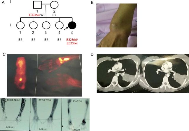
Methods: We investigated an 8-year-old girl from a nonconsanguineous Angolan kindred, who was born in France and developed disseminated E. dermatitidis disease and a 26 year-old woman from an Iranian consaguineous kindred, who was living in Iran and developed disseminated E. spinifera disease. Both patients were otherwise healthy.

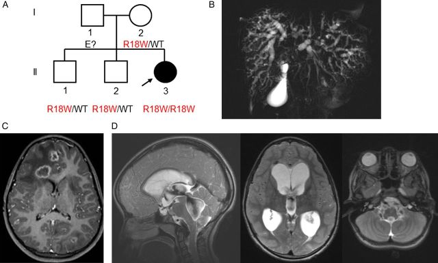
Results: We sequenced CARD9 and found both patients to be homozygous for loss-of-function mutations (R18W and E323del). The first patient had segmental uniparental disomy of chromosome 9, carrying 2 copies of the maternal CARD9 mutated allele.

Conclusions: These are the first 2 patients with inherited CARD9 deficiency and invasive Exophiala disease to be described. CARD9 deficiency should thus be considered in patients with unexplained invasive Exophiala species disease, even in the absence of other infections.

Keywords: Exophiala species; autosomal recessive CARD9 deficiency; central nervous system; invasive fungal infection; osteomyelitis; parental unidisomy.


Figures

Figure 1.
Figure 1.
A, Pedigree of the patient with Exophiala dermatitidis infection and a CARD9 mutation. Each generation is represented by a Roman numeral, and each individual is represented by an Arabic numeral. The proband with E. dermatitidis infection is shown in black and indicated by an arrow. The CARD9 genotype is indicated below each individual. B–D, Radiological features of patient 1. B, Cholangio-magnetic resonance imaging showing a dilated and irregular biliary tree with diffuse biliary duct infiltration. C, Brain magnetic resonance imaging (MRI) showing 13 cerebral lesions (diameter, 10–16 mm). D, Brain MRI showing pachymeningitis and hydrocephalus. Abbreviation: WT, wild-type.
Figure 2.
Figure 2.
A–D, Histological features of the meningeal biopsy specimen. A, Hematoxylin-eosin (H-E) staining showing fungi surrounded by an epithelioid and giant cell granuloma (original magnification ×200). B, Strong periodic acid-Schiff staining of the fungi within the granuloma (original magnification ×400). C, H-E staining revealing a huge infiltration of fungi without granuloma in another area of the biopsy sample (original magnification ×400). D, Grocott staining of this large area of fungi (original magnification ×400).
Figure 3.
Figure 3.
Impact of the CARD9 R18W mutation on protein level and function. A, Immunoblot analysis of CARD9 in whole-cell extracts of HEK-293T cells cotransfected with pcDNA3.1 V5 (C-terminally tagged), either empty or carrying the wild-type (WT) or mutant (R18W) CARD9 allele, together with a CFP plasmid, as a transfection control. Antibodies against CARD9, V5, CFP, and GAPDH (as a loading control) were used. B, Flow cytometry analysis of CARD9 in monocyte-derived macrophages from a control (left panel) or the patient (right panel). C, Interleukin 6 (IL-6) production by whole-blood cells, as measured by enzyme-linked immunosorbent assay (ELISA), after 24 hours of stimulation with zymosan (5 µg/mL), heat-killed Saccharomyces cerevisiae (106 particles/mL), heat-killed Candida albicans (106 particles/mL), heat-killed Exophiala dermatitidis (106 particles/mL), heat-killed Staphylococcus aureus (5 × 10⁸ particles/mL), vesicular stomatitis virus (VSV), lipopolysaccharide (LPS; 100 ng/mL), and PMA plus ionomycin for 6 controls, the CARD9-heterozygous (R18W/WT) mother, and the CARD9-homozygous (R18W/R18W) patient. Results are expressed as mean ± standard error of the mean (SEM) of 3 independent experiments. D, Tumor necrosis factor α (TNF-α) production by monocyte-derived dendritic cells, measured by ELISA, after 24 hours of stimulation with curdlan (25 µg/mL), zymosan (25 µg/mL), heat-killed S. cerevisiae (106 particles/mL), heat-killed C. albicans (106 particles/mL), heat-killed E. dermatitidis (107 particles/mL and 106 particles/mL), heat-killed S. aureus (5 × 10⁸ particles/mL), and LPS (100 ng/mL) for 7 controls and the CARD9-homozygous (R18W/R18W) patient. Results are expressed as mean ± SEM of 4 independent experiments. E, The CARD9 R18W allele impairs downstream NF-κB activation. NF-κB–luciferase assay in 293 HEK cells transfected with NF-κB–luciferase and pRL-SV40 vectors alone (A); with DECTIN1, SYK, and BCL10 constructs (B); with DECTIN-1, SYK, BCL10, and CARD9 WT constructs (C); and with DECTIN-1, SYK, BCL10, and CARD9 R18W constructs (D). Cells were stimulated or not stimulated with 25 µg/mL curdlan or 107 particles/mL E. dermatitidis. Results are representative of 2 independent experiments performed and are expressed as mean ± SEM of the ratio between Renilla luciferase and firefly control luciferase activities adjusted to 1. *P ≤ .05, **P ≤ .01, and ***P ≤ .001 by the Student t test. Abbreviation: RLU, relative light units.
Figure 4.
Figure 4.
A, Pedigree of the patient with Exophiala spinifera infection and a CARD9 mutation. Each generation is represented by a Roman numeral, and each individual is represented by an Arabic numeral. The proband with E. spinifera infection is shown in black and indicated by an arrow. The CARD9 genotype is indicated below the individuals sequenced. B–D, Clinical and radiological features of the patient. B, Subcutaneous skin infection. C, Hyperfixation revealing osteomyelitis on scintigraphy. D, Computed tomography of the chest. Abbreviation: WT, wild type.

Comment in

References

    1. Sudhadham M, Prakitsin S, Sivichai S, et al. The neurotropic black yeast Exophiala dermatitidis has a possible origin in the tropical rain forest. Stud Mycol 2008; 61:145–55. - PMC - PubMed
    1. Zeng JS, Sutton DA, Fothergill AW, Rinaldi MG, Harrak MJ, de Hoog GS. Spectrum of clinically relevant Exophiala species in the United States. J Clin Microbiol 2007; 45:3713–20. - PMC - PubMed
    1. Horre R, Schaal KP, Siekmeier R, Sterzik B, de Hoog GS, Schnitzler N. Isolation of fungi, especially Exophiala dermatitidis, in patients suffering from cystic fibrosis. A prospective study. Respiration 2004; 71:360–6. - PubMed
    1. de Hoog GS, Matos T, Sudhadham M, Luijsterburg KF, Haase G. Intestinal prevalence of the neurotropic black yeast Exophiala (Wangiella) dermatitidis in healthy and impaired individuals. Mycoses 2005; 48:142–5. - PubMed
    1. Patel AK, Patel KK, Darji P, Singh R, Shivaprakash MR, Chakrabarti A. Exophiala dermatitidis endocarditis on native aortic valve in a postrenal transplant patient and review of literature on E. dermatitidis infections. Mycoses 2013; 56:365–72. - PubMed

Publication types

Substances