. 2014 Jul 23;34(30):9982-94.
doi: 10.1523/JNEUROSCI.0933-14.2014.

Developmental switch of leptin signaling in arcuate nucleus neurons

Affiliations

Developmental switch of leptin signaling in arcuate nucleus neurons

Arian F Baquero et al. J Neurosci. .

Abstract

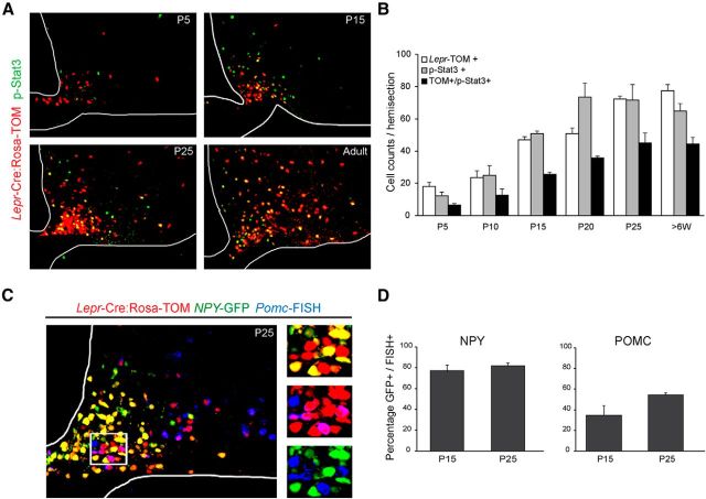
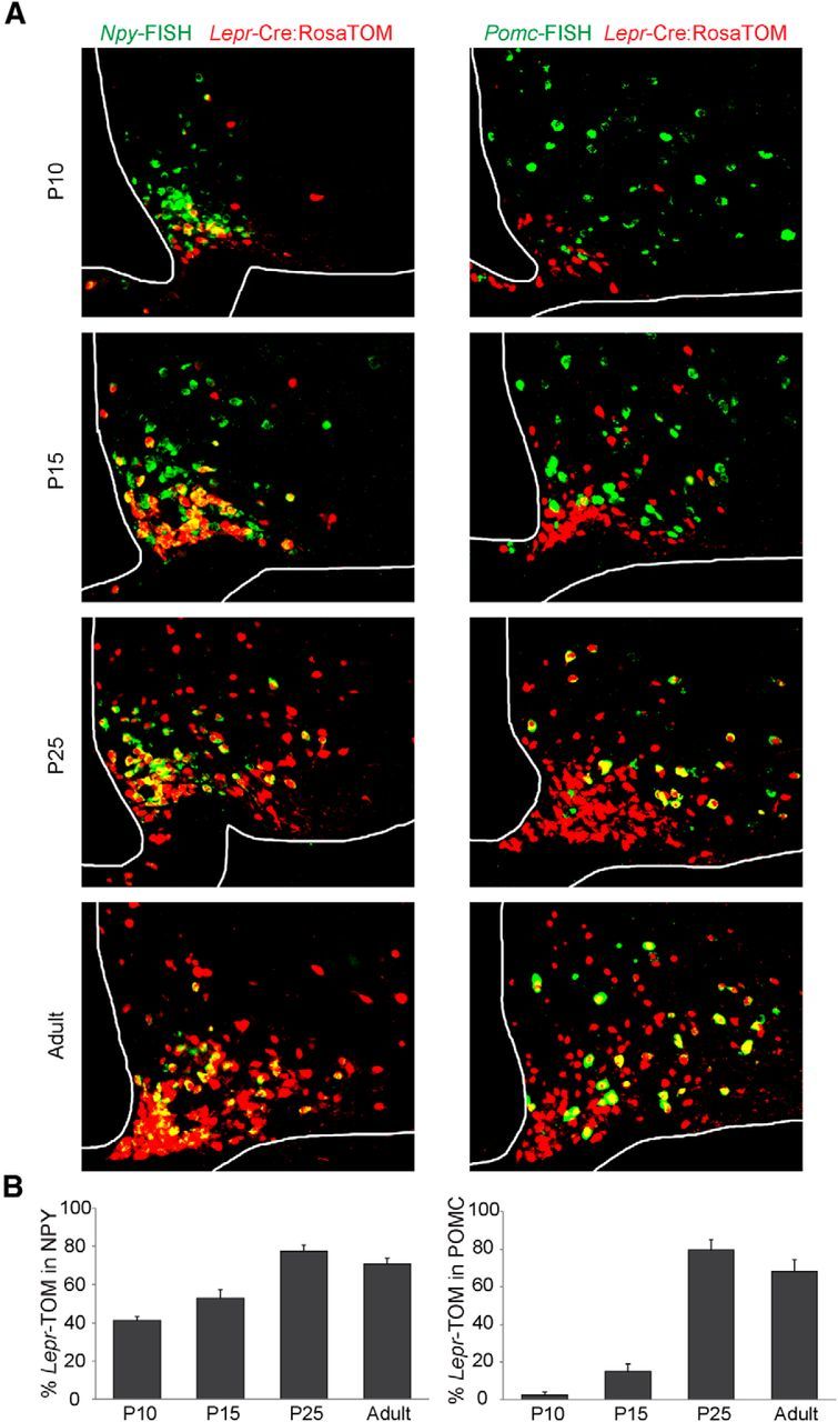
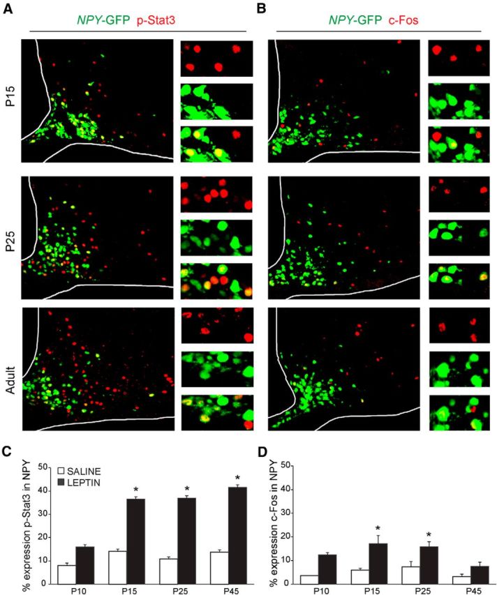
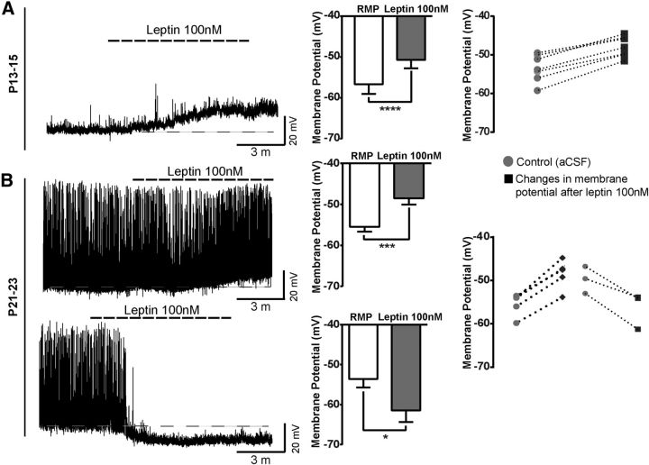
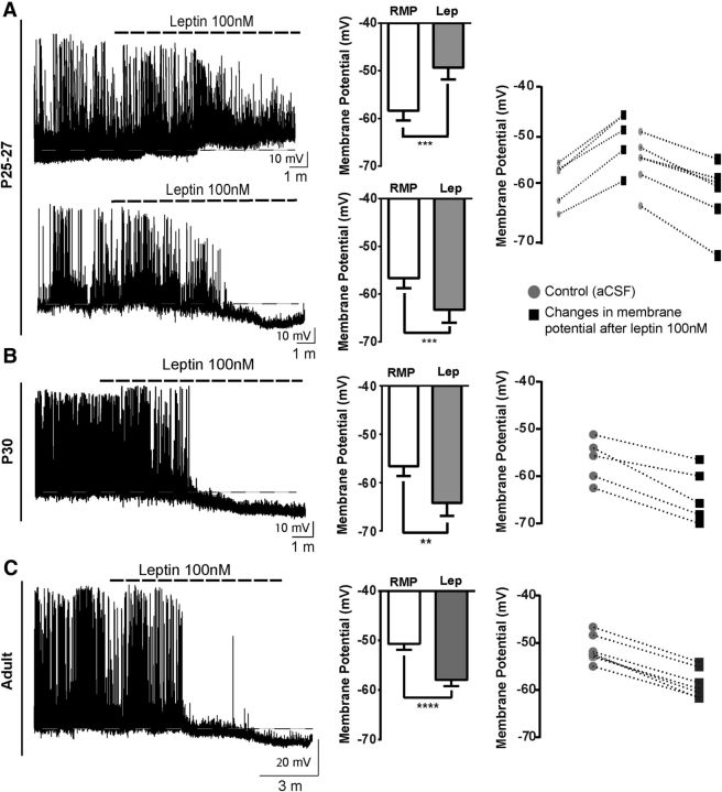
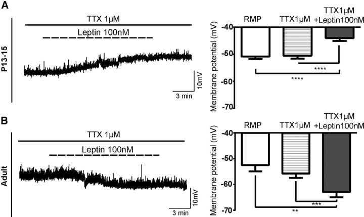
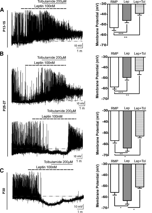
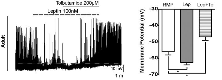
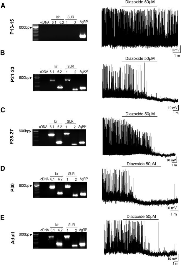
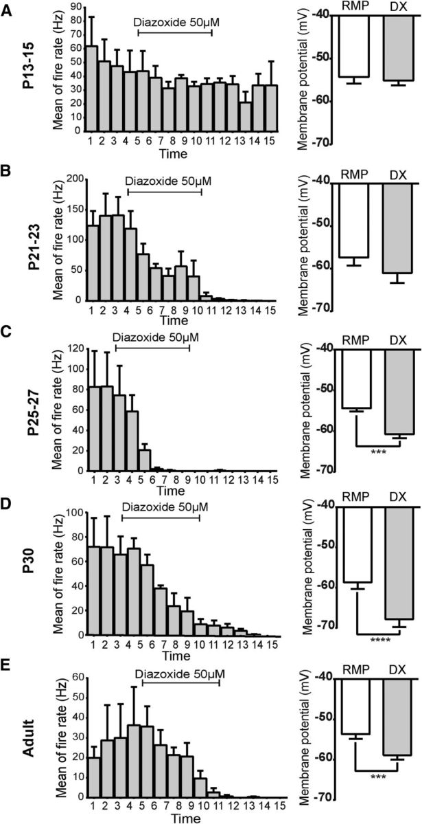
Leptin is well known for its role in the regulation of energy homeostasis in adults, a mechanism that at least partially results from the inhibition of the activity of NPY/AgRP/GABA neurons (NAG) in the arcuate nucleus of the hypothalamus (ARH). During early postnatal development in the rodent, leptin promotes axonal outgrowth from ARH neurons, and preautonomic NAG neurons are particularly responsive to leptin's trophic effects. To begin to understand how leptin could simultaneously promote axonal outgrowth from and inhibit the activity of NAG neurons, we characterized the electrochemical effects of leptin on NAG neurons in mice during early development. Here, we show that NAG neurons do indeed express a functional leptin receptor throughout the early postnatal period in the mouse; however, at postnatal days 13-15, leptin causes membrane depolarization in NAG neurons, rather than the expected hyperpolarization. Leptin action on NAG neurons transitions from stimulatory to inhibitory in the periweaning period, in parallel with the acquisition of functional ATP-sensitive potassium channels. These findings are consistent with the idea that leptin provides an orexigenic drive through the NAG system to help rapidly growing pups meet their energy requirements.

Keywords: KATP channels; NPY; arcuate nucleus; development; leptin; mouse.


Figures

Figure 1.
Figure 1.
Comparison of reagents used to characterize leptin-sensitive neurons in the ARH during postnatal development. A, Colocalization (yellow) of Lepr-Cre;Rosa-TOM (red) and leptin-induced p-Stat3+ immunoreactivity (green) in the ARH. B, Quantification of ARH neurons that are TOM and/or p-Stat3-positive. C, Representative image of NPY-GFP+ (green), Lepr-Cre;Rosa-TOM+ (red), and Pomc-FISH+ (blue) neurons at P25. Inset, Colocalizations between NPY-GFP+ and Lepr-Cre;Rosa-TOM+ (yellow), Pomc-FISH+ and Lepr-Cre;Rosa-TOM+ (magenta). There is no colocalization between NPY-GFP+ (green) and Pomc-FISH+ (blue). D, Quantification of colocalization of NPY-GFP and NPY-FISH signals (left) and POMC-GFP and Pomc-FISH signals in the postnatal period on NPY-GFP and POMC-GFP mice, respectively. Error bars indicate mean ± SEM, 4–6 sections per animal from 3 or 4 mice per age.
Figure 2.
Figure 2.
Ontogeny of leptin-sensing neurons in ARH. A, Representative images showing colocalization (yellow) of Npy-FISH (green, left panels) and Pomc-FISH (green, right panels) in ARH Lepr-TOM+ neurons (red). B, Quantification of the percentage of NPY+ (left) and Pomc+ (right) neurons that coexpress Lepr-Cre;Rosa-TOM from P5 through adult. Error bars indicate mean ± SEM; n = 3–6 sections per animal from 3 or 4 mice per age.
Figure 3.
Figure 3.
Exogenous leptin activates NAG neurons during development. Representative images of leptin-induced p-Stat3 (red) (A) and c-Fos (red) (B) expression in NPY-GFP+ neurons (green). Insets, Colocalization. Quantification of p-Stat3 (C) and c-Fos (D) immunoreactivity in ARH NPY-GFP+ neurons after intraperitoneal injection of saline or leptin (4 mg/kg) at several ages. Error bars indicate mean ± SEM from 4–6 sections per animal, 3 or 4 mice per age. *p < 0.05 (two-way ANOVA, post hoc Bonferroni correction).
Figure 4.
Figure 4.
Leptin activates NAG neurons during postnatal development. Representative traces from brain slices from NPYhrGFP mouse containing ARH. A, Neurons at P13–P15 showed membrane depolarization in the presence of leptin 100 nm; 17 of 31 neurons from 26 animals responded to leptin treatment. B, Leptin 100 nm has dual actions in the membrane potential of neurons between P21 and P23 either leads to depolarization or hyperpolarization. Leptin treatment leads to membrane depolarization in 5 of 12 neurons from 12 animals. In contrast, 3 of 12 neurons were inhibited by leptin. Black-white lines indicate the RMP (resting membrane potential). Bar graphs represent the magnitude of leptin responses in neurons for each age. Dashed line graphs represent changes in membrane potential for individual neurons that were sensitive to leptin 100 nm at each age tested. Results are mean ± SEM. *p < 0.05 (paired t test). ***p < 0.001 (paired t test). ****p < 0.0001 (paired t test).
Figure 5.
Figure 5.
NAG neurons exhibit an adult-like phenotype in the presence of leptin after postnatal day 30. Representative traces from brain slices from NPY-hrGFP mice containing ARH. A, Leptin 100 nm leads to depolarization in 5 of 19 neurons and hyperpolarization in 6 of 19 neurons from 12 animals at P25–P27. B, Leptin causes membrane hyperpolarization and inhibition of spontaneous action potentials in 5 of 8 neurons from 4 animals at P30. C, Leptin 100 nm causes membrane hyperpolarization in 12 of 21 adult neurons from 10 animals. Bar graphs represent the magnitude of leptin responses in neurons for each age. Line graphs represent changes in membrane potential for individual neurons that were sensitive to leptin 100 nm at each age tested. Results are mean ± SEM. **p < 0.01 (paired t test). ***p < 0.001 (paired t test). ****p < 0.0001 (paired t test).
Figure 6.
Figure 6.
Leptin effects, at least partially, are directly induced in NAG neurons. Representative traces of leptin effects on NAG neurons in the presence of TTX (1 μm). A, Leptin 100 nm leads to membrane depolarization in 8 of 18 neurons from 18 animals at P13–P15 under the presence of TTX (1 μm). B, Leptin 100 nm induces membrane hyperpolarization in 5 of 11 adult neurons in the presence of TTX (1 μm, 7 animals). Bar graphs represent the magnitude of the leptin-induced responses in either pup or adult neurons. Results are mean ± SEM. ****p < 0.0001 (ANOVA, post hoc Tukey's test). ***p < 0.001 (ANOVA, post hoc Tukey's test).
Figure 7.
Figure 7.
Differences in leptin signaling in NAG neurons during postnatal development. A, Tracing from a P13–P15 neuron showing that leptin-induced depolarization was not changed by a KATP channel blocker (tolbutamide). Leptin induces membrane depolarization in 7 of 12 neurons from 8 animals. B, Leptin effects on neurons were excitatory in 6 of 19 neurons or inhibitory in 5 of 19 neurons at this age (12 animals). Tolbutamide 200 μm completely reversed leptin-induced hyperpolarization. C, Leptin-mediated hyperpolarization at P30 was reverted by tolbutamide treatment. Four of 7 neurons from 4 animals responded to leptin treatment. Dashed line indicates the RMP. Bar graphs represent the magnitude of leptin responses in the absence and presence of tolbutamide. Results are mean ± SEM. *p < 0.05 (ANOVA, post hoc Tukey's). **p < 0.01(ANOVA, post hoc Tukey's). ***p < 0.001 (ANOVA, post hoc Tukey's). Lep, Leptin; Lep+Tol, leptin + tolbutamide.
Figure 8.
Figure 8.
Leptin effects in adult mouse NAG neurons are blocked by tolbutamide. Representative trace from an adult neuron showing that leptin-mediated hyperpolarization was blocked by tolbutamide treatment. Three of 7 neurons from 3 animals responded to leptin. Dashed line indicates the RMP. Bar graphs represent the magnitude of leptin responses in the absence and presence of tolbutamide. Results are mean ± SEM. *p < 0.05 (ANOVA, post hoc Tukey's test). Lep, Leptin; Lep+Tol, leptin + tolbutamide.
Figure 9.
Figure 9.
Development of KATP channels in NAG neurons. A–E, RT-PCR mRNA expression for inward rectifying potassium channels subunits Kir6.1 and Kir6.2, the sulfonylurea receptor subunits 1 and 2 (SUR1 and SUR2), and AgRP, in ARH of NPY-hrGFP mice at the following ages: P13–P15, P21–P23, P25–P27, P30, and adult (left). Amplify-stained PCR products of expected sizes (Kir6.1 448 bp, Kir 6.2 256 bp, SUR1 475 bp, SUR2 215 bp, and AgRP 247 bp). −cDNA indicates those in which cDNA was omitted from the PCR. ARH micro-punches were pooled from 4 animals for each age. These experiments were replicated three different times (60 animals). Representative traces from individual age matched neurons in current-clamp mode showing the effects of diazoxide 50 μm (right).
Figure 10.
Figure 10.
Functional characterization of KATP channels in NAG neurons during postnatal development. A, Diazoxide 50 μm was applied for 6 min and did not change the mean of fire rate or the membrane potential during the entire application in 6 neurons from 6 animals at P13–P15. B–E, Diazoxide-induced responses decreased the fire rate and hyperpolarized the membrane potential of neurons at P21–P23 (5 of 9 neurons, 2 animals), P25–P27 (5 of 7 neurons, 3 animals), P30 (7 neurons, 2 animals), and adults (6 neurons, 3 animals). Bar graphs represent the magnitude of the diazoxide-dependent hyperpolarization in neurons for each age. Results are mean ± SEM. ***p < 0.001 (paired t test). ****p < 0.0001 (paired t test). DX, Diazoxide.

References

    1. Ahima RS, Hileman SM. Postnatal regulation of hypothalamic neuropeptide expression by leptin: implications for energy balance and body weight regulation. Regul Pept. 2000;92:1–7. doi: 10.1016/S0167-0115(00)00142-7. - DOI - PubMed
    1. Ahima RS, Prabakaran D, Flier JS. Postnatal leptin surge and regulation of circadian rhythm of leptin by feeding: implications for energy homeostasis and neuroendocrine function. J Clin Invest. 1998;101:1020–1027. doi: 10.1172/JCI1176. - DOI - PMC - PubMed
    1. Aponte Y, Atasoy D, Sternson SM. AGRP neurons are sufficient to orchestrate feeding behavior rapidly and without training. Nat Neurosci. 2011;14:351–355. doi: 10.1038/nn.2739. - DOI - PMC - PubMed
    1. Ashford ML, Boden PR, Treherne JM. Glucose-induced excitation of hypothalamic neurones is mediated by ATP-sensitive K+ channels. Pflugers Arch. 1990;415:479–483. doi: 10.1007/BF00373626. - DOI - PubMed
    1. Atasoy D, Betley JN, Su HH, Sternson SM. Deconstruction of a neural circuit for hunger. Nature. 2012;488:172–177. doi: 10.1038/nature11270. - DOI - PMC - PubMed

Publication types