. 2014 Jul 23;34(30):10034-40.
doi: 10.1523/JNEUROSCI.0715-14.2014.

Mutant huntingtin affects cortical progenitor cell division and development of the mouse neocortex

Affiliations

Mutant huntingtin affects cortical progenitor cell division and development of the mouse neocortex

Maria Molina-Calavita et al. J Neurosci. .

Abstract

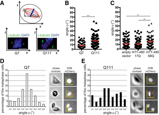
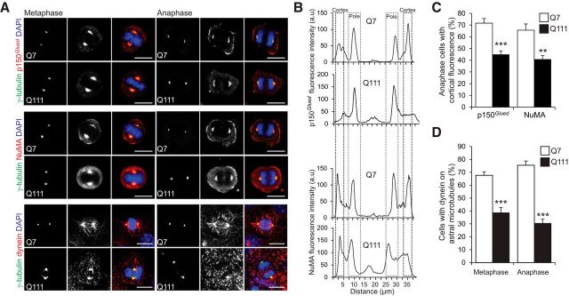
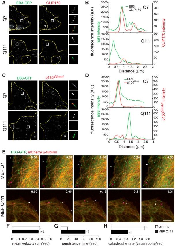
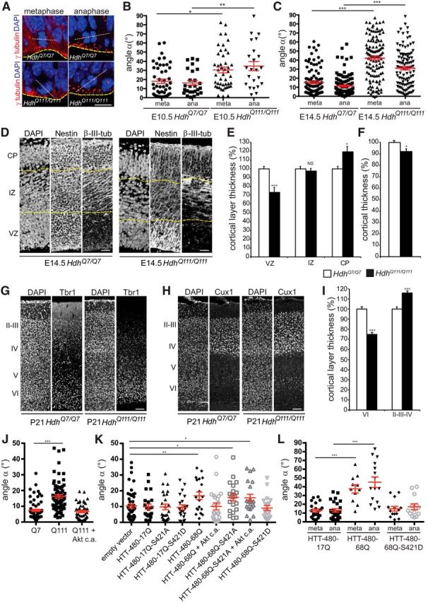
A polyglutamine expansion in huntingtin (HTT) causes the specific death of adult neurons in Huntington's disease (HD). Most studies have thus focused on mutant HTT (mHTT) toxicity in adulthood, and its developmental effects have been largely overlooked. We found that mHTT caused mitotic spindle misorientation in cultured cells by altering the localization of dynein, NuMA, and the p150(Glued) subunit of dynactin to the spindle pole and cell cortex and of CLIP170 and p150(Glued) to microtubule plus-ends. mHTT also affected spindle orientation in dividing mouse cortical progenitors, altering the thickness of the developing cortex. The serine/threonine kinase Akt, which regulates HTT function, rescued the spindle misorientation caused by the mHTT, by serine 421 (S421) phosphorylation, in cultured cells and in mice. Thus, cortical development is affected in HD, and this early defect can be rescued by HTT phosphorylation at S421.

Keywords: Huntington disease; cortical neurogenesis; mitosis; spindle orientation.


Figures

Figure 1.
Figure 1.
mHTT causes spindle misorientation. A, Measurement of the spindle angle α. STHdhQ7/Q7 (Q7) and STHdhQ111/Q111 (Q111) cells stained as indicated. B, C, Spindle angles of Q7, Q111 (B), and HeLa (C) cells. D, E, Spindle angles of Q7 (D) and Q111 (E) cells grown on micropatterns. Time-lapse pictures were used to measure the positioning of the metaphasic plate (H2B-mCherry). Scale bars, 10 μm. Error bars indicate SEM. **p < 0.01 (unpaired, one-tailed t test). ***p < 0.001 (unpaired, one-tailed t test).
Figure 2.
Figure 2.
mHTT alters the mitotic distribution of p150Glued, NuMA, and Dynein. A, STHdhQ7/Q7 (Q7) and STHdhQ111/Q111 (Q111) cells were stained as indicated. Scale bars, 10 μm. B, Representative line scan analysis showing the distribution of p150Glued and NuMA. C, Percentage of cells with cortical labeling of p150Glued and NuMA. D, Percentage of Q7 and Q111 cells with dynein on astral microtubules. C, D, At least n = 25 cells in three independent experiments. Error bars indicate SEM. **p < 0.01 (unpaired, one-tailed t test). ***p < 0.001 (unpaired, one-tailed t test).
Figure 3.
Figure 3.
+TIPs localization at microtubules plus-ends and microtubule dynamics are modified in HD. A, C, Immunostainings of EB3-GFP transfected STHdhQ7/Q7 (Q7) and STHdhQ111/Q111 (Q111) cells with anti-CLIP170 (A) and anti-p150Glued (C) antibodies. N, nucleus. B, D, Representative line scan analysis of fluorescence intensity of CLIP170 (B) or p150Glued (D), and EB3, starting at the end of the MT to 3 μm inwards. E, Time-lapse recordings illustrating the MT dynamic instability in HdhQ7/Q7 (MEF Q7) and HdhQ111/Q111 (MEF Q111) MEFs expressing mCherry-α-tubulin and EB3-GFP. Arrowheads indicate localization of the MT tip close to the leading edge. Time is expressed as min:sec. F–H, MT dynamics in Q7 and Q111 MEFs (at least 5 MTs examined in n = 10 cells in two independent experiments). F, Mean velocity of EB3-comets. G, Persistence time. H, MT catastrophe rate. Scale bars, 10 μm. Error bars indicate SEM. NS, Not significant. **p < 0.01 (unpaired, one-tailed t test). ***p < 0.001 (unpaired, one-tailed t test).
Figure 4.
Figure 4.
Spindle orientation of dividing progenitors and cerebral cortex thickness are altered in HdhQ111/Q111 embryos. A, Illustration of dividing progenitors. White dashed line indicates spindle; yellow line indicates ventricular surface. γ-tubulin marks centrosomes. Scale bar, 15 μm. B, C, Spindle angles during divisions in E10.5 (B) and E14.5 (C) embryos at metaphase (meta) and anaphase (ana). D, Nestin and β-III-tubulin immunostainings of E14.5 coronal sections. Scale bars, 50 μm. E, F, VZ, IZ, CP (E) and overall cortical (F) thicknesses (n = 3 embryos per genotype). G, H, Tbr1 (G) and Cux1 (H) immunostainings of P21 coronal sections. Scale bars, 100 μm. I, Layers VI and II-IV thicknesses (n = 3 mice per genotype). J, K, Spindle angles of STHdhQ7/Q7 (Q7), STHdhQ111/Q111 (Q111), and STHdhQ111/Q111 cells transfected with Akt c.a. (J) and of HeLa cells transfected with the indicated constructs (K). L, Spindle angles in E16.5 electroporated progenitors. Error bars indicate SEM. NS, Not significant. *p < 0.05 (unpaired, one-tailed t test). **p < 0.01 (unpaired, one-tailed t test). ***p < 0.001 (unpaired, one-tailed t test).

References

    1. Ben M'barek K, Pla P, Orvoen S, Benstaali C, Godin JD, Gardier AM, Saudou F, David DJ, Humbert S. Huntingtin mediates anxiety/depression-related behaviors and hippocampal neurogenesis. J Neurosci. 2013;33:8608–8620. doi: 10.1523/JNEUROSCI.5110-12.2013. - DOI - PMC - PubMed
    1. Caviston JP, Ross JL, Antony SM, Tokito M, Holzbaur EL. Huntingtin facilitates dynein/dynactin-mediated vesicle transport. Proc Natl Acad Sci U S A. 2007;104:10045–10050. doi: 10.1073/pnas.0610628104. - DOI - PMC - PubMed
    1. Coquelle FM, Caspi M, Cordelières FP, Dompierre JP, Dujardin DL, Koifman C, Martin P, Hoogenraad CC, Akhmanova A, Galjart N, De Mey JR, Reiner O. LIS1, CLIP-170's key to the dynein/dynactin pathway. Mol Cell Biol. 2002;22:3089–3102. doi: 10.1128/MCB.22.9.3089-3102.2002. - DOI - PMC - PubMed
    1. Dietrich P, Shanmugasundaram R, Shuyu E, Dragatsis I. Congenital hydrocephalus associated with abnormal subcommissural organ in mice lacking huntingtin in Wnt1 cell lineages. Hum Mol Genet. 2009;18:142–150. doi: 10.1093/hmg/ddn324. - DOI - PMC - PubMed
    1. Duyao MP, Auerbach AB, Ryan A, Persichetti F, Barnes GT, McNeil SM, Ge P, Vonsattel JP, Gusella JF, Joyner AL. Inactivation of the mouse Huntington's disease gene homolog Hdh. Science. 1995;269:407–410. doi: 10.1126/science.7618107. - DOI - PubMed

Publication types