. 2014 Jul 24;10(7):e1004467.
doi: 10.1371/journal.pgen.1004467. eCollection 2014 Jul.

MDRL lncRNA regulates the processing of miR-484 primary transcript by targeting miR-361

Affiliations

MDRL lncRNA regulates the processing of miR-484 primary transcript by targeting miR-361

Kun Wang et al. PLoS Genet. .

Abstract

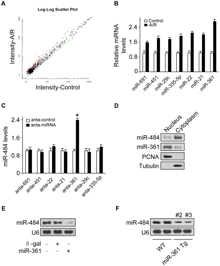
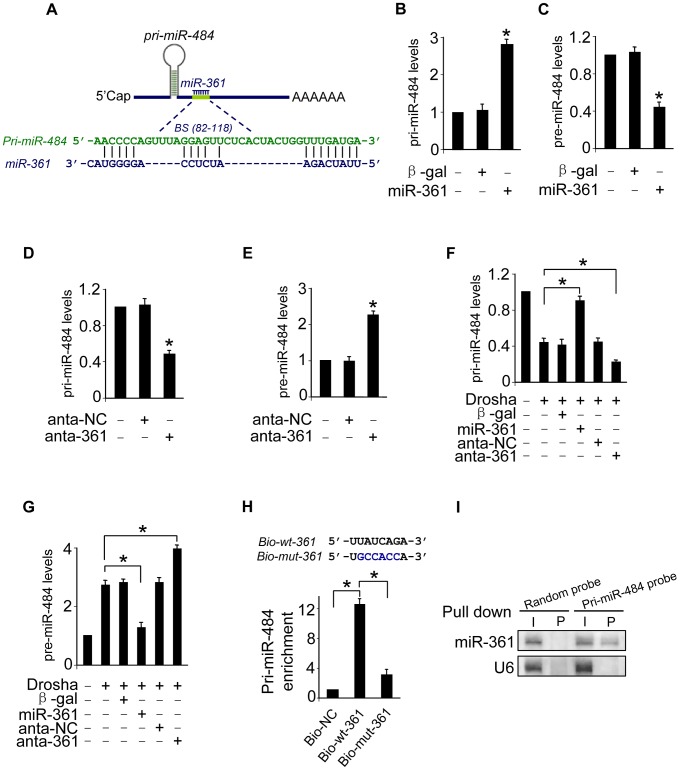
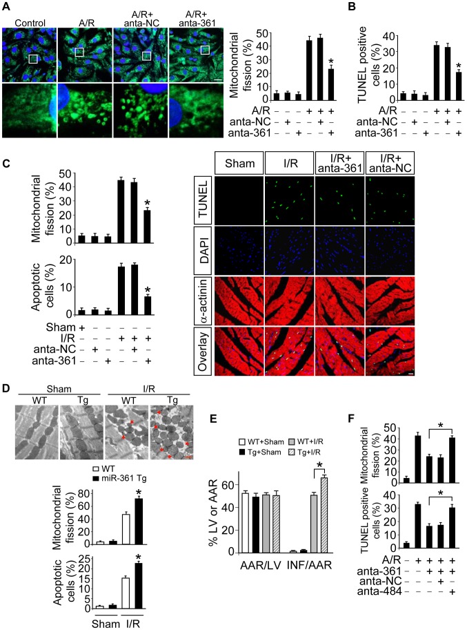
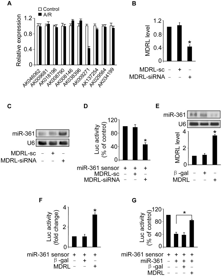
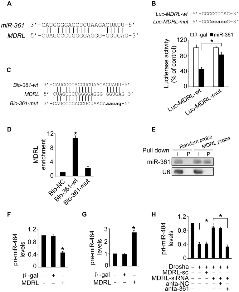
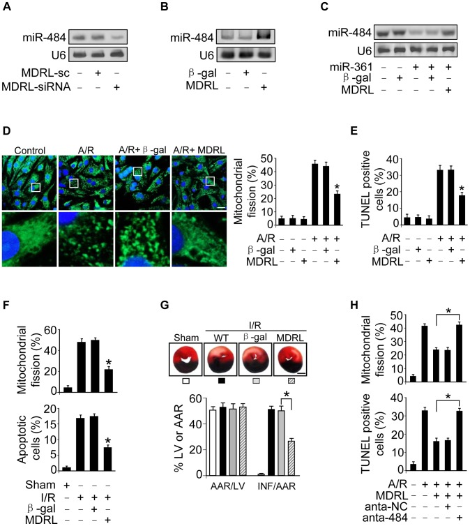
Long noncoding RNAs (lncRNAs) are emerging as new players in gene regulation, but whether lncRNAs operate in the processing of miRNA primary transcript is unclear. Also, whether lncRNAs are involved in the regulation of the mitochondrial network remains to be elucidated. Here, we report that a long noncoding RNA, named mitochondrial dynamic related lncRNA (MDRL), affects the processing of miR-484 primary transcript in nucleus and regulates the mitochondrial network by targeting miR-361 and miR-484. The results showed that miR-361 that predominantly located in nucleus can directly bind to primary transcript of miR-484 (pri-miR-484) and prevent its processing by Drosha into pre-miR-484. miR-361 is able to regulate mitochondrial fission and apoptosis by regulating miR-484 levels. In exploring the underlying molecular mechanism by which miR-361 is regulated, we identified MDRL and demonstrated that it could directly bind to miR-361 and downregulate its expression levels, which promotes the processing of pri-miR-484. MDRL inhibits mitochondrial fission and apoptosis by downregulating miR-361, which in turn relieves inhibition of miR-484 processing by miR-361. Our present study reveals a novel regulating model of mitochondrial fission program which is composed of MDRL, miR-361 and miR-484. Our work not only expands the function of the lncRNA pathway in gene regulation but also establishes a new mechanism for controlling miRNA expression.


Conflict of interest statement

The authors have declared that no competing interests exist.

Figures

Figure 1
Figure 1. miR-361 in the nucleus is able to regulate mature miR-484 levels.
A. Microarray results depicting the log-log scatter plot of intensity of miRNA expression in nuclei from control versus A/R treatment. The neonatal mouse cardiomyocytes were untreated (control) or exposed to A/R. The nuclei were purified and nuclear miRNAs were detected by microarray. The red dots and the green dots indicate 2 fold up- or down-regulated genes, respectively. B. Upregulated nuclear miRNAs upon A/R treatment. C. Knockdown of miR-361 elevates the levels of miR-484. Cardiomyocytes were transfected with indicated miRNA antagomir (anta-miRNA) or the antagomir control (anta-control). 48 h after transfection, miR-484 levels were analyzed by qRT-PCR. *p<0.05 vs anta-control. D. miR-361 predominantly localizes in the cell nucleus and miR-484 predominantly localizes in the cytoplasm. Total RNA was extracted from cardiomyocytes nucleus and cytoplasm. miR-361 and miR-484 levels were analyzed by northern blot. E. Enforced expression of miR-361 reduces the levels of miR-484. Cardiomyocytes were infected with adenoviral miR-361 or β-gal at a moi of 80. 24 h after infection, the expression of miR-484 was analyzed by northern blot. F. miR-361 suppresses the expression of miR-484 in the animal model. miR-484 levels in the hearts of WT and miR-361 transgenic mice were analyzed by northern blot.
Figure 2
Figure 2. miR-361 can directly bind to pri-miR-484 and prevents its processing by Drosha into pre-miR-484.
A. The miR-361 targeting site in pri-miR-484 is shown. B and C. Enforced expression of miR-361 increases the levels of pri-miR-484 and reduces the levels of pre-miR-484. Cardiomyocytes were infected with adenoviral miR-361 or β-gal at indicated time, the expression of pri-miR-484 (B) and pre-miR-484 (C) were analyzed by qRT-PCR. *p<0.05 vs control. D and E. Knockdown of miR-361 induces a reduction in pri-miR-484 levels and an increase in pre-miR-484. Cardiomyocytes were transfected with miR-361 antagomir (anta-361) or the antagomir negative control (anta-NC). 24 h after transfection, the expression of pri-miR-484 (D) and pre-miR-484 (E) were analyzed by qRT-PCR. *p<0.05 vs control. F and G. miR-361 prevents the processing of pri-miR-484 by Drosha into pre-miR-484. Cardiomyocytes were coinfected with the adenoviral Drosha and miR-361, transfected with anta-361 or anta-NC. 48 h after transfection, cells were harvested. pri-miR-484 (F) and pre-miR-484 (G) were analyzed by qRT-PCR. *p<0.05. H. miR-361 can directly bind to pri-miR-484 in vivo. Cardiomyocytes were transfected with biotinylated wild type miR-361 (Bio-wt-361), biotinylated mutant miR-361 (Bio-mut-361) and biotinylated negative control (Bio-NC). 48 h after transfection, cells were harvested for biotin-based pull-down assay. Only “seed” region of wt and mutant miR-361 were shown (upper panel). Pri-miR-484 were analyzed by qRT-PCR (low panel). *p<0.05. I. miR-361 is associated with pri-miR-484 in vivo. pri-miR-484 probe-coated magnetic bead was incubated with cardiomyocyte nulear lysate. After washing and enrichment of beads/RNA complex, RNA was eluted from the streptavidin beads and was analyzed by northern blot. I, input (10% samples were loaded); P, pellet (100% samples were loaded).
Figure 3
Figure 3. miR-361 provokes mitochondrial fission program.
A. Knockdown of miR-361 prevents mitochondrial fission induced by A/R. Cardiomyocytes were transfected with miR-361 antagomir (anta-361) or the antagomir negative control (anta-NC), then exposed to A/R. The cells were stained with MitoTracker Green (left panel), Bar = 20 µm. The cells with fragmented mitochondria were counted (right panel). *p<0.05 versus A/R alone. B. Knockdown of miR-361 prevents apoptosis induced by A/R. Cardiomyocytes were treated as described for (A). Apoptosis was analyzed by TUNEL assay. *p<0.05 versus A/R alone. C. Knockdown of miR-361 attenuates mitochondrial fission upon I/R. Adult male C57BL/6 mice (8 weeks old) were delivered in three consecutive days, intravenous injections of miR-361 antagomir (anta-361) or antagomir control (anta-NC) at doses of 35 mg/kg body weight. 3 days after injection the mice were exposed to 45 min of ischemia and 3 hours of reperfusion. Counting of the fragmented mitochondria was shown in upper panel. Counting of the apoptotic cell was shown in low panel and right panel. TUNEL-positive myocyte nuclei (apoptotic cells) are green. Nuclei stained by DAPI show blue. Cardiomyocytes were labeled with α-actinin. n = 6, *p<0.05 versus I/R alone. D and E. miR-361 transgenic mice exhibited increased mitochondrial fission, apoptosis and myocardial infarction sizes in response to ischemia/reperfusion (I/R). Wild type C57BL/6 mice and miR-361 transgenic mice (8 weeks old) were exposed to 45 min of ischemia and 3 hours of reperfusion. Mitochondrial fission, apoptosis (D) and myocardial infarction sizes (E) were analyzed. n = 8, *p<0.05 versus WT+I/R. F. The knockdown of miR-484 attenuates the inhibitory effect of miR-361 knockdown on mitochondria fission and apoptosis. Cardiomyocytes were transfeted with the miR-361 antagomir, miR-484 antagomir or antagomir negative control, and then treated with A/R. Mitochondria fission and apoptosis were analyzed. *p<0.05.
Figure 4
Figure 4. MDRL can regulate miR-361 expression and activity.
A. LncRNAs expression levels upon treatment with A/R. Cardiomyocytes were untreated (control) or treated with A/R. LncRNAs expressed in heart in lncRNA array from Affymetrix company (http://www.noncode.org) were analyzed by qRT-PCR. *p<0.05 vs control. B. Knockdown of MDRL induces the decrease of MDRL expression levels. Cardiomyocytes were infected with adenoviral MDRL siRNA (MDRL-siRNA) and its scramble form (MDRL-sc). 24 h after infection MDRL levels were analyzed by real time PCR. *p<0.05 vs control. C. Knockdown of MDRL upregulates miR-361 expression levels. Cardiomyocytes were infected with adenoviral MDRL siRNA (MDRL-siRNA) and its scramble form (MDRL-sc). 24 h after infection miR-361 levels were analyzed by northern blot. D. Knockdown of MDRL induces miR-361 activity. Cardiomyocytes were infected with adenoviral MDRL-siRNA and its scramble, then transfected with miR-361 sensor. Luciferase activity was analyzed. *p<0.05 vs miR-361 sensor alone. E. Enforced expression of MDRL reduces the expression levels of miR-361. Cardiomyocytes were infected with adenoviral MDRL or β-gal. 24 h after infection MDRL levels were analyzed by real time PCR (low panel), and miR-361 levels were analyzed by northern blot (upper panel). F. MDRL reduces miR-361 activity. Cardiomyocytes were infected with adenoviral MDRL or β-gal, then transfected with miR-361 sensor. Luciferase activity was analyzed. *p<0.05 vs miR-361 sensor alone. G. MDRL acts as a sponge for miR-361 activity. Cardiomyocytes were infected with adenoviral miR-361, MDRL or β-gal, then transfected with miR-361 sensor. Luciferase activity was analyzed. *p<0.05.
Figure 5
Figure 5. Interaction between MDRL and miR-361.
A. MDRL RNA contains a site complementary to miR-361. B. Luciferase assay. miR-361 binding site in MDRL RNA wild type form (Luc-MDRL-wt) and the mutated form (Luc-MDRL-mut) are shown in upper panel. HEK293 cells were infected with adenoviral miR-361 or β-gal, then transfected with the luciferase constructs of Luc-MDRL-wt or Luc-MDRL-mut. The luciferase activity was analyzed. *p<0.05. C. Wild type and the mutated form of biotin-labeled miR-361 sequence are shown. D. miR-361 can bind directly to MDRL in vivo. Cardiomyocytes were transfected with biotinylated wild type miR-361 (Bio-wt-361) or biotinylated mutant miR-361 (Bio-mut-361). A biotinylated miRNA that is not complementary to MDRL was used as a negative control (Bio-NC). 48 h after transfection, cells were harvested for biotin-based pull-down assay. MDRL expression levels were analyzed by real time PCR. *p<0.05 vs Bio-NC. E. MDRL can bind to miR-361 in vivo. Cardiomyocyte nuclear lysate was incubated with MDRL probe or random probe-coated magnetic bead. After washing and enrichment of beads/RNA complex, RNA was eluted from the streptavidin beads and was analyzed by northern blot. I, input (10% samples were loaded); P, pellet (100% samples were loaded). F and G. Enforced expression of MDRL induces the decreases of pri-miR-484 expression levels and the increases pre-miR-484 expression levels. Cardiomyocytes were infected with adenoviral MDRL or β-gal, the expression of pri-miR-484 (F) and pre-miR-484 (G) were analyzed by qRT-PCR. *p<0.05 vs control. H. Knockdown of miR-361 attenuated the inhibitory effects of MDRL knockdown on the processing of pri-miR-484 induced by Drosha. Cardiomyocytes were coinfected with the adenoviral Drosha, MDRL-siRNA and MDRL-sc, transfected with anta-361 or anta-NC. 48 h after transfection, cells were harvested. pri-miR-484 expression levels were analyzed by qRT-PCR. *p<0.05.
Figure 6
Figure 6. MDRL regulates mitochondrial fission and apoptosis through targeting miR-361 and miR-484.
A. Knockdown of MDRL reduces the expression levels of miR-484. Cardiomyocytes were infected with adenoviral MDRL-siRNA or MDRL-sc. 24 h after infection miR-484 levels were analyzed by northern blot. B. Enforced expression of MDRL induces the increase of miR-484 levels. Cardiomyocytes were infected with adenoviral MDRL or β-gal. 24 h after infection miR-484 levels were analyzed by northern blot. C. MDRL reduces the inhibitory effect of miR-361 on miR-484 expression. Cardiomyocytes were infected with adenoviral miR-361, MDRL or β-gal. miR-484 expression levels were analyzed by northern blot. D. MDRL inhibits mitochondrial fission induced by A/R. Cardiomyocytes were infected with adenoviral MDRL or β-gal, and were exposed to A/R. The cells were stained with MitoTracker Green (left panel), Bar = 20 µm. The cells with fragmented mitochondria were counted (right panel). *p<0.05 vs A/R alone. E. MDRL inhibits apoptosis induced by A/R. Cardiomyocytes were infected with adenoviral MDRL or β-gal, then were exposed to A/R. Apoptosis was assessed by TUNEL assay. *p<0.05 vs A/R alone. F. MDRL could inhibit mitochondrial fission and apoptosis upon I/R. Intracoronary delivery of adenoviral constructs of MDRL or β-gal to the hearts was described in the section of Materials and Methods. Mice were subjected to 45 min ischemia and 3 h reperfusion. Counting of fragmented mitochondria and apoptosis were shown. *p<0.05 versus I/R alone. G. MDRL attenuates myocardial infarction upon I/R. Mice were treated as described in (F). The infarct sizes were shown. *p<0.05. Bar = 2 mm. H. Knockdown of miR-484 attenuates the inhibitory effect of MDRL on mitochondria fission and apoptosis. Cardiomyocytes were infected with the adenoviral MDRL, miR-484 antagomir or antagomir-NC, and then treated with A/R. Mitochondria fission and apoptosis were analyzed. *p<0.05.

References

    1. Gong C, Maquat LE (2011) lncRNAs transactivate STAU1-mediated mRNA decay by duplexing with 3 [prime] UTRs via Alu elements. Nature 470: 284–288. - PMC - PubMed
    1. Goodrich JA, Kugel JF (2006) Non-coding-RNA regulators of RNA polymerase II transcription. Nature Reviews Molecular Cell Biology 7: 612–616. - PubMed
    1. Eis PS, Tam W, Sun L, Chadburn A, Li Z, et al. (2005) Accumulation of miR-155 and BIC RNA in human B cell lymphomas. Proceedings of the National Academy of Sciences of the United States of America 102: 3627–3632. - PMC - PubMed
    1. Khaitan D, Dinger ME, Mazar J, Crawford J, Smith MA, et al. (2011) The melanoma-upregulated long noncoding RNA SPRY4-IT1 modulates apoptosis and invasion. Cancer research 71: 3852–3862. - PubMed
    1. Ginger MR, Shore AN, Contreras A, Rijnkels M, Miller J, et al. (2006) A noncoding RNA is a potential marker of cell fate during mammary gland development. Proceedings of the National Academy of Sciences 103: 5781–5786. - PMC - PubMed

Publication types

LinkOut - more resources