. 2014 Jul 24;10(7):e1004479.
doi: 10.1371/journal.pgen.1004479. eCollection 2014 Jul.

Wnt signaling interacts with bmp and edn1 to regulate dorsal-ventral patterning and growth of the craniofacial skeleton

Affiliations

Wnt signaling interacts with bmp and edn1 to regulate dorsal-ventral patterning and growth of the craniofacial skeleton

Courtney Alexander et al. PLoS Genet. .

Abstract

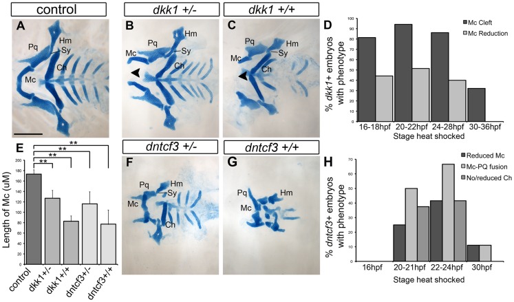
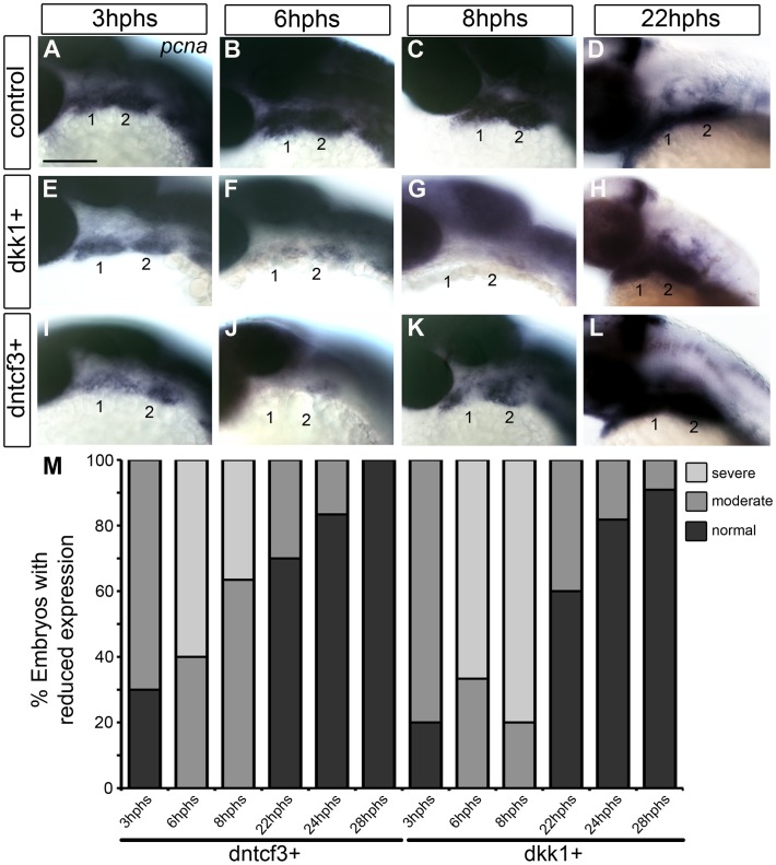
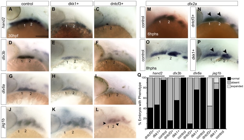
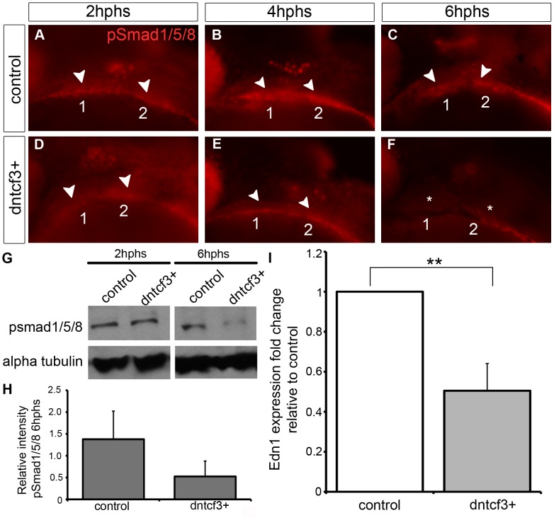
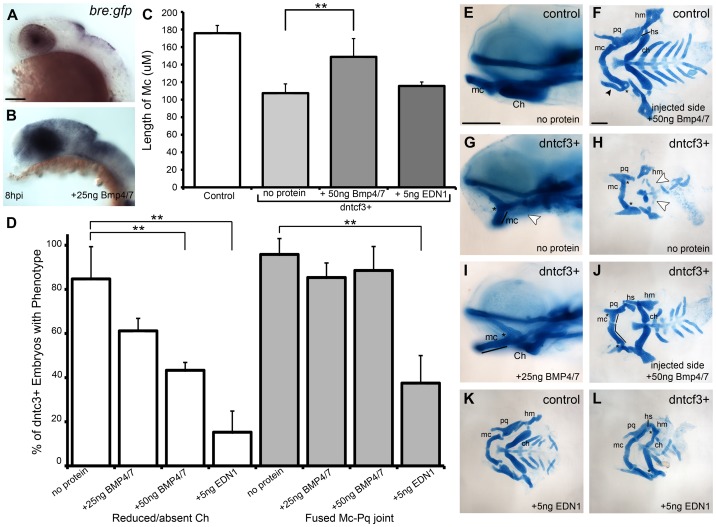
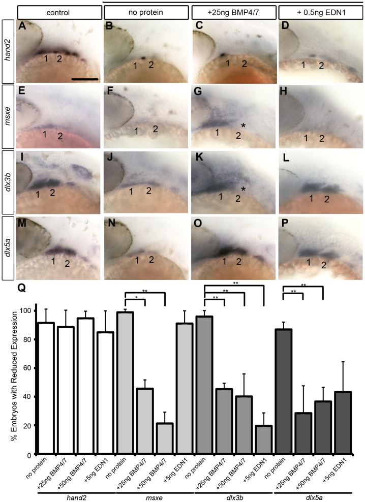
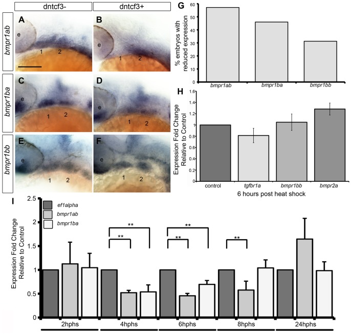
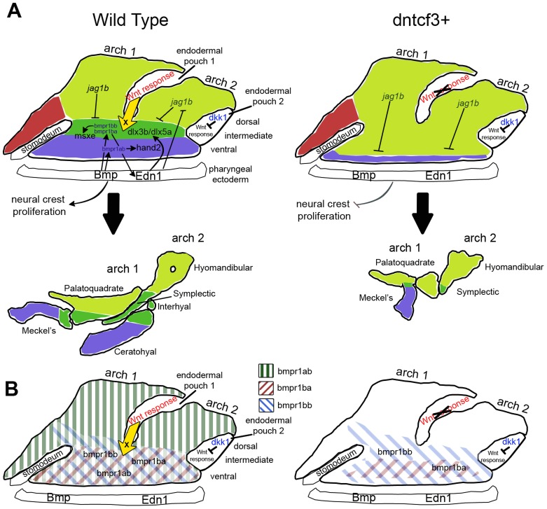
Craniofacial development requires signals from epithelia to pattern skeletogenic neural crest (NC) cells, such as the subdivision of each pharyngeal arch into distinct dorsal (D) and ventral (V) elements. Wnt signaling has been implicated in many aspects of NC and craniofacial development, but its roles in D-V arch patterning remain unclear. To address this we blocked Wnt signaling in zebrafish embryos in a temporally-controlled manner, using transgenics to overexpress a dominant negative Tcf3, (dntcf3), (Tg(hsp70I:tcf3-GFP), or the canonical Wnt inhibitor dickkopf1 (dkk1), (Tg(hsp70i:dkk1-GFP) after NC migration. In dntcf3 transgenics, NC cells in the ventral arches of heat-shocked embryos show reduced proliferation, expression of ventral patterning genes (hand2, dlx3b, dlx5a, msxe), and ventral cartilage differentiation (e.g. lower jaws). These D-V patterning defects resemble the phenotypes of zebrafish embryos lacking Bmp or Edn1 signaling, and overexpression of dntcf3 dramatically reduces expression of a subset of Bmp receptors in the arches. Addition of ectopic BMP (or EDN1) protein partially rescues ventral development and expression of dlx3b, dlx5a, and msxe in Wnt signaling-deficient embryos, but surprisingly does not rescue hand2 expression. Thus Wnt signaling provides ventralizing patterning cues to arch NC cells, in part through regulation of Bmp and Edn1 signaling, but independently regulates hand2. Similarly, heat-shocked dkk1+ embryos exhibit ventral arch reductions, but also have mandibular clefts at the ventral midline not seen in dntcf3+ embryos. Dkk1 is expressed in pharyngeal endoderm, and cell transplantation experiments reveal that dntcf3 must be overexpressed in pharyngeal endoderm to disrupt D-V arch patterning, suggesting that distinct endodermal roles for Wnts and Wnt antagonists pattern the developing skeleton.


Conflict of interest statement

The authors have declared that no competing interests exist.

Figures

Figure 1
Figure 1. Wnt signaling in the pharyngeal arches.
(A–H) In situ hybridization (ISH) and (I) quantitative, real-time PCR (qPCR) analysis of Wnt target gene expression; (A, C, E–H) lateral views, anterior to the left; (B, D) transverse sections through arch 2. (A, B) mycn mRNA is ventrally restricted (black arrowheads) in wild type (WT) embryos. Arches 1 and 2 are outlined by dotted lines. (C, D) GFP mRNA is ventrally restricted (arrowheads) in Tg(7xTCF-Xla.Siam:GFP)ia4 transgenics. (B, D) Both mycn (B) and 7xTCF;GFP (D) are expressed in ventral nc cells and pharyngeal endoderm, and excluded from pharyngeal ectoderm. (E–H) mycn and axin2 expression in controls (E, G), and dntcf3+ embryos at 26 hpf (F, H; heat shocked at 22 hpf). (I) qPCR analysis of axin2, lef1 and mycn expression in dntcf3+ embryos, normalized to nontransgenic, heat-shocked controls, with ef1alpha as an internal control. * P<0.05, ** P<0.001. Abbreviations: e, eye; D, dorsal arch; end, pharyngeal endoderm; hb, hindbrain; I, intermediate arch; mhb, mid-hindbrain boundary; nc, neural crest; pe, pharyngeal ectoderm; V, ventral arch. Scale bars: 100 µm.
Figure 2
Figure 2. Requirements for Wnt signaling in craniofacial cartilage development.
(A–C, F, G) Alcian blue stained cartilage at 96 hpf, dissected and flat mounted, ventral views, anterior to the left. (A) Wild-type control, (B) dkk1+/− heterozygote, (C) dkk1+/+ homozygote, (F) dntcf3+/− heterozygote, (G) dntcf3+/+ homozygote, heat shocked as described (see Results). Arrowheads indicate clefting of Meckel's cartilage (Mc). (D) Histogram quantifying the frequency of Mc defects in dkk1+ embryos heat shocked at different stages from 16–36 hpf. (E) Histogram quantifying average Mc length, p<0.01. (H) Histogram quantifying the frequency of cartilage defects in arches 1 and 2 in dntcf3+ embryos heat shocked at different stages from 16–30 hpf. Abbreviations: Ch, ceratohyal; Hm, hyomandibular; Mc, Meckels; Pq, palatoquadrate; Sy, symplectic. Scale bar: 100 µm.
Figure 3
Figure 3. Requirements for Wnt signaling in pharyngeal arch cell proliferation.
(A–L) Whole mount ISH for pcna in embryos fixed at 3–22 hours post-heat shock (hphs), lateral views, anterior to the left (all heat shocked at 22 hpf). (A–D) pcna is expressed throughout arches, brain and eyes. (E–H) dkk1+ embryos have reduced pcna expression at 3 hphs (E), severe reductions at 6 hphs (F) and virtually no arch expression at 8 hphs (G), before expression returns at 22 hphs (H). (I–L) dntcf3+ embryos show reduced pcna expression at 3 hphs (I), and virtually no expression at 6 hphs (J, K) before expression rebounds between 8–22 hphs (L). (M) Histogram quantifying percentages of dkk1+ and dntcf3+ embryos with moderate versus severe reductions in pcna expression. Scale bar: 100 µm.
Figure 4
Figure 4. Requirements for Wnt signaling in dorsal-ventral arch patterning.
(A–P) Whole mount ISH for genes involved in dorsal-ventral (D-V) patterning in control (A, D, G, J), dkk1+ embryos heat shocked at 20–22 hpf (B, E, H, K), and dntcf3+ embryos heat shocked at 22–24 hpf (C, F, I, L), lateral views, anterior to the left. hand2 (B, C), dlx3b (E, F), and dlx6a (H, I) expression is reduced, while jag1b (K, L) expression expands ventrally (arrowheads) particularly in dntcf3+ embryos, compared with controls. (M–P) dlx2a expression in control (M,O), dntcf3+ (N), and dkk1+ (P) embryos. The dorsal boundary of dlx2a expression (arrowheads) appears to shift ventrally in some cases, (N,P) compared to the control (M,O). (Q) Histogram quantifying percentages of dkk1+ and dntcf3+ embryos with defects in D-V patterning. Scale bar: 100 µm.
Figure 5
Figure 5. Wnt signaling regulates Bmp and Edn1 signaling in the arches.
(A–F) Anti-pSmad1/5/8 staining in control (A–C) and dntcf3+ (D–F) embryos heat shocked at 22–24 hpf, lateral views, anterior to the left. pSmad1/5/8 expression is localized ventrally in the arches (arrowheads), reduced at 2 hphs (D), severely reduced at 4 hphs (E), and absent (asterisk) by 6 hphs (F). (G) Western blot analysis of anti-pSmad1/5/8 in control and dntcf3+ embryos. Protein was extracted at 2 hphs or 6 hphs and alpha tubulin was used as a loading control. (H) Histogram quantifying pSmad1/5/8 expression at 6 hphs, based on Western blots, in control and dntcf3+ embryos, levels normalized to alpha tubulin. (I) Histogram quantifying Edn1 expression by qPCR, in control and dntcf3+ embryos, p<0.01.
Figure 6
Figure 6. BMP protein rescues craniofacial phenotypes in dntcf+ embryos.
(A,B) Whole mount ISH for gfp in Tg(Bre:gfp) embryos, lateral views, anterior to the left. (A) Wildtype and (B) following injection with 25 ng/embryo BMP4/7 into the arch region at 20 hpf. (C) Histogram quantifying Mc length (µM) in controls, dntcf3+ embryos, and dntcf3+ embryos injected with either 50 ng of BMP4/7 or 5 ng EDN1. (D) Histograms quantifying numbers of dntcf3+ embryos, alone or injected with BMP4/7 or EDN1 proteins, with reductions in Ch (left) and joint fusions between Mc and Pq (right). Numbers of embryos with rescue of Ch or Mc-Pq were averaged from three independent experiments (minimum of 10 embryos per protein injection experiment) and samples compared using a paired student t-test. (E, G, I) Whole mounted, alcian-stained 4 dpf larvae; control (E), dntcf3+ (G), and dntcf3+ injected with 25 ug BMP4/7 (I), lateral views, anterior to the left. Arrowhead in G indicates Ch loss. Black lines in G and I indicate Mc length and asterisks indicate Mc-Pq joint fusion. (F, H, J–L) Dissected, flat-mounted alcian-stained cartilages at 4 dpf, ventral views, anterior to the left; control with unilateral injection of 50 ng BMP4/7 (F), dntcf3+ (H), dntcf3+ with 25 ng BMP4/7 (J), control with 5 ng EDN1 (K) and dntcf3+ with 5 ng EDN1 (L). Arrowhead in F indicates duplicate Mc. Black lines in J indicate Mc length. Arrowheads in H indicate Sy and Ch. Asterisks in H, J and L indicate Mc-Pq joint fusions. **P<0.001. Abbreviations: Ch, ceratohyal; mc, Meckel's; mc', duplicated Meckel's; Sy, symplectic. Scale bar: 100 µm.
Figure 7
Figure 7. Bmp and Edn1 restore ventral-intermediate gene expression, but not hand2, in dntcf3+ embryos.
(A–P) Whole mount ISH for ventral-intermediate patterning genes in heat shocked controls (A, E, I, M), dntcf3+ embryos (B, F, J, N), and dntcf3+ embryos with either 25 ng BMP4/7 (C, G, K, O), or 5 ng EDN1 (D, H, L, P), lateral views, anterior to the left. Asterisks in G and K indicate cases in which rescue occurred in arch 1 but not arch 2. (Q) Histogram quantifying the number of embryos with reductions in arch expression for each gene. Quantification obtained by counting number of embryos with rescue of gene expression in three independent experiments (minimum of 10 embryos per protein injection experiment). Statistical method used is a paired student t-test. * P<0.05, ** P<0.001. Scale bar: 100 µm.
Figure 8
Figure 8. Bmp receptor expression in dntcf3+ embryos.
(A–F) Whole mount ISH for bmpr1ab (A, B), bmpr1ba (C, D), and bmpr1bb (E, F) expression, lateral views, anterior to the left. (G) Histogram quantifying the number of dntcf3+ embryos with reduced expression. (H,I) qPCR analysis of Bmp receptor expression levels in dntcf3+ embryos at different times post heat shock, normalized to nontransgenic, heat-shocked controls, with ef1alpha as an internal control. * P<0.05, ** P<0.001. Abbreviations: e, eye; nc, neural crest. Scale bar: 100 µm.
Figure 9
Figure 9. Model for the role of Wnt in dorsal-ventral arch patterning.
(A) Diagrams illustrating regulation of expression of dorsal-ventral (D-V) arch patterning genes in the skeletogenic neural crest at 24 hpf (colored regions) by Bmp and Edn1 signals from the pharyngeal ectoderm and Wnt responses in the pharyngeal endoderm. Each arch is subdivided into ventral (blue), intermediate (dark green), and dorsal (light green) domains, which correspond to eventual dorsal-intermediate-ventral cartilage identities. Arrows indicate inductive influences by each signal on target genes. The yellow arrow indicates an unknown ventralizing signal X from the endoderm induced in response to Wnt. (B) Diagrams illustrating regulation of Bmp receptor expression along the D-V axis at 24 hpf (colored diagonal bars) by Wnt, Bmp and Edn1.

References

    1. Schilling TF, Knight RD (2001) Origins of anteroposterior patterning and Hox gene regulation during chordate evolution. Philos Trans R Soc Lond B Biol Sci 356: 1599–1613. - PMC - PubMed
    1. Hunt P, Gulisano M, Cook M, Sham MH, Faiella A, et al. (1991) A distinct Hox code for the branchial region of the vertebrate head. Nature 353: 861–864. - PubMed
    1. Olsson L, Ericsson R, Cerny R (2005) Vertebrate head development: segmentation, novelties, and homology. Theory Biosci 124: 145–163. - PubMed
    1. Tucker AS, Yamada G, Grigoriou M, Pachnis V, Sharpe PT (1999) Fgf-8 determines rostral-caudal polarity in the first branchial arch. Development 126: 51–61. - PubMed
    1. Trumpp A, Depew MJ, Rubenstein JL, Bishop JM, Martin GR (1999) Cre-mediated gene inactivation demonstrates that FGF8 is required for cell survival and patterning of the first branchial arch. Genes Dev 13: 3136–3148. - PMC - PubMed

Publication types

MeSH terms