. 2014 Jul 24;10(7):e1004229.
doi: 10.1371/journal.ppat.1004229. eCollection 2014 Jul.

The frustrated host response to Legionella pneumophila is bypassed by MyD88-dependent translation of pro-inflammatory cytokines

Affiliations

The frustrated host response to Legionella pneumophila is bypassed by MyD88-dependent translation of pro-inflammatory cytokines

Seblewongel Asrat et al. PLoS Pathog. .

Abstract

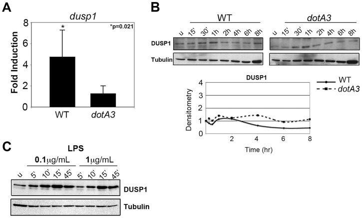
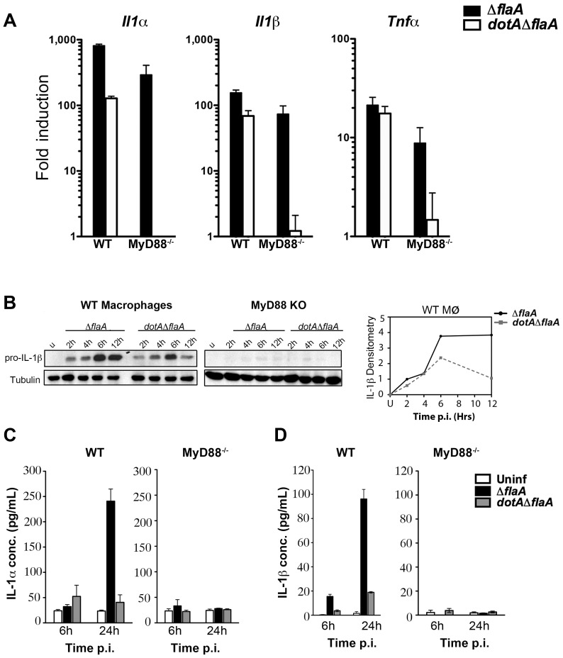
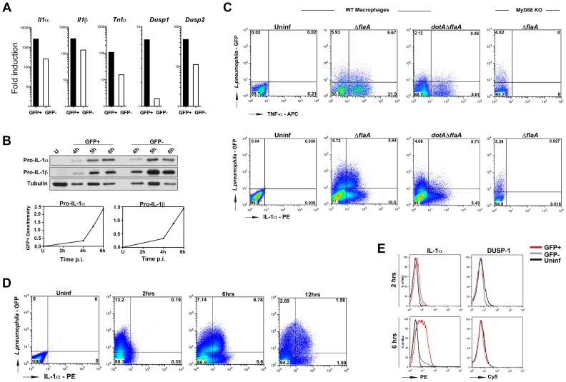
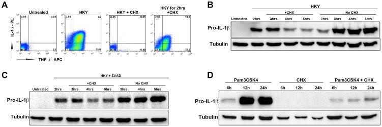
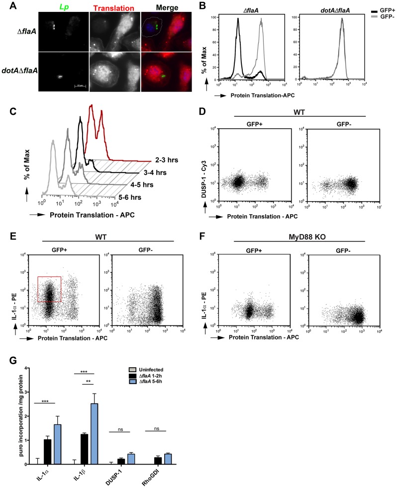
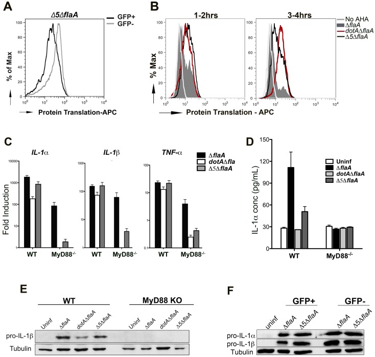
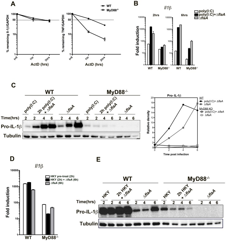
Many pathogens, particularly those that require their host for survival, have devised mechanisms to subvert the host immune response in order to survive and replicate intracellularly. Legionella pneumophila, the causative agent of Legionnaires' disease, promotes intracellular growth by translocating proteins into its host cytosol through its type IV protein secretion machinery. At least 5 of the bacterial translocated effectors interfere with the function of host cell elongation factors, blocking translation and causing the induction of a unique host cell transcriptional profile. In addition, L. pneumophila also interferes with translation initiation, by preventing cap-dependent translation in host cells. We demonstrate here that protein translation inhibition by L. pneumophila leads to a frustrated host MAP kinase response, where genes involved in the pathway are transcribed but fail to be translated due to the bacterium-induced protein synthesis inhibition. Surprisingly, few pro-inflammatory cytokines, such as IL-1α and IL-1β, bypass this inhibition and get synthesized in the presence of Legionella effectors. We show that the selective synthesis of these genes requires MyD88 signaling and takes place in both infected cells that harbor bacteria and neighboring bystander cells. Our findings offer a perspective of how host cells are able to cope with pathogen-encoded activities that disrupt normal cellular process and initiate a successful inflammatory response.


Conflict of interest statement

The authors have declared that no competing interests exist.

Figures

Figure 1
Figure 1. L. pneumophila translation inhibitors induce a frustrated MAPK response.
(A) Dusp1 transcript levels in A/J macrophages infected with wild type or dotA3 L. pneumophila for 4 hrs. Transcript levels were normalized to18S ribosomal RNA (18S) and graphed as a fold increase over uninfected controls. (B) Immunoblot analysis of DUSP-1 protein levels in A/J macrophages challenged with wild type or dotA3 strains and (C) treated with LPS (0.1 µg/mL or 1 µg/mL) for indicated time points. Data are representative of at least three independent experiments.
Figure 2
Figure 2. A subset of transcripts can bypass translation inhibition exerted by L. pneumophila effectors.
(A) C57BL/6 wild type and MyD88−/− macrophages were infected with indicated L. pneumophila strains at MOI-15. Cytokine and DUSP transcripts were analyzed at 6 hrs post infection by qRT-PCR. Results shown are pooled from at least four independent experiments and represent the mean fold induction and SEM of samples relative to uninfected controls. (B) Immunoblot analysis of IL-1β precursor in WT and MyD88−/− macrophages infected with virulent L. pneumophila (ΔflaA) or avirulent mutant dotAΔflaA for indicated time points. Graphs on the right show densitometry of IL-1β normalized to tubulin. (C and D) WT and MyD88−/− macrophages were challenged with indicated L. pneumophila strains for 6 and 24 hrs at MOI-15 and cytokine levels were measured in culture supernatants by ELISA. Data represent mean and SEM of samples from 3 independent experiments.
Figure 3
Figure 3. Cytokines are produced from both infected and bystander macrophages.
(A) B6 macrophages were challenged with ΔflaA-GFP at MOI-15 for 4 hrs and sorted by Flow Cytometry. Cytokine and Dusp transcripts were measured in both GFP+ and GFP population by qRT-PCR. (B) B6 macrophages were infected with ΔflaA-GFP at MOI-15 for indicated times and levels of IL-1β were measured by Western blot. Bottom graphs indicate densitometry of IL-1β in GFP+ cells normalized to tubulin. (C) WT and MyD88−/− macrophages were infected with indicated L. pneumophila strains at MOI-10 and intracellular cytokine levels were measured by flow cytometry. Top panels show TNF levels at 14 hrs post infection. To prevent secretion of TNF, cells were treated with Golgiplug (Brefeldin A) for 5 hrs before samples were collected. Bottom panels show intracellular IL-1α levels at 6 hrs post infection. Data shown are representative of at least 4 independent experiments. (D) Time course analysis of intracellular IL-1α levels in ΔflaA-GFP infected macrophages and (E) Comparison of intracellular IL-1α and DUSP1 levels in B6 macrophages infected with ΔflaA-GFP for 2 and 6 hrs. Infected and uninfected (bystander) cells were gated based on GFP signal and protein levels were compared to control macrophages that were left untreated. Red lines indicate GFP+ population; grey lines indicate GFP population and black lines show untreated macrophages.
Figure 4
Figure 4. The elongation inhibitor cycloheximide blocks cytokine translation independent of cell death.
(A) B6 macrophages were either left untreated (1st box), treated with heat killed Yersinia at MOI = 50 to induce cytokine expression (2nd box), treated with HKY MOI = 50 and 10 µg/mL cycloheximide (CHX) (3rd box) or treated with HKY MOI = 50 for 2 hrs followed by addition of 10 µg/mL CHX (4th box). X-axis represents intracellular TNF-α levels and Y-axis represents intracellular IL-1α levels. (B) Macrophages were pre-stimulated with heat killed Yersinia at MOI = 50 for 2 hrs. Cells were then treated with either 1 µg/mL cycloheximide or left untreated (HKY alone). IL-1β protein levels were measured by Western blot at the indicated time points. (C) B6 macrophages were pre-stimulated with heat killed Yersinia at MOI = 50 in the presence of Pan-Caspase inhibitor (Z-VAD-FMK) for 2 hrs and treated with either 0.5 µg/mL cycloheximide or left untreated. IL-1β protein levels were measured by Western blot for the next 3 hrs. (D) Macrophages were treated with 2 µg/mL Pam3CSK4 for 2 hrs in the presence of Z-VAD-FMK. CHX (0.5 µg/mL) was added to cells at 2 hrs post Pam3CSK4 treatment and IL-1β protein levels were measured at 6, 12 or 24 hrs. Data is representative of at least 3 independent experiments.
Figure 5
Figure 5. Translation of IL-1 takes place in the presence of L. pneumophila elongation inhibitors.
(A) B6 macrophages were infected with indicated L. pneumophila strains at MOI = 10 for 2 hrs and a methionine analog, L-azidohomoalanine (AHA, 50 µM) was incorporated into newly synthesized proteins for 4 hrs. Cells were fixed, permeabilized and the incorporated analog was detected by an APC conjugated phosphine and fluorescence microscopy. Nuclei were stained with Hoechst 33342 (blue) (B) Macrophages were infected with indicated L. pneumophila strains at MOI = 10 for 2 hrs and L-azidohomoalanine (AHA, 50 µM) was added to cells for additional 4 hrs. Protein translation was quantified in infected macrophages (GFP+) and uninfected bystanders (GFP) by staining with APC-labeled phosphine and flow cytometry. Experiment was performed three times. (C) B6 macrophages were infected with Dot+ L. pneumophila for 2 hrs and AHA was added for 1 hr intervals. Cells were fixed after each time point and protein translation was quantified by APC-conjugated phosphine and flow cytometry. Translation of DUSP1 (D) or IL-1α (E) was monitored in wild type macrophages infected with ΔflaA for 6 hrs by co-staining AHA with Cy3 conjugated DUSP1 antibody or PE-conjugated IL-1α antibody. (F) Translation of IL-1α in MyD88-deficient macrophages was determined by co-staining AHA with PE-conjugated IL-1α antibody. Dot plots show GFP+ (left column) and GFP (right column) gated cells. (G) Kinetics of IL-1α, IL-1β, DUSP-1 and RhoGDI translation was quantified in Dot+ infected macrophages using puromycin incorporation. B6 macrophages were infected with Dot+ L. pneumophila and 10 µg/mL of puromycin was added either between 1–2 hrs or between 5–6 hrs post infection. Cells were washed, lysed and incubated on plates coated with anti- IL-1α, IL-1β, DUSP-1 and RhoGDI antibodies. Incorporation of puromycin in the indicated samples was monitored by anti-puromycin antibody and HRP-conjugated secondary antibody. Data represent absorbance at 450 normalized to total protein levels. Values for uninfected controls were subtracted from each sample to determine the increase in puromycin incorporation upon infection. Each bar represents mean and SEM of triplicate samples.
Figure 6
Figure 6. Production of the pro-inflammatory cytokines IL-1α and IL-1β is independent of the five translocated protein synthesis inhibitors
(A) B6 macrophages were infected with Δ5Δfla-GFP+ and protein translation was measured between 2–6 hrs post infection by incorporation of the methionine analog AHA. Translation (incorporation of AHA) was compared between infected cells (GFP+, black line) and uninfected bystanders (GFP−, grey line). (B) B6 macrophages were infected with GFP expressing Dot+, Dot− and Δ5 strains of L. pneumophila and protein synthesis inhibition was compared between these strains by incorporation of AHA between 1–2 hrs (left graph) or 3–4 hrs (right graph). Graphs show translation in infected cells (GFP+). Cells incubated in the absence of the methionine analog (No AHA) were used as a negative control to show baseline staining. (C) WT and MyD88−/− macrophages were infected with the indicated strains at MOI-15 and cytokine transcripts were analyzed at 6 hrs post infection by qRT-PCR. Data represent the mean fold induction and SEM of samples relative to uninfected controls. (D) ELISA measurement of IL-1α secretion from WT and MyD88−/− macrophages infected with the indicated strains for 24 hrs. Data represent mean and SEM of triplicate samples. (E) Immunoblot analysis of pro-IL-1β in WT and MyD88−/− macrophages challenged with the indicated L. pneumophila strains for 6 hrs. (F) B6 macrophages were challenged with ΔflaA-GFP or Δ5Δfla at MOI-15 for 6 hrs and sorted by Flow Cytometry. Pro-IL-1β levels were measured in both GFP+ and GFP− population by westernblot.
Figure 7
Figure 7. Stability of Il-1β mRNA or pre-activation of the NF-kB pathway is not sufficient to induce translation bypass in MyD88−/− macrophages.
(A)WT and MyD88 deficient bone-marrow macrophages were challenged with ΔflaA for 2.5 hrs and transcription was blocked by addition of 10 µg/mL Actinomycin D. The percentage of remaining Il1β and Tnfα transcripts was measured by qRT-PCR after 1 or 2 hrs post actinomycin D treatment. Data represent mean and SEM of samples relative to GAPDH. (B) WT and MyD88−/− macrophages were pre-treated with 50 µg/mL poly(I∶C) for 2 hrs to induce NF-kB activation, after which cells were either challenged with Dot+ Legionella or left untreated (poly(I∶C) only). Macrophages that were not pre-stimulated with poly(I∶C) but were challenged with Dot+ were included for comparison. RNA was collected after 2 and 6 hrs of infection and Il1β transcript levels were analyzed by qRT-PCR. (C) B6 WT and MyD88-deficient macrophages were pre-treated with poly(I∶C) for 2 hrs or left untreated. Cells were then challenged with Dot+ Legionella for additional 2, 4 and 6 hrs and production of pro-IL-1β was examined by Western blot. Densitometry of the blot is shown on the right. (D) WT and MyD88−/− macrophages were pre-treated with HKY (MOI = 100) for 2 hrs or left untreated after which cells were challenged with Dot+ L. pneumophila. Induction of Il1β transcript was determined after 6 hrs of infection. (E) Pro-IL-1β synthesis was also examined in these cells after 2, 4 and 6 hrs of challenge.

References

    1. Akira S, Hemmi H (2003) Recognition of pathogen-associated molecular patterns by TLR family. Immunol Lett 85: 85–95. - PubMed
    1. Medzhitov R, Janeway C Jr (2000) Innate immunity. N Engl J Med 343: 338–344. - PubMed
    1. Jones JD, Dangl JL (2006) The plant immune system. Nature 444: 323–329. - PubMed
    1. Dangl JL, Jones JD (2001) Plant pathogens and integrated defence responses to infection. Nature 411: 826–833. - PubMed
    1. Ausubel FM (2005) Are innate immune signaling pathways in plants and animals conserved? Nat Immunol 6: 973–979. - PubMed

Publication types

MeSH terms

LinkOut - more resources