. 2014 Jul 24;10(7):e1004237.
doi: 10.1371/journal.ppat.1004237. eCollection 2014 Jul.

Lytic gene expression is frequent in HSV-1 latent infection and correlates with the engagement of a cell-intrinsic transcriptional response

Affiliations

Lytic gene expression is frequent in HSV-1 latent infection and correlates with the engagement of a cell-intrinsic transcriptional response

Joel Z Ma et al. PLoS Pathog. .

Erratum in

  • PLoS Pathog. 2014 Aug;10(8):e1004361

Abstract

Herpes simplex viruses (HSV) are significant human pathogens that provide one of the best-described examples of viral latency and reactivation. HSV latency occurs in sensory neurons, being characterized by the absence of virus replication and only fragmentary evidence of protein production. In mouse models, HSV latency is especially stable but the detection of some lytic gene transcription and the ongoing presence of activated immune cells in latent ganglia have been used to suggest that this state is not entirely quiescent. Alternatively, these findings can be interpreted as signs of a low, but constant level of abortive reactivation punctuating otherwise silent latency. Using single cell analysis of transcription in mouse dorsal root ganglia, we reveal that HSV-1 latency is highly dynamic in the majority of neurons. Specifically, transcription from areas of the HSV genome associated with at least one viral lytic gene occurs in nearly two thirds of latently-infected neurons and more than half of these have RNA from more than one lytic gene locus. Further, bioinformatics analyses of host transcription showed that progressive appearance of these lytic transcripts correlated with alterations in expression of cellular genes. These data show for the first time that transcription consistent with lytic gene expression is a frequent event, taking place in the majority of HSV latently-infected neurons. Furthermore, this transcription is of biological significance in that it influences host gene expression. We suggest that the maintenance of HSV latency involves an active host response to frequent viral activity.


Conflict of interest statement

The authors have declared that no competing interests exist.

Figures
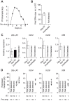
Figure 1
Figure 1. Infected DRG fulfill classical definitions of HSV latency.
(A) Plaque forming units assay of HSV-infected DRG (T5 to L1) at various time points post inoculation. (B) Copy number of viral DNA from HSV-infected DRG (day 50) was determined by quantitative PCR. (C) Relative expression of HSV 2 kb LAT and lytic genes in HSV-infected DRG (days 5 and 50) were determined by quantitative PCR. (D) Expression (Et) of HSV 2 kb LAT and lytic genes in HSV-infected DRG (> day 240) were determined by quantitative PCR. Total RNA from each sample was aliquoted into four tubes and RT and pre-amplification were done as indicated beneath each graph. Pre-amplified products were used without dilution. Data in (AC) are pooled from two independent experiments with 4–5 mice per group, (D) from one experiment with 5 mice, and plotted as mean ± S.E.M (A–C) or showing each individual result (D).
Figure 2
Figure 2. The number of infected cells remains stable during latency.
(A) Number of infected (β-galactosidase+) cells from KOS/pCMV/eGC infected DRG at the times shown post inoculation. (B) DRG explants from ROSA-YFP mice (day 20) infected with KOS0152 or WT HSV, or uninfected mice were examined under the 2 photon intra-vital microscope. Arrows identify HSV-infected cells. (C) DRG sections from KOS0152- or WT HSV-infected ROSA-YFP mice (day 50) were examined under the laser capture microscope for YFP expression and Hoechst nuclear stain. Data in (A) are pooled from two independent experiments and represented as mean ± S.E.M. Images in (B and C) are from two independent experiments.
Figure 3
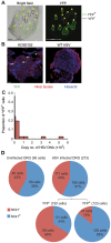
Figure 3. Latent HSV resides in a subpopulation of sensory neurons.
(A) YFP+ and YFP cells were identified and captured from DRG sections of infected ROSA-YFP mice. (B) DRG sections from KOS0152- or WT HSV-infected ROSA-YFP mice (day 20) were stained for Nissl bodies and examined under the confocal microscope. Arrows identify HSV-infected neurons. (C) Copy number of viral DNA in individual YFP+ cells (day 15) captured by LCM is shown in a histogram. (D) Number and percent of Ntrk1 + and Ntrk1 neurons from populations of infected (YFP+) and uninfected (YFP and uninfected DRG) neurons in the main set of single cell expression data are shown in pie charts. Data in (A and C) are pooled from two independent experiments. Images in (B) are from one experiment. (D) As for all single cell gene expression data, the neuron sections analysed were combined from 4 independent infections (YFP+ and YFP neurons) or 3 independent experiments (neurons from uninfected DRG). Numbers in brackets show the number of individual cells analyzed.
Figure 4
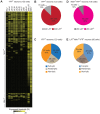
Figure 4. HSV gene expression in individual neurons during latency.
(A) Heatmap showing HSV genes determined using quantitative RT-PCR in single YFP+ neurons. (B) Number and percent of all infected YFP+ neurons containing 2 kb LAT. (C) Number and percent of all infected YFP+ neurons with different HSV lytic gene expression profiles. (D and E) As per B and C, but restricted to Ntrk1 +YFP+ neurons. Numbers in brackets show the number of individual cells analyzed. All data are from the main single cell gene expression dataset.
Figure 5
Figure 5. Single cell gene expression profiling reveals the transcriptional response of infected neurons towards latent HSV.
(A) Principal components (PC) analysis of 48 cellular genes in single Ntrk1 +neurons – uninfected, YFP and YFP+. (B) PC analysis on Ntrk1 +YFP+ neurons – YFP versus uninfected. All data are from the main single cell gene expression dataset.
Figure 6
Figure 6. Increasing viral activity is matched by progressive host neuronal transcriptional response.
(A) Violin plot representation of selected cellular gene expressions in LAT+ Ntrk1 +YFP+ neurons categorized based on their lytic gene expression profile. Log2Ex represents expression threshold (Et). Numbers in brackets show the number of individual cells analyzed. * both proportion (p<0.05) and expression levels (p<0.0167) are significant, ‡ only expression levels significant, # proportion and expression levels are not significant when comparing full-lytic to partial- and non-lytic subsets. (B) k-means clustering of Kendall tau rank correlation coefficients (τ) of every pair of 48 genes from single LAT+ Ntrk1 +YFP+ neurons. Correlation coefficient matrix of non-lytic neurons was clustered according to the optimal clustering observed in full-lytic neurons. (C) Complete-linkage clustering of Kendall tau rank correlation coefficients (τ) between expression profiles of every pair of 48 genes from single LAT+ Ntrk1 +YFP+ neurons. Correlation coefficient matrices of non-lytic and full-lytic neurons were independently clustered. All data are from the main single cell gene expression dataset.

References

    1. Jurak I, Silverstein LB, Sharma M, Coen DM (2012) Herpes Simplex Virus Is Equipped with RNA- and Protein-Based Mechanisms To Repress Expression of ATRX, an Effector of Intrinsic Immunity. J Virol 86: 10093–10102. - PMC - PubMed
    1. Wald A, Corey L, Cone R, Hobson A, Davis G, et al. (1997) Frequent genital herpes simplex virus 2 shedding in immunocompetent women. Effect of acyclovir treatment. J Clin Invest 99: 1092–1097. - PMC - PubMed
    1. Gebhardt BM, Halford WP (2005) Evidence that spontaneous reactivation of herpes virus does not occur in mice. Virol J 2: 67. - PMC - PubMed
    1. Mester JC, Rouse BT (1991) The mouse model and understanding immunity to herpes simplex virus. Rev Infect Dis 13 Suppl 11: S935–945. - PubMed
    1. Deatly AM, Spivack JG, Lavi E, Fraser NW (1987) RNA from an immediate early region of the type 1 herpes simplex virus genome is present in the trigeminal ganglia of latently infected mice. Proc Natl Acad Sci U S A 84: 3204–3208. - PMC - PubMed

Publication types