

Ocular pentastomiasis in the Democratic Republic of the Congo
- PMID: 25058608
- PMCID: PMC4109912
- DOI: 10.1371/journal.pntd.0003041
Ocular pentastomiasis in the Democratic Republic of the Congo
Abstract
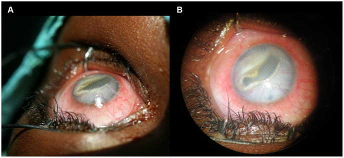
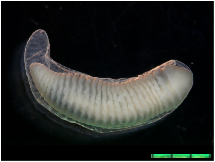
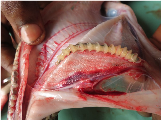
Ocular pentastomiasis is a rare infection caused by the larval stage of pentastomids, an unusual group of crustacean-related parasites. Zoonotic pentastomids have a distinct geographical distribution and utilize reptiles or canids as final hosts. Recently, an increasing number of human abdominal infections have been reported in Africa, where pentastomiasis is an emerging, though severely neglected, tropical disease. Here we describe four ocular infections caused by pentastomids from the Democratic Republic of the Congo. Two cases underwent surgery and an Armillifer grandis infection was detected by morphological and molecular approaches. Thus far, 15 other cases of ocular pentastomiasis have been reported worldwide. Twelve cases were caused by Armillifer sp., recorded almost exclusively in Africa, where such infections occur as a consequence of hunting and consuming snakes, their final hosts. Seven further cases were caused by Linguatula serrata, a cosmopolitan pentastomid whose final hosts are usually canids. Intraocular infections caused permanent visual damage in 69% and a total loss of vision in 31% of reported cases. In contrast, ocular adnexal cases had a benign clinical course. Further research is required to estimate the burden, therapeutic options and pathogenesis of this neglected disease.
Conflict of interest statement
The authors have declared that no competing interests exist.
Figures
References
-
- Pantchev N, Tappe D (2011) Pentastomiasis and other parasitic zoonoses from reptiles and amphibians. Berl Munch Tierarztl Wochenschr 124: 528–535. - PubMed
-
- Tappe D, Dijkmans AC, Brienen EA, Dijkmans BA, Ruhe IM, et al. (2014) Imported Armillifer pentastomiasis: Report of a symptomatic infection in The Netherlands and mini-review. Travel Med Infect Dis 12: 129–133.4. - PubMed
-
- Adeyekun AA, Ukadike I, Adetiloye VA (2011) Severe pentasomide Armillifer armillatus infestation complicated by hepatic encephalopathy. Ann Afr Med 10: 59–62. - PubMed
-
- Lavarde V, Fornes P (1999) Lethal infection due to Armillifer armillatus (Porocephalida): A snake-related parasitic disease. Clin Infect Dis 29: 1346–1347. - PubMed
Publication types
MeSH terms
LinkOut - more resources
Full Text Sources
Other Literature Sources
Medical
Molecular Biology Databases
